.webp&w=384&q=75)

Loading, please wait...
Loading, please wait...

Cellular survival depends on organelle integrity, and mitochondrial protein localization plays a central role in maintaining energy balance. Mitochondria possess an intricate double-membrane architecture that segregates vital metabolic reactions. When exogenous toxicants disrupt this internal architecture, severe metabolic collapse ensues. Recent advances in molecular biotechnology introduce biosensors that monitor these disruptions with nanometer precision.
Mitochondria carry out essential biological processes, including ATP generation, intracellular calcium buffering, and regulated apoptosis. However, these vital physiological actions depend strictly on the precise compartmentalization of nuclear-encoded proteins. The organelle features four distinct subcompartments, including the outer membrane, the intermembrane space, the inner membrane, and the central matrix. Consequently, each specific compartment hosts unique enzymatic cascades. Outer membrane translocases, such as TOM20 and TOM22, import cytosolic precursor polypeptides safely into the organellar interior. Meanwhile, matrix enzymes like ornithine transcarbamylase and sirtuin 3 govern urea cycle flux and post-translational deacetylation. If imported proteins fail to reach their target subcompartments, mitochondrial bioenergetics collapse rapidly. Furthermore, abnormal structural distribution triggers excessive reactive oxygen species production and promotes cellular senescence. Clinicians encounter mitochondrial breakdown across diverse conditions, ranging from neurodegenerative diseases to acute toxic metabolic injury. Therefore, tracking mitochondrial protein localization inside living cells helps investigators identify molecular failures before macroscopic tissue damage manifests. Understanding these compartmental dynamics provides essential mechanistic insight for clinical toxicology and metabolic medicine.
Historically, researchers evaluated organellar architecture through cell fractionation or conventional confocal microscopy. Unfortunately, these traditional methods frequently disrupt delicate organellar membranes or lack sufficient spatial resolution in real time. To overcome these technological challenges, investigators engineered bioluminescence resonance energy transfer biosensors. This sophisticated molecular platform measures non-radiative energy transfer between a luminescent donor and a fluorescent acceptor in living cells. Specifically, the team generated targeted sensor fusions to observe key resident proteins, including VDAC1, MICU1, and ATP5F1C. Because bioluminescent resonance transfer occurs exclusively across distances below ten nanometers, these reporters track precise protein coordinates accurately. In addition, the assay functions noninvasively in intact cells without requiring mechanical organellar isolation. As a result, pharmacologists can now monitor real-time spatial redistributions during acute chemical stress. This technological breakthrough transforms how laboratory scientists detect subtle organellar toxicity. Moreover, it permits direct quantitative evaluation of transient molecular translocations that older imaging modalities routinely overlook. Consequently, biosensor technology accelerates mechanistic toxicology studies.
Beyond identifying static resident proteins, these biosensors dynamically detect the stress-induced migration of cytosolic factors. During severe metabolic stress, signaling cascades direct mobile cytoplasmic proteins toward the outer mitochondrial membrane. In particular, the pro-apoptotic executioner BAX and the fission regulator dynamin-related protein 1 translocate rapidly during lethal injury. When BAX accumulates at the outer membrane, it oligomerizes to form large permeabilizing pores. Consequently, cytochrome c escapes into the cytosol and triggers downstream executioner caspases. Similarly, Drp1 recruitment accelerates mitochondrial fission, dividing elongated networks into fragmented organelles. The newly engineered biosensors recorded these translocation events with high temporal fidelity. Furthermore, the bioluminescent signal increased predictably as cellular stress intensified. Thus, these biosensors deliver an early warning of impending organellar failure. By recording initial structural shifts, investigators can study how environmental chemical insults trigger programmed cell death cascades before irreversible organ destruction develops. Accordingly, the platform provides invaluable data regarding stress kinetics.
Environmental xenobiotics represent a severe hazard in clinical medicine, particularly across agricultural regions in India. Clinicians frequently encounter severe poisonings caused by the bipyridyl herbicide paraquat, which carries extremely high mortality. When investigators exposed living cells to paraquat, the biosensors revealed significant disturbance of mitochondrial protein localization. Paraquat undergoes continuous redox cycling, generating destructive bursts of superoxide anions. Consequently, acute oxidative stress damages translocase complexes and impairs protein import pathways across both membranes. In addition, the investigators tested di-tert-butyl-4-methylphenol, a common antioxidant food additive known as butylated hydroxytoluene. Surprisingly, this ubiquitous dietary preservative provoked noticeable disorganization of mitochondrial subcompartments. Furthermore, 4-hydroxynonenal, a toxic lipid peroxidation product, severely compromised inner mitochondrial membrane integrity. These observations prove that common synthetic compounds directly harm vital organellar machinery. Therefore, toxic chemical exposures can initiate organelle breakdown even at concentrations previously deemed harmless by conventional toxicity screenings.
These experimental observations carry profound clinical relevance for internists and critical care physicians managing toxic exposures. Paraquat poisoning remains a lethal medical emergency across rural healthcare facilities due to rapid pulmonary and multiorgan failure. Because paraquat damages mitochondrial structures directly, supportive medical therapy often fails to halt disease progression. Recognizing that herbicide exposure causes rapid mislocalization of protective proteins reinforces the necessity for emergent antioxidant administration and hemoperfusion. Furthermore, the demonstrated toxicity of food additives like butylated hydroxytoluene raises concerns regarding chronic dietary consumption. Chronic mitochondrial disruption contributes to insulin resistance, hepatic steatosis, and cardiovascular disorders. Accordingly, clinicians must evaluate environmental mitochondrial toxicants when investigating patients with unexplained cellular damage. In addition, these biosensors establish a robust high-throughput platform for discovering novel cytoprotective antidotes. As molecular toxicology evolves, clinicians may eventually use targeted therapies to preserve mitochondrial protein localization during toxic insults.
BRET biosensors monitor mitochondrial protein localization by measuring non-radiative energy transfer between a luminescent donor enzyme and a fluorescent acceptor protein. When tagged target proteins assemble within ten nanometers of each other inside mitochondrial compartments, this proximity generates a quantifiable optical signal. Consequently, researchers can monitor dynamic protein trafficking and spatial orientation in real time without destroying fragile cellular membranes or organelle structures.
Paraquat is exceptionally lethal because it enters cells rapidly and undergoes relentless redox cycling, which generates massive amounts of superoxide free radicals. These reactive oxygen species induce severe lipid peroxidation and damage structural translocase complexes within mitochondrial membranes. As a direct result, vital proteins fail to localize properly, bioenergetic production halts, and rapid pro-apoptotic cascades trigger irreversible tissue necrosis and multiorgan failure.
Although food manufacturers widely incorporate butylated hydroxytoluene as a synthetic antioxidant preservative, excessive exposure disrupts normal mitochondrial subcompartment organization. Specifically, novel biosensor assays show that the compound impairs protein distribution across inner and outer mitochondrial membranes. This disruption promotes membrane destabilization, impairs metabolic enzyme functions, and increases oxidative stress, highlighting the urgent need for comprehensive regulatory reassessments of ubiquitous food additives.
Disclaimer: This content is for informational and educational purposes only... Refer to the latest local and national guidelines for clinical practice.
References
Abdulrahman AO et al. BRET-Based Mitochondrial Subcompartment Localization Biosensors Reveal the Impact of Pesticides and Food Additives on Mitochondrial Protein Localization. ACS Sens. 2026 Sep 27. doi: 10.1021/acssensors.6c01115. PMID: 42801737.
Ahmed A, Prasad A, Bhattacharjee A. Management of Paraquat Poisoning—The Way Forward. Indian J Crit Care Med 2024;28(8):722–723.
Dinis-Oliveira RJ, Duarte JA, Sánchez-Navarro A, Remião F, Bastos ML, Carvalho F. Paraquat poisonings: mechanisms of lung toxicity, clinical features, and treatment. Crit Rev Toxicol. 2008;38(1):13-71.

Read summarized clinical updates, watch expert medical content, and earn CME certifications right from your smartphone.
Open your smartphone camera to scan and install.





Novel BRET biosensors reveal how common environmental toxicants, including the herbicide paraquat and food additive BHT, disrupt mitochondrial protein localization. This breakthrough provides new molecular insights into cellular stress, organ failure, and metabolic disease mechanisms.
Today
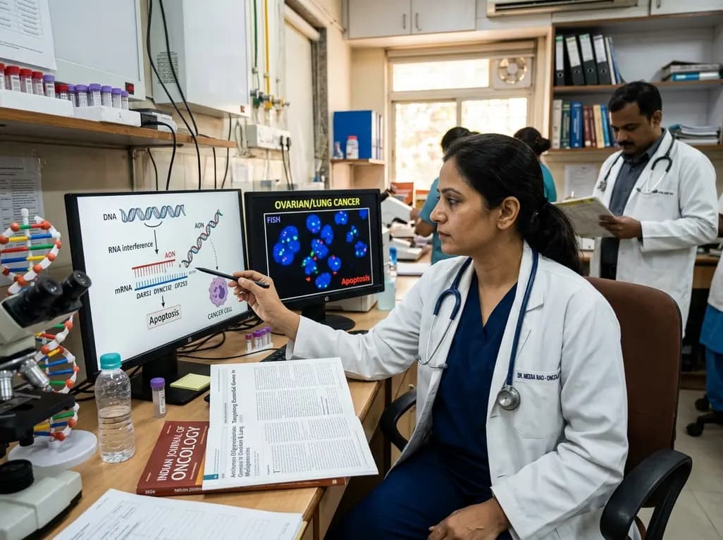
Novel preclinical research demonstrates that antisense oligonucleotides targeting essential genes such as DARS1, DYNC1I2, and EIF2S3 selectively induce apoptosis in ovarian and lung cancer cells. Conditional biomarker activation provides a viable strategy to spare normal tissue while overcoming therapy resistance.
Today
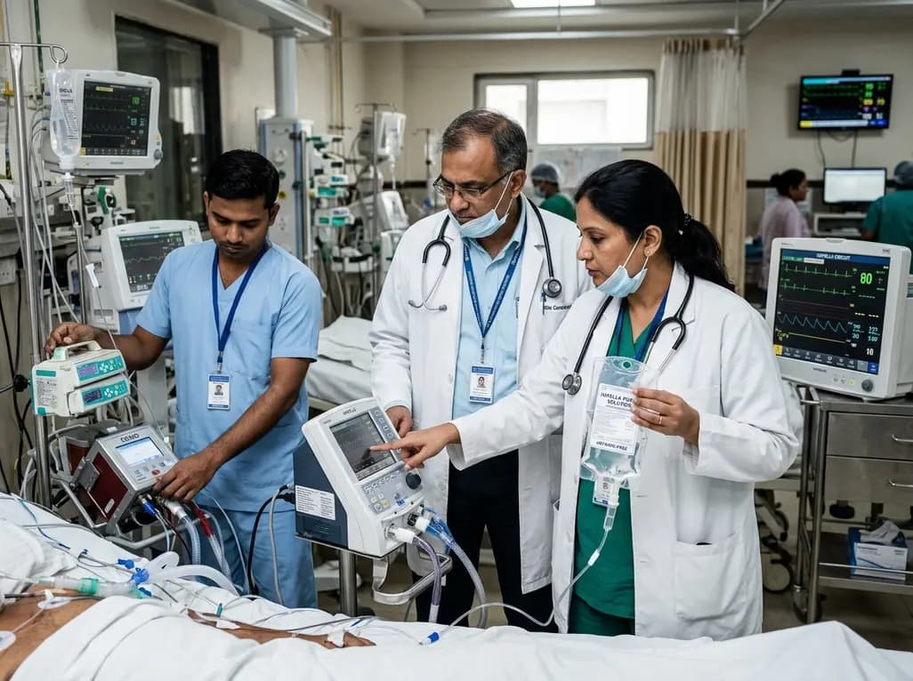
This clinical analysis examines heparin-free versus heparin-containing Impella purge solutions in patients receiving concomitant ECMO support (ECPELLA). Evidence suggests anticoagulant-free purges maintain device patency while reducing bleeding risks, offering a viable strategy alongside systemic anticoagulation.
Today

A 10-year Markov state-transition model shows minimally invasive tubular decompression dominates open laminectomy for pure lumbar spinal stenosis, yielding 5.95 vs 5.91 QALYs and saving $4,167 per patient through reduced complications and lower index costs.
Today

A preclinical study demonstrates that naringin-loaded phytosomes significantly enhance oral bioavailability and provide neuroprotective efficacy in diabetic neuropathy by dampening inflammation, reducing oxidative stress, and relieving neuropathic pain.
Today
