.webp&w=384&q=75)

Loading, please wait...
Loading, please wait...
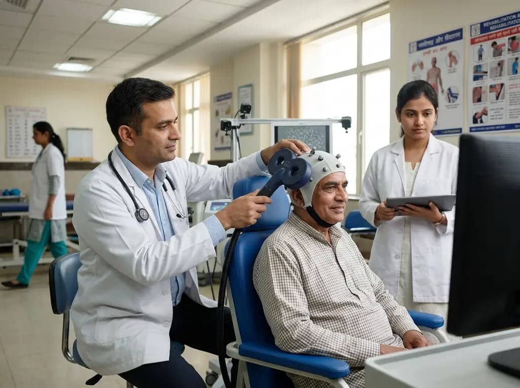
Stroke frequently injures retrochiasmal visual pathways, which leaves many individuals with homonymous hemianopia. Consequently, patients suffer from debilitating visual field loss that impairs daily living, navigation, and reading abilities. While traditional rehabilitation approaches primarily emphasize compensatory strategies, clinicians increasingly investigate methods to achieve genuine post-stroke visual recovery. Recent breakthroughs in neuromodulation offer promising therapeutic pathways to stimulate neuroplasticity. By targeting residual connections within the cortical visual network, non-invasive neuromodulation protocols can potentially restore lost visual processing capabilities.
Homonymous hemianopia occurs after vascular insults to the occipital cortex or optic radiations, causing contralateral field deficits. Importantly, patients lose conscious perception across the affected hemifield, which severely limits their visual independence. However, subcortical and residual cortical connections frequently survive the initial ischemic injury. Specifically, reciprocal pathways connecting the primary visual cortex (V1) and the motion-sensitive middle temporal area (MT or V5) often remain partially intact. These bidirectional circuits play an indispensable role in motion perception, visual awareness, and spatial orientation.
When an occipital stroke occurs, communication between these interconnected visual regions becomes disrupted. Because the timing of neuronal oscillations degrades, signal transmission across surviving visual fibers deteriorates significantly. Standard rehabilitation protocols require extensive, repetitive perceptual training over many months to elicit modest gains. Therefore, researchers seek active neuromodulatory interventions that directly reinforce residual synaptic connections. By strengthening these latent circuits, clinicians can help patients harness surviving pathways and improve visual motion discrimination within previously blind visual fields.
To reactivate dormant visual networks, investigators utilize cortico-cortical paired associative stimulation (ccPAS). This innovative transcranial magnetic stimulation paradigm relies directly on Hebbian principles of spike-timing-dependent plasticity. Specifically, ccPAS delivers paired magnetic pulses to two anatomically linked cortical nodes with millisecond-level precision. When the inter-stimulus interval matches the physiological conduction delay between targets, it selectively modulates synaptic efficacy along the targeted pathway.
In clinical trials, researchers applied bidirectional ccPAS across the ipsilesional V1 and MT visual hubs. They compared feedforward stimulation (V1-to-MT) against feedback stimulation (MT-to-V1) in chronic stroke patients with homonymous hemianopia. Interestingly, the direction of paired stimulation fundamentally determines the functional outcome. The top-down MT-to-V1 protocol selectively induced long-term potentiation-like plasticity within feedback visual projections. Consequently, this targeted associative stimulation enhanced the patient's capacity to process visual motion stimuli presented within the blind hemifield. This physiological mechanism confirms that precise timing is essential to engage surviving visual circuits effectively.
The experimental findings demonstrated that MT-to-V1 paired stimulation significantly improved motion direction discrimination within the blind visual field. In contrast, V1-to-MT stimulation failed to elicit equivalent behavioral recovery in the cohort. Electrophysiological analyses using electroencephalography and Granger causality revealed that effective behavioral improvement was coupled with enhanced top-down information transfer. Specifically, patients who achieved measurable visual recovery displayed a distinct increase in MT-to-V1 directional connectivity.
Furthermore, successful neuromodulation extended beyond the stimulated local pathway. Patients showing robust clinical gains also demonstrated improved functional coupling across wider visual processing networks. This widespread re-orchestration indicates that enhancing feedback signals from secondary visual areas can re-engage suppressed primary visual networks. Therefore, re-establishing top-down signaling represents a crucial mechanism for restoring visual motion awareness after occipital stroke. These discoveries underscore the therapeutic importance of prioritizing feedback visual projections during neurorehabilitation protocols.
Although neuromodulation yields encouraging outcomes, therapeutic responses vary substantially among stroke survivors. Recent evidence highlights critical neuroanatomical and functional biomarkers that predict post-stroke visual recovery following associative stimulation. Specifically, structural magnetic resonance imaging revealed that individuals with preserved structural integrity along the ipsilesional V1-MT tract achieved the greatest visual improvements. Conversely, extensive destruction of these connecting white matter pathways prevented stimulation-induced recovery.
Moreover, baseline functional connectivity and perilesional cortical activity strongly correlate with positive treatment outcomes. Patients who possess viable residual neural substrates exhibit enhanced electrophysiological responsiveness to paired associative stimulation protocols. Thus, structural tractography and baseline electroencephalography serve as reliable screening tools for clinical stratification. By identifying favorable anatomical profiles early, clinicians can accurately select suitable candidates for intensive neuromodulatory therapies. This biomarker-driven stratification ensures that clinical resources focus on patients who possess the structural framework necessary for functional neuroplastic recovery.
Integrating pathway-specific paired associative stimulation into clinical neurorehabilitation offers major advantages for stroke care. Currently, conventional visual therapy relies on saccadic eye-movement exercises or optical aids like prisms. Although these compensatory tools improve environmental navigation, they rarely restore damaged sensory processing pathways. Combining targeted non-invasive brain stimulation with active visual perceptual training presents a powerful, synergistic paradigm.
Clinicians can use neuromodulation to prime surviving cortical networks immediately before patients undergo structured perceptual learning tasks. This multimodal strategy accelerates synaptic remodeling and shortens the lengthy training timelines typically required for visual restoration. Furthermore, identifying intact visual pathways allows therapists to tailor perceptual stimuli to a patient's preserved capacities. Consequently, patients achieve greater visual gains with fewer rehabilitation sessions. In the future, adapting brain stimulation protocols to individual neuroanatomy will transform post-stroke visual management from passive compensation into active neural restoration.
Cortico-cortical paired associative stimulation is a non-invasive neuromodulation technique that delivers synchronized magnetic pulses to two interconnected brain regions. By precisely timing these pulses to match neural conduction delays, the protocol induces Hebbian synaptic plasticity, effectively strengthening or modulating functional communication between targeted cortical networks.
Homonymous hemianopia causes loss of the same half of the visual field in both eyes following retrochiasmal brain injury. This condition impairs reading, spatial orientation, and navigation while increasing fall risks. Consequently, affected individuals often experience reduced functional independence and diminished quality of life during daily activities.
Current brain stimulation protocols transiently enhance specific visual functions, such as motion discrimination, rather than providing complete visual field restoration. However, combining non-invasive stimulation with structured perceptual training can promote sustained neuroplasticity, maximize residual visual pathway function, and significantly improve functional visual performance in selected patients.
Disclaimer: This content is for informational and educational purposes only and is intended solely for healthcare professionals. It should not be used as a substitute for professional clinical judgment, diagnosis, or treatment. Medical knowledge and clinical guidelines evolve continuously; therefore, independent verification of diagnoses, drug dosages, and treatment protocols is strongly recommended. The authors and publishers assume no liability for any injury, loss, or damage resulting from the application of the information presented. Refer to the latest local and national guidelines for clinical practice.
References

Read summarized clinical updates, watch expert medical content, and earn CME certifications right from your smartphone.
Open your smartphone camera to scan and install.





A clinical study demonstrates that cortico-cortical paired associative stimulation (ccPAS) over MT-to-V1 pathways improves visual motion discrimination in stroke survivors with homonymous hemianopia, highlighting biomarkers that predict therapeutic response and visual network recovery.
last month

A meta-analysis of 14 RCTs reveals that dietary fiber significantly increases bowel movements and stool consistency in functional constipation while modulating gut-brain axis biomarkers, including short-chain fatty acids, serotonin, and inflammation, highlighting benefits beyond simple stool bulking.
Today
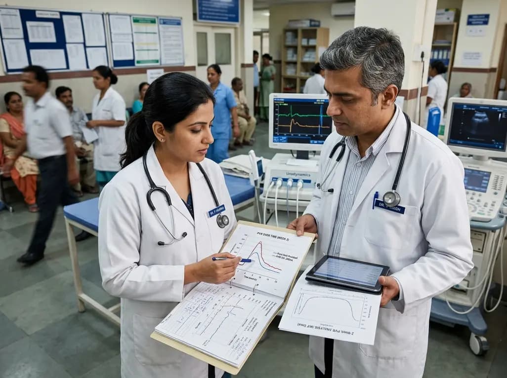
A retrospective study of 824 patients undergoing over 2,200 intradetrusor onabotulinumtoxinA injections identifies sex- and etiology-specific predictors of 2-week post-void residual, highlighting the influence of baseline residual volumes, toxin dosage, and distinct urodynamic parameters on urinary retention risk.
Today
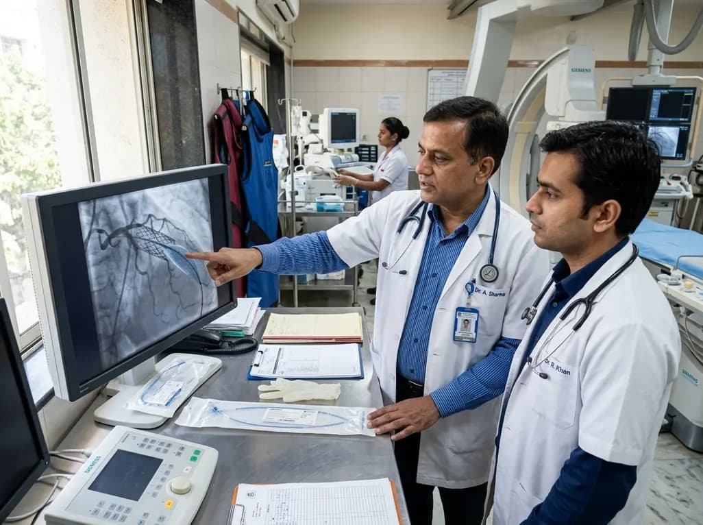
The US FDA has granted approval to the SELUTION SLR drug-eluting balloon for treating coronary in-stent restenosis. This milestone introduces a sustained-release sirolimus platform that eliminates the need for additional permanent metallic stents while maintaining robust vascular healing and luminal patency.
Today

The Thumbs-Out Classification (TOC) is a reliable screening tool for detecting thumb-in-palm deformity in children with cerebral palsy. Utilizing traffic-light tiers for radial abduction, TOC enables clinicians to monitor upper limb function and initiate timely therapeutic or surgical interventions.
Today

A comprehensive review analyzes periconceptional GLP-1 receptor agonist exposure, revealing reassuring data on congenital anomalies and adverse perinatal outcomes while emphasizing the need for cautious patient counseling.
Today
