.webp&w=384&q=75)

Loading, please wait...
Loading, please wait...

With the global population aging rapidly, bone diseases pose a significant socioeconomic burden. Traditionally, X-ray-based techniques served as the primary clinical standard for bone assessment. However, these methods involve ionizing radiation exposure and provide limited soft-tissue contrast. Consequently, AI in bone MRI has emerged as a revolutionary, radiation-free alternative. Modern MRI sequences now allow for a detailed evaluation of tissue properties, microstructure, and pathological changes that were previously difficult to capture.
Historically, the intrinsically low proton density and rapid transverse relaxation of bone tissue made MRI a technical challenge. Engineers and clinicians initially relied on conventional T1-weighted and T2-weighted imaging. Recently, researchers developed advanced sequences such as ultrashort echo time (UTE) and zero echo time (ZTE) to address these limitations. These innovations overcome technical barriers, providing clearer images of cortical bone structures. Furthermore, metabolic imaging like magnetic resonance spectroscopy offers new insights into the functional status of bone tissue.
Artificial intelligence significantly enhances the capabilities of modern musculoskeletal assessments. Deep learning models reduce scanning times while simultaneously improving overall image quality. Specifically, AI in bone MRI enables the creation of synthetic CT scans, which offer high-resolution bone details without the risks of radiation. Therefore, clinicians can achieve higher diagnostic accuracy for conditions like osteoporosis, osteoarthritis, and osteosarcoma. Moreover, these tools facilitate precise therapeutic monitoring and personalized management plans for patients.
The introduction of ultra-high-field MRI, including 7T and 14T systems, marks a new era in bone research. These high-power magnets provide unprecedented resolution for studying bone microstructure at a microscopic level. Although challenges remain regarding clinical translation and high costs, the trajectory of this technology remains promising. In addition, the synergy between hardware and software continues to bridge the gap between preclinical research and daily clinical practice in India and worldwide.
AI enhances bone MRI by reducing scan times, improving signal-to-noise ratios, and generating synthetic CT images for better visualization of cortical structures without ionizing radiation.
Radiation-free imaging via MRI eliminates the health risks associated with repeated X-ray exposure, making it safer for pediatric patients and long-term therapeutic monitoring.
Disclaimer: This content is for informational and educational purposes only. It does not constitute medical advice or a professional recommendation. Readers should consult with qualified healthcare professionals for specific medical concerns. Refer to the latest local and national guidelines for clinical practice.
References
He Y et al. Bone Magnetic Resonance Imaging: From Conventional Methods to AI-Driven Solutions. J Magn Reson Imaging. 2026 Apr 12. doi: 10.1002/jmri.70330. PMID: 41968381.
Bhave NM et al. Synthetic CT generation from MRI for bone-related applications. Br J Radiol. 2021;94(1124):20210214.
Guermazi A et al. Artificial Intelligence in Musculoskeletal Imaging: Current Status and Future Directions. AJR Am J Roentgenol. 2020;215(6):1317-1329.

Read summarized clinical updates, watch expert medical content, and earn CME certifications right from your smartphone.
Open your smartphone camera to scan and install.





A review of bone MRI evolution, from conventional T1/T2 sequences to AI-driven synthetic CT, enhancing diagnosis for osteoporosis and osteoarthritis....
6 months ago
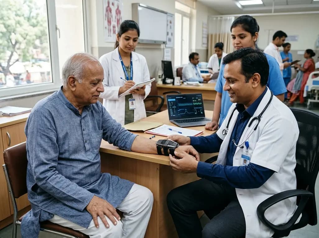
The TREM-AB open-label pilot study evaluated a wearable passive nonlinear vibration absorber for essential tremor. The Nonlinear Energy Sink bracelet reduced clinical Fahn-Tolosa-Marin tremor scores by 32.1% and electrophysiological tremor power by 49.2% with no adverse effects reported.
Today

A study in JAMIA Open shows lightweight open-source reasoning LLMs achieve up to 95% sensitivity and accelerate evidence synthesis screening by nearly twentyfold, presenting a cost-effective, scalable AI solution for biomedical research and systematic reviews.
Today
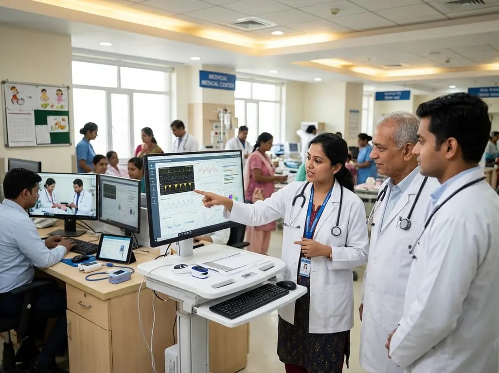
A landmark study details how an enterprise pediatric remote patient management model expanded from a single cardiology pilot to 16 diverse specialty programs by combining centralized digital operations with specialty clinical leadership.
Today
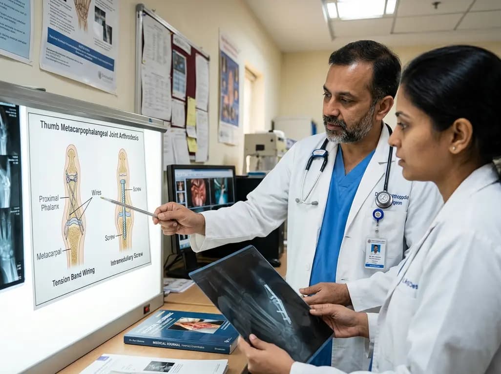
A comparative review of tension band wiring and intramedullary screw fixation in thumb metacarpophalangeal arthrodesis, evaluating union rates, hardware complications, and clinical outcomes.
Today
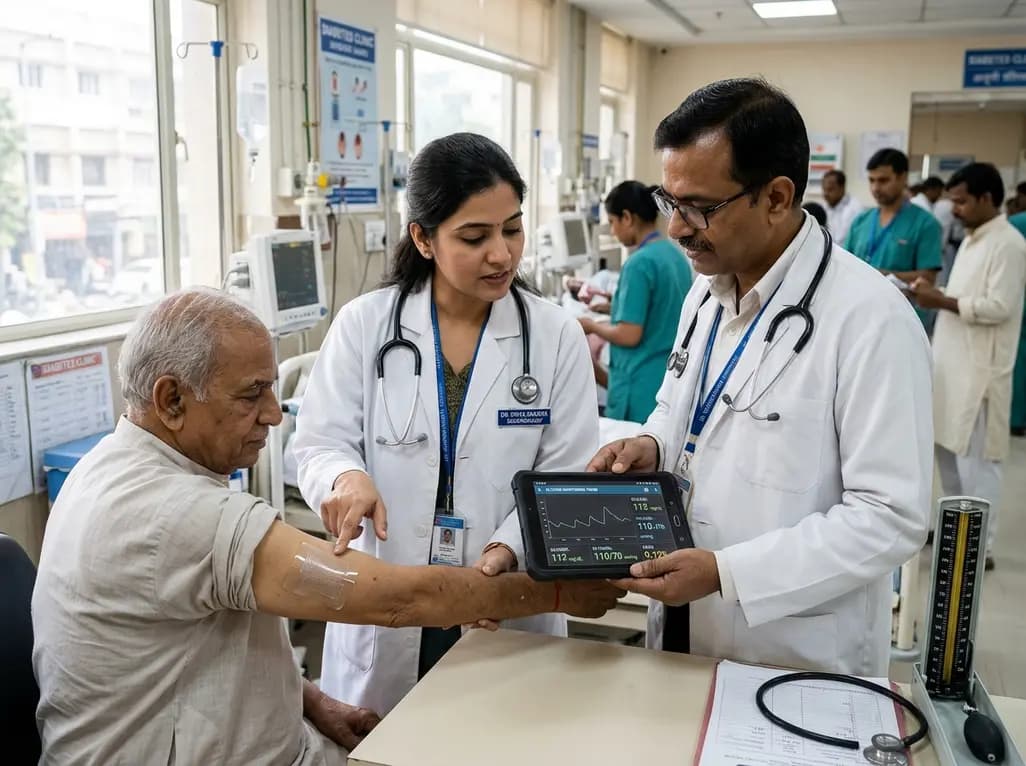
A breakthrough ultrathin multimodal sensor decouples physical hemodynamic monitoring from disposable electrochemical calibration, achieving long-term wearable glucose prediction with 81.36% accuracy and a 9.12% MARD. This offers a robust, drift-free solution for outpatient metabolic surveillance.
Today
