

Loading, please wait...
Loading, please wait...
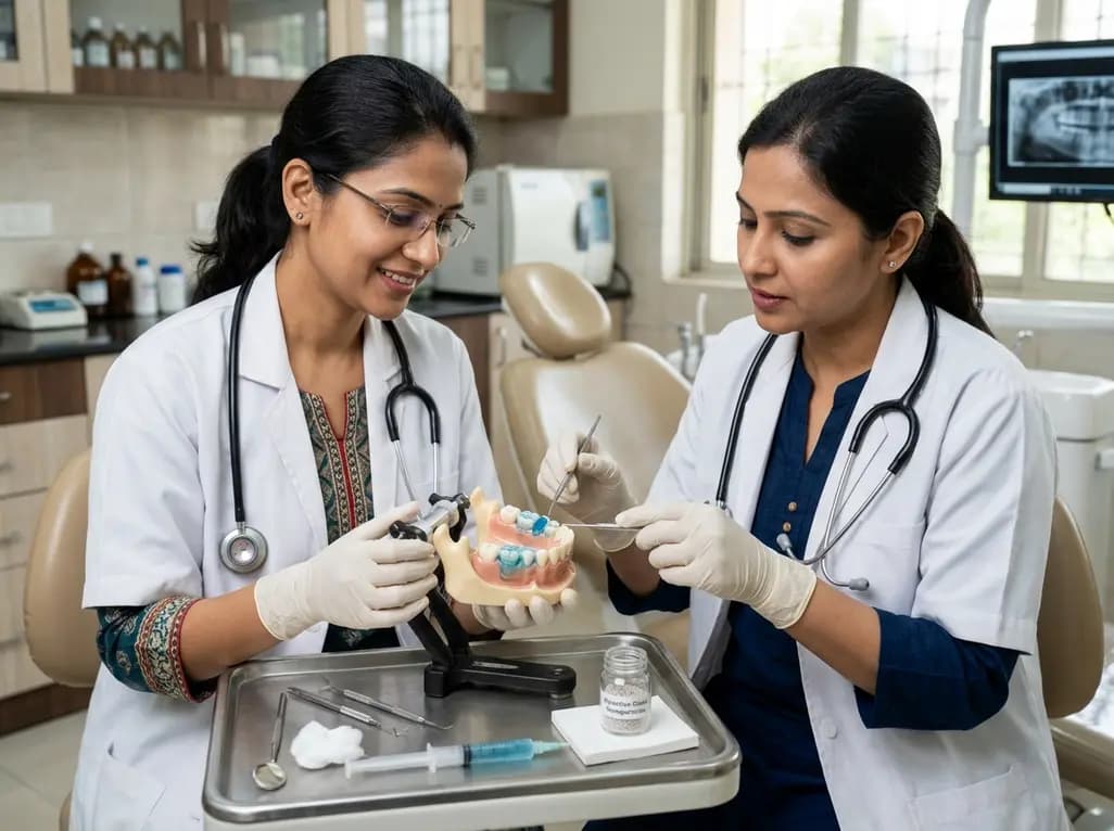
Dental erosion represents an escalating oral health challenge driven by acidic dietary patterns and gastric reflux. While standard topicals provide temporary relief, rapid salivary washout severely impairs durable dental erosion protection across dynamic oral interfaces. Researchers have recently engineered novel bio-adhesive hydrogels that incorporate bioactive glass nanoparticles to overcome this limitation. Consequently, these innovative biomaterials create a resilient therapeutic barrier on exposed tooth surfaces, thereby offering clinicians an advanced translational strategy for counteracting chemical demineralization.
Dental professionals worldwide frequently encounter patients suffering from progressive enamel thinning and dentin hypersensitivity. In clinical practice, non-carious chemical dissolution occurs through frequent contact with extrinsic dietary acids or intrinsic gastric secretions. However, standard desensitizing and remineralizing varnishes face critical retention obstacles in the mouth. Dynamic salivary flow, tongue friction, and chewing forces rapidly dislodge therapeutic compounds before meaningful remineralization can occur. Consequently, conventional aqueous suspensions demonstrate poor substantivity at the fluid-flushed tooth interface. Furthermore, repeated acid challenges degrade the newly precipitated superficial layers, which leaves underlying dentinal tubules exposed and vulnerable. This therapeutic gap underscores the need for robust formulations that resist mechanical clearance and enzymatic degradation. Clinicians therefore require proactive biomaterials capable of forming an intimate, long-lasting bond with both organic collagen and inorganic hydroxyapatite. In addition, addressing this retention problem is especially critical in diverse outpatient populations where continuous dietary counseling often yields variable patient adherence. Specifically, without continuous protection, acidic drinks erode exposed cervical dentin, triggering painful pulpal responses and costly restorative interventions. Thus, bio-adhesive platforms provide a promising route to circumvent these persistent clearance barriers.
To overcome rapid wash-off, investigators successfully synthesized bio-adhesive hydrogel carriers using two distinct polymer matrices: sodium carboxymethyl cellulose and carbomer. The team systematically loaded bioactive glass nanoparticles at an eight percent concentration into these polymeric frameworks. Notably, rheological testing demonstrated approximately a one-thousand-fold viscosity increase compared to free aqueous suspensions. This substantial thickening provides adequate rheological resistance against vigorous oral shear stresses. Furthermore, both formulations exhibited exceptional colloidal stability, maintaining high absolute zeta potential values of at least negative thirty millivolts. Such electrostatic repulsion prevents unwanted particle aggregation during prolonged storage and intraoral delivery. Importantly, cytotoxicity evaluations confirmed that the hydrogel matrices remained entirely safe, achieving relative growth rates around one hundred percent in biological assays. Because biocompatibility is non-negotiable in oral therapies, these results confirm favorable mucosal and cellular safety. Moreover, the crosslinked polymer chains physically entrap the bioactive nanoparticles while facilitating steady ion exchange. Therefore, this formulation design maintains a hydrated, protective reservoir directly against the tooth surface, ensuring that therapeutic ions remain concentrated where acid challenges occur.
During rigorous in vitro erosive cycling, the bio-adhesive hydrogels demonstrated remarkable efficacy compared to established clinical controls. Specifically, profilometry revealed that both hydrogel formulations reduced enamel and dentin surface loss by approximately eighty percent relative to deionized water. In stark contrast, standard sodium fluoride solutions failed to achieve comparable tissue preservation under aggressive acidic challenges. Confocal laser scanning microscopy corroborated these profilometric findings, showing negligible rhodamine dye penetration across hydrogel-treated substrates. Conversely, unprotected controls exhibited extensive subsurface demineralization bands. Furthermore, field emission-scanning electron microscopy coupled with energy-dispersive spectroscopy revealed a dense, silicon-rich mineralized layer coating the treated dental interfaces. This uniform mineral precipitation effectively occluded patent dentinal tubules, sealing pathways that transmit hydrodynamic pain stimuli. Consequently, the hydrogel barrier prevented fluid movement within dentinal channels, explaining its robust protective capacity. In addition, the demineralized organic matrix retained significantly greater thickness in hydrogel groups than in fluoride cohorts. Accordingly, these outcomes confirm that adhesive hydrogel delivery outperforms conventional aqueous remineralizing regimens by combining continuous physical coverage with reactive mineral synthesis.
To uncover the fundamental science behind tissue preservation, investigators explored interfacial interactions through molecular dynamics simulations and biochemical marker assessments. Notably, the study monitored type I collagen C-terminal telopeptide release to evaluate organic matrix breakdown. Untreated dentin exposed to acid and endogenous matrix metalloproteinases suffered extensive collagen degradation. However, dentin treated with bioactive glass hydrogels preserved structural collagen integrity and curtailed peptide cleavage. Moreover, molecular dynamics simulations clarified that the polymeric chains form strong electrostatic and hydrogen bonds with exposed collagen fibrils. Specifically, carboxylate groups along the hydrogel backbones coordinate directly with calcium ions and positive amino acid residues on collagen triple helices. This dense molecular network shields susceptible cleavage sites from host proteases and external acidic attack. Furthermore, the local dissolution of bioactive glass elevates microenvironmental pH, which naturally suppresses acid-activated endogenous enzymes. Simultaneously, the steady release of calcium and silicate ions induces biomimetic apatite nucleating directly upon the collagen scaffold. Thus, this synergistic interplay between adhesive polymer immobilization and bioceramic mineral deposition stabilizes both the organic matrix and the mineral phase simultaneously.
The clinical translation of bio-adhesive bioactive hydrogels holds substantial promise for contemporary preventive dentistry. In regions like India, dietary changes involving increased soft drink consumption, packaged juices, and spicy foods contributing to acid reflux have accelerated tooth wear rates. Clinicians frequently struggle to manage extensive erosive lesions using short-acting varnishes alone. Therefore, introducing bio-adhesive hydrogels into routine prophylaxis or professional chairside protocols could significantly reduce the incidence of non-carious cervical defects. Furthermore, applying these formulations prior to restorative procedures could reinforce degraded hybrid layers and improve adhesive bond longevity. Dental practitioners could also prescribe home-use formulations to patients exhibiting severe bulimia, gastroesophageal reflux, or medication-induced xerostomia. Nevertheless, transitioning from bench to chairside requires standardized randomized clinical trials to evaluate intraoral retention under complex salivary shearing forces. Regulatory approvals and commercial manufacturing scalability will also dictate clinical adoption speeds. Ultimately, incorporating bioactive glass hydrogels represents a paradigm shift from passive observation to active interfacial defense, empowering dental practitioners to intercept erosive wear before irreversible structural destruction occurs.
Bio-adhesive hydrogels utilize polymeric networks such as carbomer and carboxymethyl cellulose that establish multivalent hydrogen bonds and electrostatic interactions with dental surfaces. Unlike aqueous fluoride solutions that wash away quickly in dynamic saliva, these hydrogels exhibit a thousand-fold increase in viscosity. Consequently, they resist mechanical shear and salivary clearance, maintaining a stable reservoir of bioactive glass nanoparticles that release remineralizing ions directly at the tooth interface over extended periods.
Yes, bioactive glass hydrogels offer potent therapy for dentin hypersensitivity. When exposed to oral fluids, bioactive glass dissolves locally, rapidly releasing calcium and phosphate ions that precipitate as a dense, silicon-rich apatite layer. In addition, the bio-adhesive hydrogel securely occludes patent dentinal tubules and limits fluid movement triggered by thermal or tactile stimuli. Furthermore, preserving the collagen matrix protects underlying nerve fibers, delivering profound and sustained desensitizing relief for vulnerable patients.
Bioactive glass hydrogels protect the organic dentin matrix through both physical and biochemical mechanisms. The polymer chains bind intimately to type I collagen fibrils, forming a protective shield against proteolytic cleavage. Concurrently, the alkaline dissolution products of bioactive glass elevate local pH, thereby neutralizing acid and inhibiting host-derived matrix metalloproteinases. Consequently, telopeptide release drops substantially, preserving the structural integrity and thickness of the demineralized organic matrix during subsequent acidic erosive challenges.
Disclaimer: This content is for informational and educational purposes only. It is not intended to replace professional medical advice, diagnosis, or treatment. Refer to the latest local and national guidelines for clinical practice.
References

Read summarized clinical updates, watch expert medical content, and earn CME certifications right from your smartphone.
Open your smartphone camera to scan and install.





A novel bio-adhesive hydrogel incorporating bioactive glass nanoparticles reduces enamel and dentin surface loss by 80%. By resisting salivary washout and occluding tubules, this formulation establishes an effective barrier against erosive tooth wear.
Today

While India achieved a 38% reduction in tuberculosis mortality since 2015, public health groups flag persistent disease transmission, delayed epidemiological reports, and drug supply gaps. Clinicians must navigate these systemic challenges to safeguard treatment adherence and prevent drug resistance across communities.
Today

Maternal codeine therapy during breastfeeding sparks debate regarding neonatal opioid toxicity. This review re-evaluates the Toronto case, explores PBPK modeling, and highlights how maternal and neonatal pharmacogenomics converge to inform safer postpartum pain management.
Today

Novel intranasal simvastatin transbilosomes demonstrate superior bioavailability and cardioprotective efficacy compared to oral suspensions in experimental heart failure induced by doxorubicin and ischemia/reperfusion injury.
Today

A longitudinal cohort study highlights how baseline anxiety impairs long-term pelvic fracture recovery. Tracking PROMIS scores across physical and psychological domains reveals critical windows for early psychosocial intervention.
Today

A multi-modal machine learning model integrating ultrasound radiomics, tumor morphology, TPO-Ab, and BRAF V600E accurately predicts lymph node metastasis in papillary thyroid carcinoma with concurrent Hashimoto's thyroiditis, achieving 0.879 accuracy.
Today
