

Loading, please wait...
Loading, please wait...
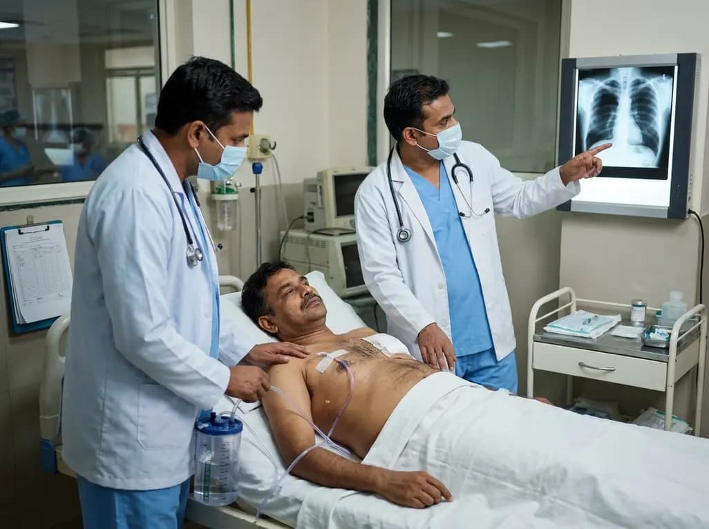
Liposuction remains one of the most frequently performed cosmetic procedures worldwide. Although surgeons view it as generally safe, severe thoracic complications can emerge rapidly. In particular, the development of pneumothorax after liposuction represents a life-threatening emergency that clinicians must identify and treat without delay. While pleural perforation typically presents unilaterally following chest wall suctioning, atypical bilateral presentations following upper extremity and back contouring demand heightened vigilance across surgical teams.
Aesthetic contouring of the upper extremities and back rarely leads to thoracic trauma. However, practitioners must recognize that complex anatomy increases vulnerability during aggressive tissue manipulation. For example, a recent case involved a healthy 42-year-old woman who underwent bilateral upper limb and upper back liposuction. Approximately eleven hours after surgery, she developed acute dyspnea, left-sided pleuritic chest pain, and significant upper-body edema. On clinical examination, the patient exhibited marked tachycardia and signs of acute respiratory distress. Consequently, the medical team prioritized an immediate cardiorespiratory workup. Notably, severe subcutaneous emphysema obscured initial physical findings across the cervicothoracic region. Diminished breath sounds were audible bilaterally, which elevated clinical suspicion for pleural compromise. Although bilateral pleural entry is extraordinarily rare during arm contouring, forceful cannula passes near the axillary apex can easily transgress thin musculoskeletal planes. Therefore, emergency teams must never dismiss progressive postoperative dyspnea as routine post-surgical discomfort. Clinicians must maintain an aggressive diagnostic stance when patients report pleuritic pain following any upper-body suctioning.
Direct mechanical penetration represents the primary mechanism behind pleural injury during aesthetic surgery. Specifically, surgical cannulas can breach the intercostal spaces when surgeons direct instruments toward the thoracic cage. In fibrous regions like the back and axilla, dense tissue resists smooth passage. As a result, the surgeon may apply excess forward pressure to advance the cannula. This axial force can inadvertently propel the instrument through the serratus anterior and intercostal musculature. Furthermore, flexible infiltration needles and thin cannulas carry higher risks because their tips deflect unpredictably under force. Stiffer instruments provide better tactile feedback and lower perforation rates. Once the cannula tears the parietal and visceral pleura, atmospheric air rushes into the pleural space. Additionally, high-flow tumescent infiltration can dissect along fascial planes, which drives air and fluid into the mediastinum and adjacent hemithorax. Bilateral involvement usually occurs when the operator mirrors aggressive passes across both upper extremities and thoracic boundaries. Consequently, both lungs lose negative intrapleural pressure, triggering rapid functional collapse and ventilation-perfusion mismatch.
Rapidly detecting bilateral pneumothorax after liposuction presents unique clinical hurdles in ambulatory and emergency settings. Clinicians often observe that supplemental oxygen masks the true severity of underlying lung collapse. For instance, a patient receiving low-flow oxygen may demonstrate normal pulse oximetry despite significant bilateral volumes of trapped intrapleural air. Therefore, clinicians must not interpret an adequate oxygen saturation as evidence of normal pulmonary function. Instead, providers should evaluate persistent tachypnea, tachycardia, and asymmetric chest excursion as reliable indicators of mechanical compromise. Furthermore, extensive subcutaneous emphysema often produces diffuse crepitus across the neck and chest wall. This palpable crackling should immediately trigger urgent diagnostic protocols. Delayed recognition carries disastrous risks, including tension physiology and catastrophic hemodynamic collapse. If positive pressure ventilation begins before pleural decompression, intra-thoracic pressure spikes precipitously, which impairs venous return to the heart. Thus, attending staff must maintain continuous monitoring during the entire postoperative recovery window. Prompt clinical suspicion remains the cornerstone of preventing fatal respiratory arrest in these surgical patients.
Definitive diagnosis requires prompt radiographic imaging whenever patients demonstrate unexplained respiratory distress. Clinicians typically initiate the evaluation with an upright posteroanterior chest radiograph. This initial study reveals pleural lines, mediastinal shifting, and deep sulcus signs. However, portable supine films in postoperative recovery units often underestimate the true extent of bilateral pneumothorax. Therefore, emergency physicians should utilize bedside point-of-care ultrasonography to accelerate triage. Ultrasonography rapidly demonstrates absent lung sliding, absent B-lines, and distinctive lung point signs with high diagnostic accuracy. Furthermore, contrast-enhanced computed tomography of the chest represents the gold standard for comprehensive evaluation. Computed tomography clearly defines the size of bilateral hydropneumothorax and identifies underlying pulmonary parenchymal lacerations. Additionally, CT pulmonary angiography effectively excludes acute pulmonary thromboembolism and fat embolism syndrome, which share overlapping symptoms with pleural perforation. Early axial imaging also maps the precise distribution of soft-tissue emphysema across cervical and retroperitoneal planes. Consequently, comprehensive thoracic imaging guides urgent interventions and rules out alternative life-threatening diagnoses.
Management of bilateral pneumothorax requires rapid decompression and rigorous hemodynamic stabilization. Surgical teams should insert bilateral tube thoracostomies without delay to evacuate intrapleural air and restore vital lung expansion. Following chest tube placement, patients typically experience immediate symptomatic relief and normalization of physiological parameters. However, clinicians must also maintain acute vigilance for concurrent hemorrhage. During aggressive liposuction passes, cannulas can lacerate chest wall perforators, intercostal vessels, or muscular arterial branches. Notably, significant blood loss can accumulate silently within expansive subcutaneous cavities rather than draining freely through the thoracostomy tubes. For example, patients may display a sharp drop in hemoglobin levels despite minimal pleural drainage. In such scenarios, clinicians must quickly recognize occult soft-tissue bleeding and initiate volume resuscitation. Transfusion of packed red blood cells stabilizes circulatory volume while teams rule out ongoing active extravasation. Furthermore, continuous serial blood counts and hemodynamic assessments ensure early identification of hidden hematomas. Multidisciplinary collaboration between thoracic surgeons, intensivists, and plastic surgeons ensures complete recovery and optimal patient safety.
Preventing thoracic injury during body contouring requires meticulous technical adherence and structural vigilance. First, surgeons must always maintain complete spatial awareness of the cannula tip throughout every stroke. Operators should keep their non-dominant hand placed flat over the target area to continuously guide instrument depth. Moreover, practitioners must avoid pointing cannulas toward intercostal spaces when treating fibrous back tissue and axillary boundaries. Using blunt-tipped, rigid cannulas measuring at least 3.5 millimeters reduces the risk of unintentional pleural penetration. In contrast, narrow and flexible needles bend unpredictably and penetrate deep muscular fascial planes without warning. Additionally, surgical teams should avoid excessive positive-pressure ventilation during anesthesia, which expands lung margins toward the chest wall. Preoperative planning must identify prior surgical scars, because adhesive tissue alters normal resistance patterns. Finally, comprehensive informed consent should explicitly address thoracic risks, even when contouring peripheral zones. Standardized clinical pathways and heightened perioperative awareness significantly minimize catastrophic surgical misadventures.
Upper-limb and back contouring involves vigorous cannula strokes near the axillary apex and fibrous thoracic boundaries. When surgeons apply substantial axial force against resistant tissues, thin or flexible instruments can deflect inward. The cannula inadvertently traverses intercostal spaces and punctures the parietal and visceral pleura. If the surgeon performs reciprocal aggressive passes across both upper arms, bilateral mechanical perforations occur, permitting atmospheric and subcutaneous air to collapse both pleural spaces simultaneously.
Patients in postoperative recovery units frequently receive supplemental oxygen via nasal cannula or face masks. This oxygen therapy artificially elevates arterial oxygen saturation, which creates a false impression of stability. Consequently, severe mechanical ventilation impairment remains concealed while resting oximetry readings appear reassuring. Clinicians must therefore look beyond digital pulse oximetry, actively evaluating subtle tachypnea, tachycardia, pleuritic chest discomfort, and extensive subcutaneous emphysema to detect tension physiology or bilateral lung collapse promptly.
Cannula strikes can lacerate intercostal vessels and subcutaneous arterial branches during aesthetic contouring. Because dissected soft-tissue planes accommodate substantial blood volumes, hemorrhage frequently accumulates within deep operative cavities without draining outward. Clinicians should monitor serial hemoglobin measurements, vital signs, and skin turgor closely. When a sharp hemoglobin decline occurs alongside tachycardia, medical teams must initiate immediate packed red blood cell transfusions, intravenous fluid resuscitation, and computed tomography imaging to achieve hemodynamic stability.
Disclaimer: This content is for informational and educational purposes only. It is not intended to be a substitute for professional medical advice, diagnosis, or treatment. Always seek the advice of your physician or other qualified health provider with any questions you may have regarding a medical condition. Refer to the latest local and national guidelines for clinical practice.
References

Read summarized clinical updates, watch expert medical content, and earn CME certifications right from your smartphone.
Open your smartphone camera to scan and install.





Bilateral pneumothorax after upper-limb liposuction is a rare surgical complication. Clinicians must recognize dyspnea despite normal pulse oximetry, evaluate for occult bleeding, and perform prompt tube thoracostomy to ensure complete patient recovery.
Today

A longitudinal feasibility pilot study highlights that combining polygenic risk score disclosure with in-person lifestyle counseling significantly enhances genomic literacy in individuals at risk for type 2 diabetes, overcoming educational disparities and reinforcing patient motivation.
Today

AIIMS researchers suggest yoga may alleviate gut dysbiosis in Polyendocrine Metabolic Ovarian Syndrome (PMOS) by modulating the gut-brain axis, lowering chronic inflammation, and enhancing parasympathetic tone to support metabolic and hormonal equilibrium.
Today

A prospective cohort study demonstrates that feeding tubes, formula exposure, aortic cross-clamp duration, and socioeconomic deprivation disrupt the neonatal gut microbiome in congenital heart disease, emphasizing the need for targeted nutritional and perioperative gut-protective strategies.
Today

Deep intramuscular vascular malformations in pediatric Klippel-Trenaunay syndrome can mimic Maffucci syndrome. Multimodality imaging with Doppler ultrasound and MRI, along with histopathology, ensures prompt differential diagnosis, targeted surgical debulking, and optimized conservative surveillance.
Today

Modern management of adrenal incidentalomas is shifting away from universal testing toward risk-stratified biochemical evaluation. Guidelines suggest omitting metanephrine testing in lipid-rich adenomas (≤10 HU), while maintaining autonomous cortisol testing to prevent cardiometabolic complications.
Today
