.webp&w=384&q=75)

Loading, please wait...
Loading, please wait...

Acute vision loss in young adults requires immediate triage and diagnostic clarity. When a 23-year-old male presents with worsening headaches, retrobulbar eye pain, and rapidly declining vision, emergency clinicians must act swiftly. Ophthalmologic examination often reveals bilateral optic neuritis, marked by diminished visual acuity and impaired color discrimination. Patients frequently report that ocular movements aggravate their retrobulbar pain. Although typical optic neuritis predominantly affects young females unilaterally, bilateral presentations represent atypical forms that demand comprehensive diagnostic evaluation. Clinicians must immediately assess pupillary reflexes, confrontational visual fields, and funduscopic findings. Papillitis presents with evident disc swelling, whereas retrobulbar neuritis often shows a normal baseline fundus. Consequently, clinicians should not exclude optic neuropathy simply because the initial optic disc appearance remains normal. Furthermore, early bedside evaluation must identify potential constitutional red flags. Clinicians should specifically evaluate patients for fever, meningismus, cutaneous eruptions, and focal neurological signs. Therefore, an accurate clinical history establishes the foundation for separating typical demyelinating episodes from rare inflammatory, infectious, or toxic etiologies. Recognizing these critical signs ensures prompt neuroimaging and prevents catastrophic delays in definitive medical management.
Clinicians evaluating optic neuropathy must distinguish between primary demyelinating disease, direct infectious invasion, and post-infectious inflammatory reactions. Demyelinating disorders such as multiple sclerosis, neuromyelitis optica spectrum disorder (NMOSD), and myelin oligodendrocyte glycoprotein antibody-associated disease (MOGAD) represent primary autoimmune culprits. In contrast, direct infectious optic neuropathies arise from pathogens like syphilis, herpesviruses, tuberculosis, Bartonella, and Lyme disease. However, para-infectious optic neuritis emerges following an acute systemic illness via immune-mediated molecular mimicry rather than direct pathogen invasion of neural tissue. In this clinical scenario, identifying the precise mechanism dictates immediate therapeutic decisions. If an active microbial pathogen directly invades the anterior visual pathways, high-dose immunosuppression alone could worsen the underlying infection. Conversely, clinicians who delay corticosteroid therapy during immune-mediated attacks risk permanent axonal loss and irreversible vision reduction. Therefore, clinicians must thoroughly investigate the timeline between antecedent viral symptoms and the onset of visual compromise. Furthermore, systemic signs like lymphadenopathy, pharyngitis, or mild transaminitis frequently suggest a post-viral or para-infectious immune cascade. Consequently, balancing these diagnostic possibilities requires careful clinical judgment, urgent paraclinical testing, and close interdisciplinary communication between ophthalmologists, neurologists, and infectious disease specialists.
A rigorous diagnostic pathway isolates the underlying etiology in atypical optic neuropathy. Contrast-enhanced magnetic resonance imaging (MRI) of the brain and orbits with fat suppression serves as the gold standard investigation. MRI reveals hyperintensity and enhancement along the optic nerves while identifying demyelinating plaques or leptomeningeal inflammation. In addition, lumbar puncture provides essential cerebrospinal fluid (CSF) analysis to exclude elevated opening pressure, persistent pleocytosis, and intrathecal oligoclonal bands. Serological testing must simultaneously screen for autoantibodies, including aquaporin-4 (AQP4-IgG) and MOG-IgG. Moreover, extensive microbiological panels must evaluate treponemal antibodies, human immunodeficiency virus, and relevant viral pathogens. In this illustrative young patient, routine blood investigations revealed moderately elevated liver transaminases alongside high cytomegalovirus (CMV) immunoglobulin G titers. Importantly, the absence of CMV IgM and undetectable viral DNA indicated past exposure rather than an acute invasive viremia. Clinicians frequently encounter elevated IgG titers in the general population, which can create diagnostic confusion during an acute crisis. Therefore, physicians must carefully correlate laboratory results with the overall clinical picture. Thus, advanced neuroimaging combined with targeted serology reliably excludes invasive central nervous system infections while confirming an inflammatory optic nerve process.
Encountering elevated transaminases and positive viral serologies often creates significant clinical hesitation regarding high-dose steroid administration. Physicians worry that intense immunosuppression might trigger fulminant hepatitis or accelerate latent viral dissemination. However, untreated inflammatory optic neuritis rapidly leads to irreversible retinal ganglion cell apoptosis and permanent blindness. In this patient, negative viral PCR assays and undetectable acute-phase IgM antibodies provided reassurance against active cytomegalovirus replication. Consequently, the medical team promptly initiated intravenous methylprednisolone at a standard pulse dose of one gram daily for three consecutive days. Rapid anti-inflammatory therapy halts destructive lymphocyte infiltration within the optic nerve sheath and suppresses pathogenic cytokine cascades. As a result, the patient experienced dramatic improvement in visual acuity and resolution of retrobulbar eye pain within forty-eight hours. Furthermore, serial laboratory testing confirmed stable hepatic enzymes throughout corticosteroid administration, illustrating that transient transaminitis should not delay urgent sight-saving therapy. Subsequently, patients transition to an oral prednisone taper depending on their clinical response and suspected autoimmune etiology. Therefore, clinicians must weigh the verifiable risk of permanent visual disability against the theoretical risk of systemic infection before withholding pulse steroids.
Long-term clinical monitoring remains essential after managing acute bilateral optic neuropathy. Most patients who receive prompt intravenous corticosteroids demonstrate substantial functional visual recovery within several weeks. However, ophthalmologists must conduct serialized examinations using optical coherence tomography (OCT) to quantify retinal nerve fiber layer thinning. In addition, automated visual field perimetry and formal contrast sensitivity testing detect subtle lingering visual deficits that standard Snellen charts miss. Clinicians should also schedule repeat neuroimaging within three to six months to assess for asymptomatic demyelinating lesions. For instance, the emergence of subclinical white matter changes or persistent antibody positivity alters the long-term prognosis significantly. If serological testing demonstrates persistent MOG or AQP4 antibodies, clinicians must evaluate patients for maintenance immunotherapy to prevent disabling future relapses. Similarly, patients diagnosed with clinically isolated syndrome require ongoing neurological surveillance for potential conversion to multiple sclerosis. Furthermore, educating patients on reporting subtle neurological symptoms ensures early detection of recurrent disease. Therefore, comprehensive post-acute care bridges emergency stabilization and durable neurological preservation through diligent outpatient follow-up.
This intriguing case provides crucial educational pearls for primary physicians, emergency doctors, and consulting specialists alike. First, clinicians should maintain a low threshold for neuroimaging when young adults present with acute vision loss and retrobulbar pain. Second, physicians must recognize that bilateral involvement represents an atypical presentation requiring thorough multi-organ evaluation. Furthermore, incidental positive IgG titers reflect widespread community exposure and should never substitute for evidence of active viral infection. Clinicians must rapidly verify PCR assays and IgM markers rather than delaying urgent pulse corticosteroid therapy unnecessarily. In addition, multidisciplinary collaboration between neurology, ophthalmology, and internal medicine ensures seamless diagnostic integration and avoids therapeutic indecision. When clinical teams execute prompt diagnostic workups, they prevent unnecessary morbidity and preserve irreplaceable visual function. Moreover, clinicians must remain vigilant during follow-up, ensuring that patients receive timely subspecialty evaluation for underlying autoimmune diseases. Ultimately, disciplined clinical reasoning empowers physicians to navigate conflicting laboratory anomalies with confidence and precision. Thus, mastering these core diagnostic and management principles directly optimizes clinical outcomes across diverse clinical settings.
Unilateral optic neuritis typically presents in young women and strongly suggests multiple sclerosis. In contrast, bilateral optic neuritis occurs less frequently and suggests atypical etiologies, including myelin oligodendrocyte glycoprotein antibody-associated disease, neuromyelitis optica spectrum disorder, systemic autoimmune illnesses, or para-infectious immune responses. Consequently, bilateral cases demand broader diagnostic investigations.
Clinicians can safely initiate intravenous corticosteroids once clinical evaluation and molecular testing effectively rule out direct central nervous system infection. Specifically, the absence of active fever, normal cerebrospinal fluid opening pressure without neutrophilic pleocytosis, and negative viral polymerase chain reaction assays allow clinicians to start high-dose pulse methylprednisolone promptly.
Cytomegalovirus IgG antibodies merely indicate prior lifetime exposure and immunological memory rather than acute tissue infection. High IgG seroprevalence exists among healthy immunocompetent adults. Clinicians confirm active cytomegalovirus disease only when patients exhibit compatible systemic features alongside positive viral IgM antibodies, rising paired titers, or detectable CMV DNA in serum or CSF.
Disclaimer: This content is for informational and educational purposes only and should not be considered as medical advice. Always consult a qualified healthcare professional regarding any medical condition or treatment. Refer to the latest local and national guidelines for clinical practice.
References

Read summarized clinical updates, watch expert medical content, and earn CME certifications right from your smartphone.
Open your smartphone camera to scan and install.





A 23-year-old male presenting with acute vision loss highlights the diagnostic challenges of bilateral optic neuritis. Learn to differentiate demyelinating, infectious, and para-infectious etiologies, evaluate laboratory anomalies, and initiate timely corticosteroid therapy safely.
Today
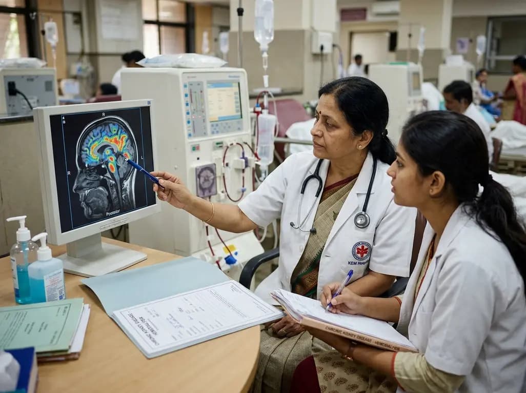
A retrospective clinical analysis reveals that hemodialysis-associated cognitive dysfunction arises from synergistic pathological mechanisms, including severe chronic anemia, worsening renal clearance, abnormal regional brain iron deposition, and an imbalance between neuroinflammatory and neurotrophic factors.
Today

A multiparametric MRI approach combining quantitative T2 shading, apparent diffusion coefficient (ADC) measurements, and morphological signs provides superior diagnostic accuracy over subjective evaluation to differentiate ovarian endometriomas from benign hemorrhagic cysts.
Today

Discover a novel laparoscopic approach for iatrogenic diaphragmatic hernia repair following pedicled omentoplasty. This modified Sugarbaker technique integrates dorsal pedicle lateralization and round ligament reinforcement to secure the diaphragmatic defect while preserving vital omental vascularity.
Today

Evaluating Hill-Sachs lesion size and the distance to dislocation is critical in recurrent shoulder instability. As bone loss concepts shift from binary on-track classifications to continuous risk assessment, clinicians must identify when isolated Bankart repair is insufficient and supplementary procedures are needed.
Today

New research reveals that M1 macrophage-derived exosomes aggravate diabetic nephropathy by transferring WTAP to stabilize S1PR2 mRNA. Silencing WTAP in these vesicles attenuates endothelial injury and renal fibrosis, pointing toward innovative nanomedicine therapies.
Today
