.webp&w=384&q=75)

Loading, please wait...
Loading, please wait...
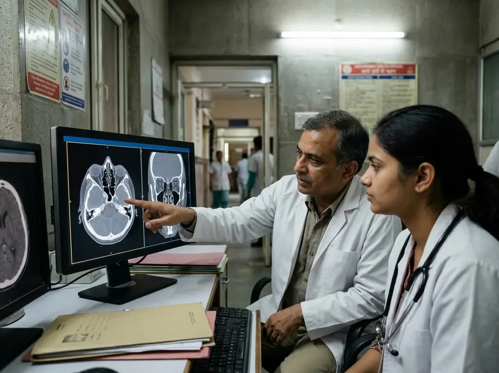
Anatomical variations of the lateral nasal wall present unique challenges for otolaryngologists and radiologists during routine diagnostic workups. Among these rare structural anomalies, a bifid inferior turbinate represents an unusual developmental variant that clinicians can easily overlook during routine nasal endoscopy. Because this structure often mimics abnormalities of the uncinate process or the middle meatus, accurate identification is essential for successful surgical planning. In this comprehensive review, we examine a recently documented case of an inferomedially oriented bifid inferior turbinate alongside co-occurring nasal pathologies, evaluating its clinical implications, diagnostic methods, and surgical considerations.
The embryological development of the nasal cavity involves a complex sequence of invaginations and ossifications. While the middle, superior, and supreme turbinates derive from the ethmoid bone, the inferior nasal turbinate develops independently from the maxilloturbinal ossification center. Disruption during this delicate embryological process can produce structural duplication or splitting, resulting in a bifid configuration.
Although pneumatization of the middle turbinate, known as concha bullosa, occurs frequently in clinical practice, bifid inferior turbinate structures remain extraordinarily rare. Clinicians encounter this anomaly in a tiny fraction of patients undergoing sinonasal evaluation. Consequently, understanding its anatomical foundation helps prevent diagnostic errors.
Furthermore, embryonic variations can cause the duplicated inferior turbinate to project in different directions within the nasal vault. Some cases present with superior or lateral extensions, whereas others show inferomedial orientation into the main nasal airway. When an accessory turbinate extends inferomedially, it alters local airflow mechanics and reduces the effective cross-sectional area of the nasal passage. Moreover, this spatial displacement can obscure typical anatomical landmarks during routine anterior rhinoscopy. Therefore, surgeons must recognize how subtle embryological alterations alter normal endonasal architecture before embarking on therapeutic interventions.
Patients with a bifid inferior turbinate rarely present due to the anomaly alone. Instead, clinical symptoms usually arise because the structural variant coexists with inflammatory or mechanical obstructive conditions. For instance, a recent case report highlighted a 24-year-old female who presented with long-standing right-sided nasal obstruction, frontal headache, rhinorrhea, snoring, sleep disturbance, and allergic rhinitis.
In many cases, the duplicated turbinate operates silently until mucosal inflammation or secondary anatomical deviations narrow the airway further. Additionally, chronic allergic rhinitis often exacerbates mucosal swelling across both normal and duplicated turbinate tissue. This tissue enlargement severely compromises the nasal valve area, impairing normal laminar airflow and inducing chronic nasal congestion.
Furthermore, clinical evaluations frequently identify concurrent structural abnormalities alongside the bifid turbinate. In the documented patient, diagnostic workups revealed bilateral inferior turbinate hypertrophy, an S-shaped septal deviation, and a prominent concha bullosa. These combined features created significant mechanical resistance within the sinonasal passages. Consequently, patients suffer from persistent respiratory discomfort, sleep disruption, and secondary facial headaches. Recognizing that a bifid inferior turbinate often forms part of a complex anatomical constellation allows otolaryngologists to tailor comprehensive treatment plans rather than focusing solely on isolated findings.
Accurate pre-operative diagnosis of lateral nasal wall variants depends heavily on cross-sectional imaging modalities. Although flexible and rigid nasal endoscopy serve as routine tools in rhinologic practice, endoscopic visualization alone has clear limitations when evaluating a bifid inferior turbinate.
During nasal endoscopy, the mucosal surface of a duplicated turbinate may closely resemble an aberrant uncinate process, an accessory middle turbinate, or a polypoid mass. Because the endoscope offers a two-dimensional view of complex three-dimensional cavities, clinicians can easily misinterpret the inferomedial duplication as an abnormality of the middle meatus. This misclassification creates surgical risk during functional endoscopic sinus surgery, as mistaking a turbinate variant for uncinate tissue might lead to unnecessary bone resection or inadvertent orbital entry.
Conversely, non-contrast paranasal sinus computed tomography (CT) provides definitive anatomical clarity. Coronal and axial CT sections precisely display the osseous core and mucosal covering of the duplicated turbinate. In the featured case, coronal CT imaging demonstrated an inferomedially oriented duplication of the left inferior turbinate alongside bilateral soft-tissue enlargement. Moreover, CT multiplanar reformations allow clinicians to trace the attachment of each turbinate limb directly to the lateral nasal wall. Therefore, thorough CT assessment remains the gold standard for differentiating true turbinate duplication from adjacent paranasal structures.
To avoid intraoperative missteps, surgical teams must understand the distinct embryological origins of lateral nasal wall structures. The uncinate process originates from the first ethmoturbinal, whereas the inferior turbinate originates independently from the maxilloturbinal. When an inferomedially oriented bifid inferior turbinate exists, its superior component can project toward the hiatus semilunaris, superficially resembling an uncinate process anomaly.
Rhinologic literature classifies these variations into distinct anatomical categories. A "true" bifid inferior turbinate contains its own bony lamella that attaches directly to the inferior turbinate bone on the maxilla, coexisting with a normal uncinate process. In contrast, cases where an uncinate process undergoes severe inferior displacement and hypoplasia are sometimes mislabeled as inferior turbinate duplications.
Additionally, secondary or accessory middle turbinates can project into the middle meatus, causing further diagnostic confusion. Careful review of high-resolution computed tomography scans helps clinicians differentiate these entities by confirming the presence or absence of a distinct uncinate process. Furthermore, preserving the normal uncinate process during surgery prevents disruption of the ostiomeatal complex. Indeed, identifying the exact insertion site of the accessory turbinate limb ensures that surgeons preserve key anatomical landmarks, thereby maintaining normal mucociliary clearance mechanisms throughout the paranasal sinuses.
Managing patients with a bifid inferior turbinate requires a thoughtful, tailored surgical strategy. Because the bifid component itself may not always cause primary obstruction, surgeons must determine whether the anomaly or co-existing structural deviations drive the patient's symptoms. In many cases, addressing adjacent obstructive lesions resolves airway resistance without requiring aggressive resection of the duplicated turbinate tissue.
In the highlighted clinical case, the surgical team performed a combination of septoplasty, bilateral inferior turbinoplasty, and concha bullosa resection. The septoplasty corrected the severe S-shaped septal deviation, restoring midline alignment. Meanwhile, inferior turbinoplasty reduced the bulky hypertrophied mucosa while preserving functional respiratory epithelium. Additionally, partial resection of the middle turbinate concha bullosa decompressed the ethmoid infundibulum and restored sinus drainage.
Furthermore, surgeon preference and mucosal preservation principles guide whether the accessory limb of a bifid inferior turbinate requires partial excision or radiofrequency ablation. Complete excision is usually reserved for cases where the duplicated limb physically blocks the nasal valve or middle meatus. By focusing on targeted mucosal preservation and correcting septal asymmetry, surgeons achieve dramatic symptom relief, resolve chronic headaches, and improve long-term sleep quality for affected patients.
Comprehensive evaluation of lateral nasal wall variations requires close collaboration between diagnostic radiologists and sinus surgeons. Recognizing a bifid inferior turbinate on pre-operative computed tomography prevents misclassification and protects patients from inappropriate surgical resections or inadvertent damage to surrounding structures.
When reviewing paranasal sinus CT scans, radiologists should systematically trace the bony attachment of all turbinate structures. This careful tracking confirms whether a duplicated bone arises from the maxilloturbinal or ethmoid origins. Furthermore, operative reports should clearly detail the spatial relationship between the bifid turbinate, the uncinate process, and the ostiomeatal complex to guide future endoscopic procedures.
Overall, surgical intervention should prioritize relieving symptomatic airflow resistance while conserving functioning respiratory mucosa. Combining septoplasty, targeted inferior turbinoplasty, and concha bullosa excision provides optimal relief for complex obstructive cases. By maintaining high clinical suspicion and conducting precise radiological reviews, surgical teams safely navigate complex endonasal anatomy, avoid intraoperative complications, and deliver superior patient outcomes.
A bifid inferior turbinate is a rare anatomical variant where the inferior nasal turbinate splits or duplicates into two distinct structures along the lateral nasal wall. During embryological development, maxilloturbinal ossification variations create this double configuration. While many patients remain asymptomatic, the duplicated structure can narrow the nasal cavity or obstruct normal airflow, especially when accompanied by allergic rhinitis, septal deviation, or secondary mucosal hypertrophy.
Accurate diagnosis requires non-contrast paranasal sinus computed tomography (CT). Although nasal endoscopy visualizes endonasal mucosa, it can easily mistake a duplicated turbinate for an abnormal uncinate process or a middle meatus lesion. High-resolution coronal and axial CT scans reveal the inner bony core of the duplicated turbinate, allowing radiologists and surgeons to trace its exact attachment to the lateral nasal wall and differentiate it from uncinate pathology.
Surgical treatment is not mandatory for an incidental, asymptomatic bifid inferior turbinate. However, if the patient suffers from long-standing nasal obstruction, chronic headaches, or refractory rhinosinusitis due to co-occurring conditions like septal deviation or concha bullosa, surgery becomes necessary. Surgical management typically focuses on restoring the airway through septoplasty, inferior turbinoplasty, and treating adjacent sinus pathology while preserving functional respiratory mucosa.
Disclaimer: This content is for informational and educational purposes only and does not constitute medical advice. Consult qualified healthcare professionals for diagnosis and treatment. Refer to the latest local and national guidelines for clinical practice.
References

Read summarized clinical updates, watch expert medical content, and earn CME certifications right from your smartphone.
Open your smartphone camera to scan and install.





Bifid inferior turbinate is a rare lateral nasal wall anomaly that can be misidentified on endoscopy. This clinical overview explores CT diagnostic findings, distinction from uncinate process abnormalities, co-occurring obstructive conditions, and individualized surgical strategies including turbinoplasty.
last month

A multicenter study validates a hybrid clinical decision support system combining rule-based logic and machine learning to optimize anticoagulant prescription reviews, reducing alert fatigue and intercepting prescribing errors.
Today

The ClinGen Prenatal Gene Curation Expert Panel evaluated 63 disease relationships across 61 genes, establishing clinical validity for severe fetal phenotypes like hydrops and stillbirth to enhance prenatal genomic interpretation and clinical care.
Today

A metataxonomic study reveals distinct gut bacteriome biomarkers in type 2 diabetes, obesity, and cardiovascular complications, identifying specific bacterial shifts that pave the way for precision metabolic medicine.
Today

A psychometric validation study confirms that the Turkish Copenhagen Hip and Groin Outcome Score (HAGOS-T) offers excellent reliability, construct validity, and interpretability for patients with hip osteoarthritis, providing clinicians with a robust tool for functional assessment.
Today

Clinical guidelines rely heavily on isolated biomarkers like IGF-1 and HbA1c. However, portal insulin delivery fundamentally gates hepatic growth hormone sensitivity. This physiological continuum unites type 1 and type 2 diabetes, obesity, cirrhosis, and acromegaly, challenging conventional treatment strategies.
Today
