.webp&w=384&q=75)

Loading, please wait...
Loading, please wait...
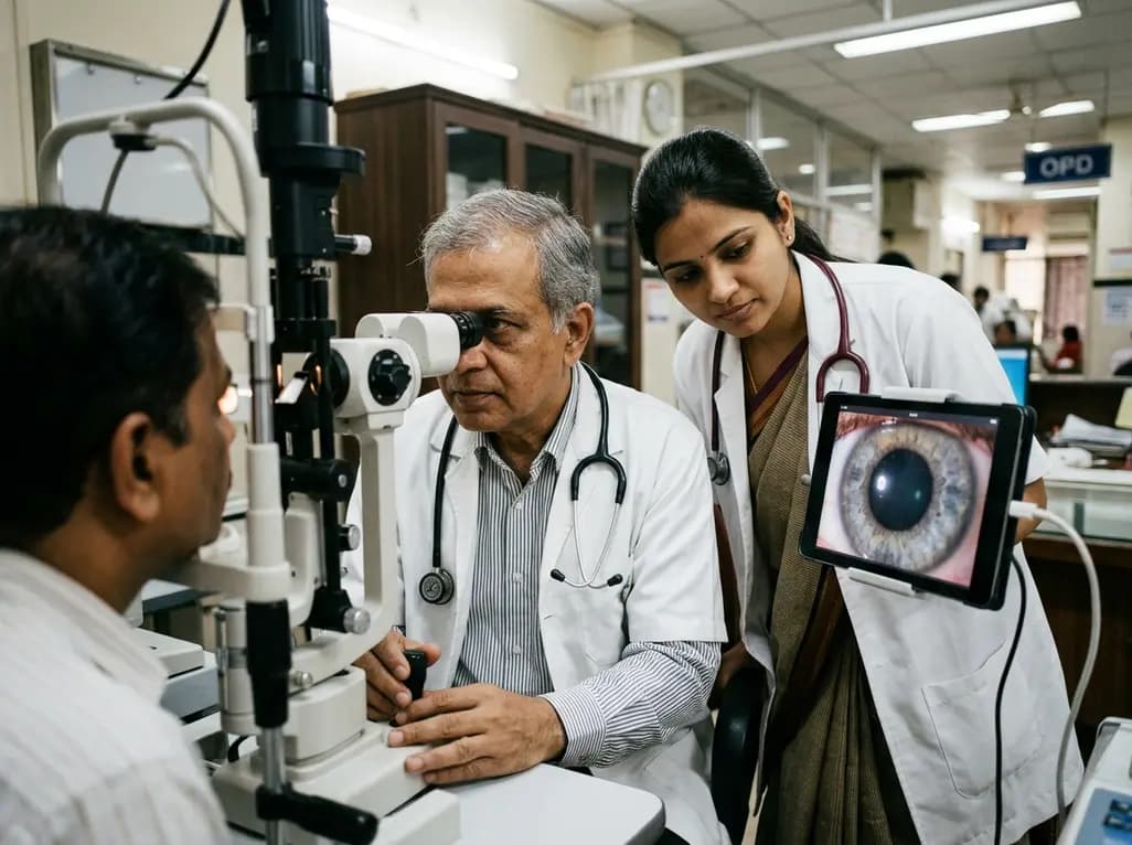
Clinical researchers and neuroscientists traditionally interpret changes in pupil diameter as a direct window into autonomic arousal. When an individual focuses on an engaging task, experiences emotional excitement, or undergoes cognitive strain, the pupils dilate reliably. Consequently, clinicians and investigators have long relied on spontaneous pupil fluctuations to index locus coeruleus noradrenergic activity and central nervous system alertness. However, attributing every pupillary variation solely to arousal oversimplifies complex ocular and neurophysiological dynamics. Recent investigations present a comprehensive four-mechanism framework that expands our understanding beyond this unitary model. This framework reveals that pupillary dynamics actively influence sensory intake and shape visual perception across various physiological states.
For decades, pupillometry literature emphasized the ascending reticular activating system and sympathetic-parasympathetic balance. Whenever autonomic tone shifts, downstream pathways adjust the sphincter and dilator pupillae muscles in the iris. Therefore, clinicians frequently use baseline pupil dynamics to monitor cognitive workload, vigilance decrements, and neurocritical status.
However, viewing pupillary changes strictly as passive markers of central arousal overlooks essential sensory feedback loops. Spontaneous pupil fluctuations occur continuously even under stable environmental conditions and resting states. When researchers treat the pupil merely as an autonomic readout, they disregard direct interactions between ocular mechanics and sensory encoding. Neurological networks do not operate in complete isolation from the peripheral sensory organs that capture incoming light. Furthermore, autonomic shifts and visual processing happen simultaneously in interconnected cortical and subcortical pathways. Therefore, clinicians and neuroscientists must evaluate how pupillary size adjustments actively participate in sensory modulation rather than assuming a purely reflective role.
Specifically, modern neuroimaging and electrophysiological studies demonstrate that pupillary diameter fluctuates across multiple frequency bands during quiet rest. These spontaneous diameter shifts correlate with cortical state transitions, yet they also directly alter the physical parameters of retinal illumination. Thus, treating pupil dynamics as a single physiological index restricts our ability to interpret subtle neurological signals accurately.
The first pillar of the expanded framework focuses on fundamental optical effects. Pupil diameter directly governs optical aberrations, depth of field, and retinal illuminance. When the pupil constricts, depth of focus expands, and spherical aberrations diminish significantly. Consequently, a constricted pupil optimizes visual acuity and enhances spatial resolution for detailed viewing tasks.
Conversely, pupillary dilation increases retinal illuminance, allowing more photons to reach photoreceptor outer segments. This expanded aperture markedly enhances visual sensitivity in dim environments, although it introduces higher-order optical aberrations. Therefore, spontaneous changes in pupil size continuously modulate the physical quality of the retinal image. When neural networks detect subtle changes in visual input, those signals often reflect altered ocular optics rather than fluctuations in central drive. Moreover, the visual cortex must constantly process sensory data that vary in sharpness and brightness due to these optical shifts. As a result, pupillary dynamics actively balance visual resolution against contrast sensitivity during natural behavior.
Additionally, ophthalmologists and optometrists recognize that individual variations in corneal shape and refractive status influence these optical outcomes. Therefore, assessing baseline optical properties remains essential when interpreting physiological pupillary responses in both clinical and experimental settings.
The second core mechanism involves brightness constancy and rapid neural adaptation. When pupil diameter alters light entry into the eye, one might expect perceived scene brightness to oscillate wildly. Nevertheless, humans maintain a stable perceptual experience of external illumination across continuous pupil movements.
To achieve this perceptual stability, the visual system engages rapid compensation mechanisms across the retina and primary visual cortex. Electrophysiological findings indicate that early retinal responses undergo instantaneous normalization relative to spontaneous pupil changes. Consequently, neural circuits discount self-generated shifts in retinal illuminance, preserving accurate representations of external surface reflectance. Furthermore, cortico-thalamic loops adjust their gain control to prevent optical luminance fluctuations from distorting sensory interpretation. Without such brightness constancy mechanisms, spontaneous pupil fluctuations would disrupt contrast discrimination and visual perception. Therefore, neural activity correlated with pupillary shifts often represents compensatory processing rather than changing autonomic arousal states.
Moreover, this adaptive gain control operates over remarkably brief timescales, demonstrating the tight temporal coupling between ocular aperture adjustments and sensory coding. Clinicians must account for these adaptive computations when evaluating visual evoked potentials or electroretinographic signals.
The third mechanism addresses self-generated visual stimulation produced by pupillary movements. When the iris constricts or dilates, it generates an internal change in retinal illuminance independent of external lighting. This intrinsic luminance change acts as a self-induced visual stimulus that triggers downstream neural responses throughout the visual pathway.
Recent investigations reveal that pupil constriction evokes measurable electrophysiological activity in human visual cortex. Specifically, rapid constriction reduces light flux, stimulating off-pathway retinal ganglion cells and modulating cortical evoked potentials. Thus, spontaneous pupil fluctuations generate their own afferent sensory signals that propagate toward higher cortical areas. These internally generated signals interact directly with ongoing sensory processing and behavioral response times. Consequently, researchers who observe neural fluctuations coincident with pupillary changes may observe sensory feedback rather than neuromodulatory arousal. Furthermore, the brain must continuously differentiate between external environmental changes and internal pupillary-induced luminance shifts.
Accordingly, future diagnostic protocols must isolate these self-generated visual inputs during functional neuroimaging and cognitive electrophysiology. Recognizing this feedback mechanism provides deeper clarity regarding the bi-directional communication between ocular dynamics and cortical circuits.
Integrating these four mechanisms offers transformative implications for clinical practice across neurology, neuro-ophthalmology, and optometry. Clinicians routinely utilize pupillometry to detect autonomic dysregulation, evaluate brainstem reflexes, and monitor neurodegenerative disorders like Parkinson's disease. However, recognizing that pupil dynamics involve optics, brightness constancy, and sensory feedback refines diagnostic precision.
For example, when assessing pupillary abnormalities in autonomic neuropathy or traumatic brain injury, clinicians should account for baseline optical properties and luminance adaptation. Furthermore, psychiatric and cognitive research exploring attention-deficit disorders, autism spectrum conditions, and schizophrenia often uses pupillometry as a biomarker. Incorporating the four-mechanism framework ensures that investigators do not misattribute sensory normalization processes to mental effort or affective dysregulation. Moreover, ophthalmic surgeons and optometrists evaluating multifocal intraocular lenses or refractive interventions can better anticipate how altered pupil dynamics influence functional vision.
Ultimately, moving beyond the unitary arousal model allows clinicians to appreciate the multifaceted nature of pupillometry. By considering both sensory and autonomic contributions, medical professionals can achieve richer insights into brain-eye interactions in health and disease.
Although autonomic arousal modulates iris musculature, spontaneous pupil fluctuations also arise from optical requirements, brightness constancy mechanisms, and self-generated visual stimulation. When the pupil changes size, it alters light flux and optical clarity, prompting the retina and visual cortex to normalize luminance changes and generate internal sensory signals. Therefore, pupillary shifts reflect a combination of autonomic regulation, active optical tuning, and compensatory sensory processing rather than a single unitary arousal state.
Pupil diameter directly regulates fundamental optical parameters within the eye. A constricted pupil increases depth of field and minimizes spherical aberrations, thereby enhancing high-contrast visual acuity and spatial detail. Conversely, a dilated pupil expands retinal illumination, significantly improving contrast sensitivity in dimly lit environments at the cost of optical sharpness. Consequently, spontaneous pupillary variations continuously adjust the balance between spatial resolution and light sensitivity during active visual perception.
Pupillometry serves as an indispensable non-invasive tool for tracking autonomic tone, brainstem integrity, and central neuromodulation. Clinicians routinely evaluate pupillary dynamics to monitor traumatic brain injury, autonomic neuropathy, and neurodegenerative disorders. Furthermore, understanding the four underlying pupillary mechanisms prevents erroneous interpretations of cognitive effort or arousal. By distinguishing optical compensation from central autonomic drive, medical practitioners can improve diagnostic precision and refine therapeutic monitoring across diverse clinical neuro-ophthalmic conditions.
Disclaimer: This content is for informational and educational purposes only and does not constitute medical advice, diagnosis, or treatment. Always consult a qualified healthcare provider for specific health concerns or clinical decision-making. Refer to the latest local and national guidelines for clinical practice.
References

Read summarized clinical updates, watch expert medical content, and earn CME certifications right from your smartphone.
Open your smartphone camera to scan and install.





A new neuroscientific framework demonstrates that spontaneous pupil fluctuations reflect optical dynamics, brightness constancy, and self-generated visual stimulation alongside arousal, reshaping our understanding of visual processing and clinical pupillometry.
last month

A systematic review of 41 preclinical studies reveals that baicalin dampens systemic inflammation, inhibits NF-κB and NLRP3 pathways, and improves organ preservation in sepsis models. However, substantial study heterogeneity and a complete lack of human clinical trials highlight the need for further translational research.
Today
Discover the crucial distinctions between mesonephric and mesonephric-like proliferations of the female genital tract. Learn the key histomorphologic, immunohistochemical, and molecular differences essential for accurate diagnosis and clinical management.
Today

A large UK Biobank study reveals that plasma proteomic signatures capture preclinical organ damage and improve multiorgan risk prediction across early CKM syndrome stages 0 to 2 beyond traditional PREVENT clinical models.
Today
A narrative review investigates whether the articularis genus muscle functions as an independent anatomical entity or blends with the vastus intermedius. We evaluate its morphology, role in retracting the suprapatellar bursa, and direct clinical significance in anterior knee pain and arthroplasty.
Today

A Bayesian multilevel meta-analysis reveals that aerobic training combined with moderate carbohydrate restriction modestly lowers HbA1c in type 2 diabetes. However, sparse data and very low certainty leave incremental benefits over exercise or diet alone unproven, highlighting the need for individualized care.
Today
