.webp&w=384&q=75)

Loading, please wait...
Loading, please wait...
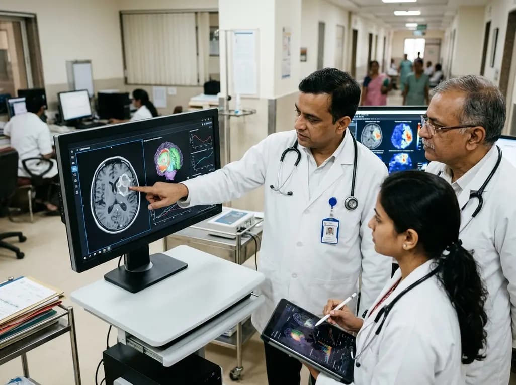
Tracking disease progression over time is essential for managing neuro-oncology patients. Specifically, longitudinal brain tumor response assessment guides critical treatment choices and defines progression-free survival in clinical practice and trials. Currently, clinicians evaluate treatment efficacy by reviewing serial magnetic resonance imaging (MRI) scans acquired at multi-week intervals. However, manual response evaluation presents formidable operational hurdles. Radiologists must measure perpendicular lesion diameters across multiple sequences. Consequently, this manual process demands substantial time and introduces notable interobserver variability. In busy clinical workflows, measurement discrepancies can cause premature treatment cessation or delayed detection of recurrence. Furthermore, post-radiation effects and pseudoprogression frequently confound visual assessments by mimicking true tumor progression on contrast-enhanced scans. As novel targeted therapies and immunotherapies expand, clinicians require objective, reproducible response metrics. Automated computational frameworks offer an effective solution by replacing subjective manual measurements with algorithmic precision.
Recent developments in computational neuroscience have expanded the role of artificial intelligence in oncology. Specifically, machine learning for brain tumor response assessment enables automated segmentation of complex intracranial lesions. Deep neural networks process multi-parametric MRI sequences, isolating enhancing tumor cores, non-enhancing infiltration, and necrotic cavities with high fidelity. Rather than relying on simplified bidimensional calipers, machine learning algorithms calculate continuous 3D volumetric changes across time. A comprehensive systematic review analyzed twenty studies evaluating automated response pipelines in adult brain tumors, including glioblastoma and metastases. Most evaluated algorithms emulated established criteria, such as the Response Assessment in Neuro-Oncology (RANO) framework. When compared against expert neuroradiologists, automated systems demonstrated strong diagnostic accuracy and segmentation performance. Furthermore, volumetric algorithms detected subtle tumor progression earlier than traditional two-dimensional metrics. Therefore, machine learning pipelines provide a more reliable foundation for tracking longitudinal treatment efficacy.
Although artificial intelligence algorithms demonstrate promising technical performance, systematic analysis reveals significant methodological shortcomings across published literature. Researchers utilized the QUADAS-2 tool and the CLAIM checklist to assess the quality of twenty longitudinal studies. Crucially, the investigators identified a high or unclear risk of bias in the majority of evaluated papers. Patient selection bias was common, as most investigations evaluated retrospective single-center cohorts with strict exclusion criteria. Consequently, these curated datasets fail to represent the noisy imaging data encountered in routine clinical practice. Furthermore, index test pipelines lacked standardized image preprocessing, including skull stripping and intensity calibration. Reference standard validation also showed notable weaknesses, often relying on unblinded local radiologist consensus rather than multi-expert review or histological confirmation. Because real-world generalizability remains unproven across diverse scanner vendors, clinicians must interpret published performance metrics with appropriate caution.
The introduction of updated RANO 2.0 criteria represents a pivotal evolution in neuro-oncology response standardization. RANO 2.0 unifies response definitions across adult low-grade and high-grade gliomas, establishing strict guidelines for measurable disease. Nevertheless, RANO 2.0 still utilizes bidimensional product measurements for routine use because manual volumetry remains unfeasible during standard clinical workflows. Automated machine learning pipelines effectively eliminate this operational bottleneck. Algorithms can rapidly extract total 3D tumor volume and generate longitudinal subtraction maps within seconds. However, machine learning models must strictly mirror RANO 2.0 rules. For example, algorithms must reliably distinguish non-enhancing tumor progression on T2-FLAIR imaging from benign radiation-induced changes. Additionally, post-operative cavity collapse and blood degradation products often confuse naive segmentation algorithms. Therefore, developers must train next-generation models on longitudinal datasets annotated according to RANO 2.0 standards, ensuring smooth integration into clinical trials.
Translating automated neuro-imaging tools into everyday hospital practice requires addressing notable practical and logistical hurdles. In real-world clinics, artificial intelligence cannot operate as an opaque black box. Instead, algorithms must integrate directly into Picture Archiving and Communication Systems (PACS) and electronic health records. Clinicians require intuitive interfaces that display automated segmentations, confidence metrics, and quantitative volumetric trajectories. Moreover, automated tools must maintain robust performance despite real-world imaging artifacts, motion degradation, and varying slice thicknesses. In diverse healthcare environments, such as medical centers across India, MRI protocols and hardware vary considerably between institutions. Machine learning models trained solely on homogeneous academic datasets often suffer performance degradation in community settings. Therefore, multicenter validation across diverse patient populations remains essential before widespread clinical adoption. Regulatory compliance and algorithmic transparency will further ensure these tools deliver practical clinical utility.
To advance automated longitudinal response tracking into standard practice, future investigations must prioritize prospective multicenter research designs. First, the scientific community must establish open-access longitudinal imaging repositories containing standardized multi-parametric MRI scans linked to clinical outcomes. These public datasets will enable transparent benchmarking of novel machine learning architectures. Second, future studies must adhere strictly to standardized reporting guidelines, including CLAIM and STARD-AI protocols. Third, clinical trial investigators should include automated volumetry as secondary endpoints alongside standard manual RANO assessments. Evaluating automated algorithmic predictions against overall survival in phase II and phase III trials will confirm the true clinical utility of digital volumetry. Furthermore, researchers must assess how automated response notifications affect oncologists' clinical decisions. Through rigorous prospective validation and open scientific collaboration, automated response tracking can evolve into an essential pillar of precision neuro-oncology.
Automated tools reduce interobserver variability and save clinical time. While manual methods rely on simple bidimensional measurements, machine learning algorithms calculate precise three-dimensional volumetric tumor burden across sequential MRI scans. Consequently, this technology captures irregular lesion morphology and identifies subtle disease progression earlier than conventional radiologist reviews.
Most included studies analyzed retrospective, single-center cohorts with restrictive selection criteria, limiting real-world applicability. In addition, many investigations lacked standardized image preprocessing, varied substantially in test pipelines, and relied on unblinded local radiologist consensus as the reference standard rather than multi-expert panels or histological validation.
RANO 2.0 unifies response evaluation for adult gliomas while updating measurement rules. Automated machine learning models must adapt to these guidelines by reliably distinguishing true tumor progression from post-treatment pseudoprogression, radiation necrosis, and surgical cavity changes across multi-parametric MRI sequences in both clinical trials and routine practice.
Disclaimer: This content is for informational and educational purposes only and should not be considered medical advice. Healthcare professionals should make clinical decisions based on their independent judgement and individual patient assessments. While we strive to provide accurate and up-to-date information, medical knowledge is constantly evolving, and we cannot guarantee that all information provided is entirely accurate, complete, or current. Readers are encouraged to verify information with other reliable sources. Medical practices and regulations may vary across different regions. Refer to the latest local and national guidelines for clinical practice.
References

Read summarized clinical updates, watch expert medical content, and earn CME certifications right from your smartphone.
Open your smartphone camera to scan and install.





A systematic review evaluates machine learning models for longitudinal brain tumor response assessment. While AI demonstrates high accuracy in volumetric tracking, study biases and limited real-world validation highlight the need for prospective studies aligned with RANO 2.0.
last month

A systematic review of 41 preclinical studies reveals that baicalin dampens systemic inflammation, inhibits NF-κB and NLRP3 pathways, and improves organ preservation in sepsis models. However, substantial study heterogeneity and a complete lack of human clinical trials highlight the need for further translational research.
Today
Discover the crucial distinctions between mesonephric and mesonephric-like proliferations of the female genital tract. Learn the key histomorphologic, immunohistochemical, and molecular differences essential for accurate diagnosis and clinical management.
Today

A large UK Biobank study reveals that plasma proteomic signatures capture preclinical organ damage and improve multiorgan risk prediction across early CKM syndrome stages 0 to 2 beyond traditional PREVENT clinical models.
Today
A narrative review investigates whether the articularis genus muscle functions as an independent anatomical entity or blends with the vastus intermedius. We evaluate its morphology, role in retracting the suprapatellar bursa, and direct clinical significance in anterior knee pain and arthroplasty.
Today

A Bayesian multilevel meta-analysis reveals that aerobic training combined with moderate carbohydrate restriction modestly lowers HbA1c in type 2 diabetes. However, sparse data and very low certainty leave incremental benefits over exercise or diet alone unproven, highlighting the need for individualized care.
Today
