.webp&w=384&q=75)

Loading, please wait...
Loading, please wait...

Drug-resistant focal epilepsy in young children presents profound neurodevelopmental and cognitive challenges when standard pharmacotherapy fails to control seizures. Pediatric epilepsy teams rely heavily on advanced magnetic resonance imaging to detect underlying pathologies such as focal cortical dysplasia and low-grade epilepsy-associated tumors. However, conventional structural sequences do not always demonstrate the entire epileptogenic network driving recurrent paroxysmal activity. Recent neuroimaging research highlights non-invasive arterial spin labeling as an essential functional modality to overcome this diagnostic obstacle. By accurately capturing regional microvascular alterations, arterial spin labeling reveals physiological disturbances extending beyond macroscopic anatomical lesion boundaries. Consequently, incorporating perfusion sequences into presurgical planning offers significant promise for improving surgical outcomes and securing lasting seizure freedom.
Children suffering from refractory focal lesional epilepsy experience recurrent seizures that severely compromise cognitive maturation and social development. Among surgical candidates, focal cortical dysplasia (FCD) and low-grade epilepsy-associated tumors (LEATs) represent the most frequent structural etiologies. Therefore, early and complete surgical resection provides the most definitive opportunity to achieve cure. Nevertheless, delineating the true boundary of the epileptogenic zone remains exceptionally demanding using standard anatomical magnetic resonance sequences alone. Many cortical malformations present subtle blurring of the gray-white matter junction that defies clear visual demarcation. In addition, dysplastic neural tissue and epileptogenic networks frequently spread several millimeters into adjacent, visually normal cortex. If neurosurgeons resect solely the visible structural lesion, residual epileptogenic cortex often continues generating debilitating seizures postoperatively. Consequently, a notable proportion of pediatric patients fail to attain seizure freedom despite undergoing extensive craniotomy. Furthermore, invasive intracranial monitoring entails procedural risks, elevated costs, and specialized institutional resources that are not universally accessible. Because of these persistent limitations, pediatric epilepsy teams urgently require non-invasive functional imaging tools capable of defining true pathological margins.
Arterial spin labeling delivers non-invasive quantitative evaluation of cerebral perfusion without requiring exogenous gadolinium contrast agents. Instead of using intravenous chemicals, the sequence applies radiofrequency pulses to magnetically label water protons in cervical arterial blood. These labeled protons travel into the cerebral microvasculature, providing a completely endogenous hemodynamic tracer. Consequently, pediatric patients avoid repeated intravenous cannulation, nephrotoxicity risks, and intracranial gadolinium accumulation. In epileptic disorders, localized microvascular perfusion changes directly reflect underlying interictal and ictal neuronal hyper-synchrony. Paroxysmal network activity typically manifests as focal hypoperfusion or localized hyperperfusion during neuroimaging evaluation. Therefore, arterial spin labeling successfully detects functional microcirculatory abnormalities in cortical regions that appear normal on conventional T1- or T2-weighted scans. Furthermore, modern pseudo-continuous labeling protocols provide exceptional signal-to-noise ratios and spatial resolution on standard clinical scanners. Multidisciplinary teams can subsequently coregister these hemodynamic maps directly with volumetric anatomical datasets. As a result, clinicians obtain an integrated multiparametric roadmap that combines structural architecture with functional hemodynamic boundaries before entering the operating suite.
A recent pilot investigation by Dr. Antonio Giulio Gennari and colleagues evaluated 18 pediatric patients undergoing surgery for pharmacoresistant lesional epilepsy. The cohort had a median age of 4.8 years, with confirmed histological diagnoses of FCD or LEATs. Every participant completed presurgical arterial spin labeling alongside high-resolution structural MRI, followed by at least one year of postoperative clinical monitoring. To investigate whether perfusion margins predicted surgical outcomes, the researchers employed advanced computational image postprocessing. Specifically, they segmented and coregistered presurgical anatomical lesions, arterial spin labeling perfusion alterations, and postoperative resection cavities. The investigators utilized DICE similarity coefficient scores to objectively quantify the geometric alignment between presurgical target volumes and the resected cavity. In addition, they calculated the residue ratio to measure the exact percentage of presurgical perfusion abnormality remaining postoperatively. By relying on rigorous volumetric quantification rather than subjective visual interpretation, the researchers minimized operator bias. Consequently, this computational framework generated robust objective metrics to evaluate whether residual perfusion abnormalities directly correlated with persistent postsurgical seizures.
The study yielded remarkable findings that challenge conventional assumptions regarding epilepsy surgery planning. Overall, 14 of the 18 children (78%) achieved complete seizure freedom, while 13 patients (72%) achieved complete structural lesion resection. Crucially, qualitative analysis demonstrated that complete resection of the anatomical lesion did not correlate significantly with postsurgical seizure freedom. In contrast, complete inclusion of arterial spin labeling perfusion abnormalities within the surgical resection cavity strongly predicted seizure freedom. Quantitative volumetric metrics corroborated these qualitative conclusions with remarkable statistical clarity. Specifically, higher DICE similarity scores between perfusion alterations and the resection cavity significantly correlated with seizure-free outcomes. Conversely, anatomical lesion alignment with the resection cavity showed no statistically significant relationship with seizure control. Furthermore, patients with larger residual volumes of abnormal perfusion exhibited a statistically significant rate of seizure recurrence. These objective data confirm that the epileptogenic zone frequently exceeds visible anatomical lesion boundaries. Therefore, removing the surrounding functionally abnormal tissue remains essential for achieving lasting seizure control.
These findings provide clear guidance for neurosurgical teams seeking to refine resection margins in pediatric epilepsy. Traditionally, surgeons restrict resections to obvious structural margins to avoid damaging adjacent functional brain networks. However, un-resected perilesional dysplastic tissue frequently continues to trigger postoperative seizures if functional boundaries remain unaddressed. By integrating perfusion maps into stereotactic neuronavigation platforms, neurosurgeons can visualize both structural lesions and abnormal microvascular zones intraoperatively. Consequently, surgical teams can optimize cortical resection boundaries safely, especially when perfusion anomalies occupy non-eloquent territory. Furthermore, these imaging principles hold substantial practical value for tertiary epilepsy care centers across India. While advanced metabolic modalities like positron emission tomography are expensive and geographically restricted, standard 3.0 Tesla or 1.5 Tesla MRI scanners are widely available. Healthcare facilities can readily add non-contrast arterial spin labeling sequences to existing epilepsy imaging protocols without incurring extra consumable costs. Ultimately, adopting this accessible functional sequence can optimize surgical precision, reduce invasive monitoring needs, and improve clinical outcomes for pediatric epilepsy patients nationwide.
Arterial spin labeling magnetically tags water protons in cervical arterial blood before imaging the brain, using endogenous blood as a tracer. In pediatric focal epilepsy, localized neuronal hypersynchrony alters regional microvascular autoregulation. This sequence detects focal interictal hypoperfusion or peri-ictal hyperperfusion, accurately identifying dysfunctional epileptogenic networks extending beyond structural MRI margins.
Focal cortical dysplasias and low-grade epilepsy-associated tumors frequently possess microscopic dysplastic boundaries and perilesional epileptogenic networks extending beyond visible MRI abnormalities. Consequently, resecting solely the visible anatomical lesion often leaves active epileptogenic cortex intact. Resecting the perfusion-defined abnormality ensures comprehensive removal of the functional epileptogenic zone, securing superior postoperative seizure freedom.
Yes, tertiary Indian medical centers with standard 1.5 Tesla or 3.0 Tesla MRI systems can readily implement arterial spin labeling protocols. The sequence requires neither intravenous contrast agents nor expensive cyclotron-dependent radiotracers. Therefore, it offers an accessible, non-invasive, and cost-effective functional imaging solution for pre-surgical pediatric epilepsy evaluation nationwide.
Disclaimer: This content is for informational and educational purposes only and does not constitute medical advice, diagnosis, or treatment. Healthcare professionals should use their clinical judgment and review complete prescribing information and guidelines. The views expressed do not necessarily reflect the official policy or position of any institution. Refer to the latest local and national guidelines for clinical practice.
References

Read summarized clinical updates, watch expert medical content, and earn CME certifications right from your smartphone.
Open your smartphone camera to scan and install.





A pilot study demonstrates that integrating non-invasive arterial spin labeling perfusion MRI into presurgical planning predicts seizure freedom in pediatric focal lesional epilepsy, showing that complete resection of perfusion abnormalities significantly outperforms conventional structural lesion margins.
Today

Survival analyses often report hazard ratios, but mathematical non-collapsibility creates crucial differences between conditional and marginal estimates. Clinicians must account for unmeasured confounding and model selection when interpreting trial endpoints and real-world cardiovascular outcomes.
Today

A cross-sectional study of 700 college students highlights significant contraceptive knowledge disparities between sexually active and inactive young adults. The findings demonstrate a critical need for proactive, comprehensive sexual health counseling before sexual debut.
Yesterday
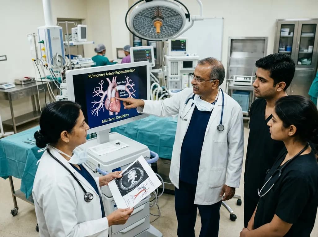
Autologous tissue-engineered vascular graft patches created via in-body tissue architecture show excellent mid-term durability and safety for pulmonary artery reconstruction in congenital heart surgery, particularly for peripheral branch augmentation.
Today

A novel cluster analysis of real-world datasets reveals four distinct CPPD disease phenotypes, validating existing EULAR categories while uncovering previously unrecognized monoarticular and axial presentations to refine patient management.
Today

Recent pharmacokinetic research demonstrates that trace dermal exposure to microgram quantities of trenbolone and metenolone produces detectable urinary metabolites for days to over a week, providing critical objective data for evaluating unintentional contamination claims in sports drug testing.
Today
