.webp&w=384&q=75)

Loading, please wait...
Loading, please wait...
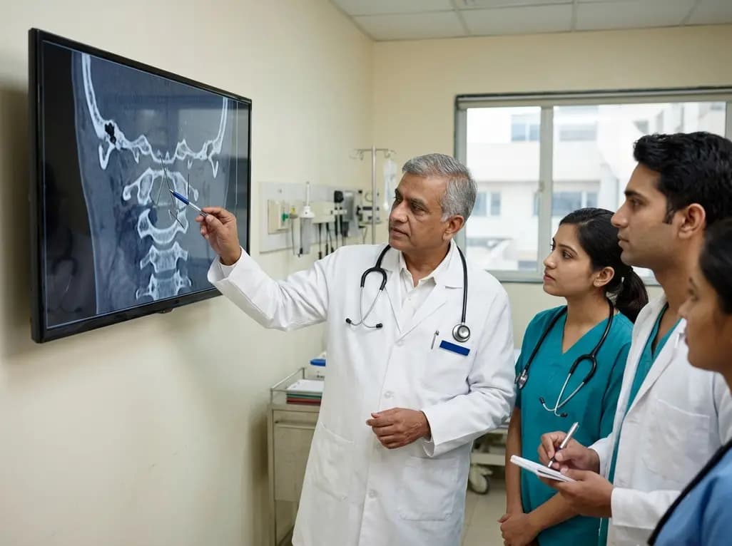
Managing an upper cervical spine injury presents intricate challenges for trauma surgeons, spine specialists, and critical care teams worldwide. The craniocervical junction and high cervical spine exhibit unique biomechanical complexity, mobility, and distinct anatomical architecture compared to subaxial segments. Historically, spine clinicians relied on isolated, eponymous classification schemes for separate anatomical parts, such as occipital condyle fractures, atlas fractures, and axis fractures. However, these fragmented systems lacked unified prognostic value and failed to provide a continuous scale of injury severity. To overcome these limitations, the AO Spine Knowledge Forum Trauma designed a comprehensive, unified taxonomy. This system systematically standardizes how clinicians characterize craniocervical and upper cervical trauma. Recent global validation studies have critically appraised the hierarchical progression of this classification scheme. Specifically, researchers evaluated whether perceived severity scores align consistently across international geographical regions, diverse surgical subspecialties, and differing levels of clinical experience. Consequently, establishing this hierarchical integrity provides a reliable foundation for future treatment algorithms and international surgical trials.
The AO Spine Upper Cervical Injury Classification divides trauma into three distinct anatomical regions based on structural localization. Region I encompasses fractures involving the occipital condyle and the craniocervical junction. Region II covers trauma to the atlas ring and the atlantoaxial joint complex. Region III pertains to injuries affecting the axis body, dens, and the C2-C3 articular joint. Within each distinct anatomical region, fractures follow a clear hierarchical morphology graded as Type A, Type B, or Type C. Type A represents isolated bony fractures that typically spare the primary ligamentous stabilizers. Furthermore, Type B designates severe tension band or ligamentous disruption without major displacement. Type C represents severe translational, displaced, or multidirectional instability patterns. Therefore, as morphology progresses from Type A through Type C, biomechanical disruption and intrinsic instability increase dramatically. In addition, this predictable progression helps clinicians rapidly determine if conservative bracing or emergent operative stabilization is warranted during trauma triage.
In addition to structural injury morphology, the classification system incorporates an objective neurological assessment alongside patient-specific modifiers. The neurological grading designates patients from N0, indicating an intact neurological status, through N1 for transient deficits, N2 for radicular signs, N3 for incomplete spinal cord injury, and N4 for complete spinal cord compromise. Clinicians assign NX when patients cannot undergo adequate examination due to intoxication or severe traumatic brain injury. Furthermore, the system includes four crucial clinical modifiers denoted as M1 through M4. Modifier M1 identifies injury patterns carrying a significant risk of bony nonunion. Modifier M2 highlights severe ligamentous injury or latent instability patterns that might fail conservative management. In contrast, Modifier M3 flags patient-specific risk factors, such as ankylosing spondylitis or severe osteoporosis, whereas Modifier M4 designates traumatic vertebral artery injury. Consequently, these comprehensive variables capture both the immediate neurovascular threat and long-term structural vulnerability.
To validate the hierarchical integrity of the system, a multicenter international study surveyed 151 AO Spine members across diverse practice environments. Participating orthopaedic spine surgeons and neurosurgeons scored the perceived severity of each classification component on an injury severity scale ranging from zero to one hundred. Notably, the study revealed a significant step-wise increase in median perceived severity as fractures advanced from Type A to Type B and ultimately to Type C across all anatomical regions. Similarly, neurological status scores showed a clear upward trajectory in perceived severity from intact function to complete paralysis, although transient deficits and radiculopathies received comparable ratings. Moreover, surgeons rated the instability modifier M2 significantly more severe than the patient-specific modifier M3. Most importantly, perceived injury severity scores remained highly consistent regardless of surgeon years in practice. Thus, these findings validate the innate hierarchical nature of the system across diverse clinical backgrounds.
The survey results demonstrated remarkable consensus across global regions and surgical disciplines, confirming broad international applicability. However, subtle differences emerged among specific geographic territories and surgical subspecialties during statistical analysis. For instance, respondents from North America, Central America, and South America perceived displaced craniocervical disruptions and complex tension-band injuries somewhat more severely than surgeons from other regions. Furthermore, neurosurgeons perceived craniocervical ligamentous injuries and complex C2 tension-band disruptions as slightly more critical compared to orthopaedic colleagues. In addition, these nuances likely reflect differences in regional trauma protocols, training pathways, and health resource availability. Nevertheless, the overarching injury severity ranking remained remarkably robust across all international cohorts. Consequently, the classification provides a universal common language, facilitating seamless multidisciplinary communication between emergency physicians, radiologists, neurosurgeons, and orthopaedic trauma specialists.
Validating the hierarchical nature of this system carries vital clinical implications for contemporary spine trauma management. Standardized injury stratification allows multidisciplinary trauma teams to quickly triage patients and predict treatment failure. For example, lower-tier Type A injuries without neurological deficit typically achieve successful healing with nonoperative rigid immobilization. Conversely, Type B and Type C injuries with concomitant neurovascular compromise or M1/M2 modifiers frequently require urgent surgical decompression and posterior instrumented stabilization. Furthermore, integrating these validated parameters into electronic medical records streamlines clinical documentation and clinical quality registries worldwide. As trauma centers adopt this standardized framework, collaborative research initiatives can more effectively compare conservative versus surgical outcomes. Ultimately, implementing this robust hierarchical classification enhances diagnostic precision, standardizes clinical communication, and supports evidence-based therapeutic decisions in complex cervical spine trauma.
The system evaluates three core components: anatomical injury morphology across regions I to III graded into types A through C, neurological status ranging from N0 to N4 or NX, and case-specific modifiers designated as M1 to M4 addressing nonunion, instability, patient comorbidities, and vascular injuries.
Hierarchical validation confirms that assigned injury scores correspond directly to perceived clinical severity and biomechanical instability. Consequently, this structured hierarchy provides clinicians with a reliable, standardized prognostic tool to guide clinical decision-making, surgical indications, and future international multicenter comparative research.
Overall concordance between surgical specialties is remarkably high. However, neurosurgeons tend to score craniocervical ligamentous disruptions and complex C2-C3 tension-band injuries slightly higher in perceived severity than orthopaedic surgeons, reflecting distinct subspecialty training emphases on craniocervical junction kinematics and neurovascular protection.
Disclaimer: This content is for informational and educational purposes only and should not be considered medical advice. Always consult a qualified healthcare professional regarding any medical condition or treatment. Refer to the latest local and national guidelines for clinical practice.
References

Read summarized clinical updates, watch expert medical content, and earn CME certifications right from your smartphone.
Open your smartphone camera to scan and install.





A global validation study confirms the hierarchical integrity of the AO Spine Upper Cervical Spine Injury Classification. Perceived injury severity increases systematically across anatomical subtypes and neurological grades regardless of surgeon experience or geographic setting.
4 weeks back

A Bayesian multilevel meta-analysis reveals that aerobic training combined with moderate carbohydrate restriction modestly lowers HbA1c in type 2 diabetes. However, sparse data and very low certainty leave incremental benefits over exercise or diet alone unproven, highlighting the need for individualized care.
Today

Findings from Project Viva indicate that sustained postpartum weight retention, rather than excessive gestational weight gain alone, directly worsens women's mid-life cardiovascular health, degrading blood pressure and glycemic parameters decades after delivery.
Today

A large UK Biobank study reveals that plasma proteomic signatures capture preclinical organ damage and improve multiorgan risk prediction across early CKM syndrome stages 0 to 2 beyond traditional PREVENT clinical models.
Today

A retrospective CBCT study reveals that superior TMJ space asymmetry correlates moderately with occipital condyle-atlas-axis asymmetry in unilateral TMJ osteoarthritis, highlighting an imaging correlate between jaw biomechanics and upper cervical alignment rather than a standalone diagnostic marker.
Today
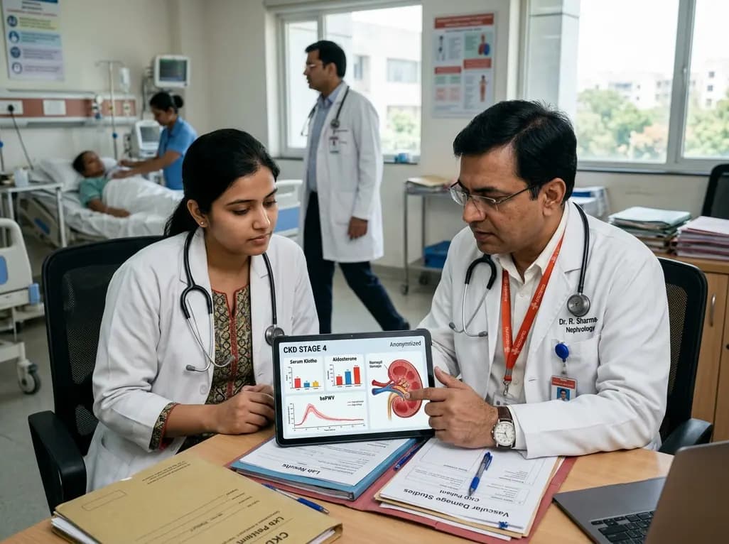
A cross-sectional investigation highlights the complex pathophysiological links between serum Klotho deficiency, secondary hyperaldosteronism, and vascular damage in CKD. Discover key insights into non-invasive vascular stiffness markers and targeted therapeutic pathways for cardiorenal protection.
Today
