.webp&w=384&q=75)

Loading, please wait...
Loading, please wait...
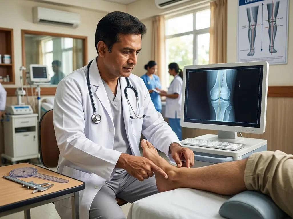
Total knee arthroplasty (TKA) requires precise bone resections to ensure long-term implant survival. Obtaining optimal tibial coronal alignment remains a major debate among orthopedic surgeons worldwide. Although modern robotic systems and computer navigation assist alignment, they increase operative costs and complexity. Recent radiological research demonstrates that easily palpable ankle landmarks offer reproducible references. These surface boundaries allow surgeons to safely execute personalized alignment strategies using standard extramedullary instrumentation.
Achieving proper alignment during total knee replacement remains critical for preventing early mechanical failure. Misalignment often creates asymmetrical stress distribution across the polyethylene insert. Consequently, excessive tibial component varus or valgus tilt accelerates polyethylene wear and risks aseptic loosening. Historically, surgeons aimed for a neutral mechanical axis of zero degrees to maximize implant longevity. However, recent trends favor personalized alignment strategies, such as kinematic or functional alignment, to improve clinical satisfaction and natural knee kinematics.
Despite these advancements, defining safe surgical boundaries without expensive technology remains challenging. Advanced robotic platforms calculate patient-specific alignment, yet they demand extra surgical time, costly capital investment, and extensive training. Furthermore, digital navigation systems still rely on manual identification of distal bony landmarks by the surgeon. Therefore, orthopaedic researchers evaluated whether direct palpation of distal ankle structures could offer the same precision during standard extramedullary cutting guide setup. By clarifying the exact geometrical relationship between the tibial mechanical axis and surface anatomical landmarks, clinicians can establish reliable reference lines without requiring computerized tracking units during routine procedures.
To evaluate the clinical utility of distal ankle landmarks, investigators conducted a rigorous radiological trial. The team analyzed long-leg weight-bearing radiographs from a representative cohort of 119 patients, examining 167 distinct lower extremity X-rays. Two independent orthopedic surgeons performed all measurements at two separate time points spaced eight weeks apart to minimize recall bias. This standardized protocol ensured thorough evaluation of intra-rater and inter-rater reliability.
Specifically, the researchers projected the long mechanical axis of the tibia and measured three key anatomical angles. First, they evaluated the angle between the tibial mechanical axis and the lateral border of the lateral malleolus. Second, they measured the angle extending to the lateral border of the talus. Third, they determined the angular offset to the medial border of the medial malleolus. By systematically evaluating these distinct reference points on weight-bearing images, the study established precise numerical baselines for standard intraoperative alignment guides. In addition, statistical testing using intraclass correlation coefficients provided strong verification of measurement consistency across different clinical observers and evaluation intervals.
The quantitative results demonstrated remarkable consistency across all evaluated radiographic parameters. Specifically, the mean angular measurement for the lateral malleolus border was 4.8 degrees, with a narrow ninety-five percent confidence interval ranging between 4.7 and 4.8 degrees. Similarly, the medial malleolus border yielded a mean angle of 4.2 degrees. In contrast, the lateral border of the talus produced a smaller mean angular deviation of 2.6 degrees from the primary tibial mechanical axis.
Furthermore, statistical analysis confirmed exceptional reproducibility for these physical reference points. Inter-rater reliability reached an intraclass correlation coefficient of 0.84 for the lateral malleolus measurements and 0.80 for the medial malleolus readings. Although the lateral talar border demonstrated a slightly lower inter-rater agreement at 0.67, intra-rater reliability remained consistently high across all parameters, exceeding 0.80 for every measured index. These robust values confirm that surface ankle landmarks provide predictable anatomical trajectory lines. As a result, surgeons can confidently reference these palpable points during surgical exposure and extramedullary rod positioning to guide precision cuts without technological assistance.
The clinical implications of these radiological findings provide actionable guidance for modern knee arthroplasty surgeons. By recognizing these consistent anatomical relationships, clinicians can establish definitive safe boundaries when performing personalized surgical cuts. For example, referencing the lateral malleolus border establishes a predictable safe threshold corresponding to 4.8 degrees of tibial component varus. Conversely, aligning the extramedullary rod toward the medial malleolus border defines a safe limit of 4.2 degrees of tibial valgus cut.
Additionally, checking alignment against the lateral border of the talus provides a precise landmark equivalent to a 2.6-degree varus cut. Utilizing these tangible anatomical landmarks empowers surgeons to customize coronal alignment based on individual patient anatomy. Consequently, operating teams can avoid dangerous mechanical extremes that predispose components to early loosening or edge loading. Most importantly, surgeons can execute these personalized alignment protocols using standard, widely available extramedullary alignment jigs. Therefore, this straightforward anatomical method delivers high clinical precision, reduces financial expenditures, and avoids the added intraoperative setup time required by robotic or computer-guided systems.
These findings hold immense practical significance for orthopaedic practice in India and other developing healthcare ecosystems. High-volume joint replacement centers in India perform thousands of knee arthroplasties annually, yet access to costly robotic systems remains restricted to premier private hospitals. By utilizing simple, palpable ankle landmarks, orthopedic surgeons working in tier-two and tier-three centers can deliver personalized alignment with superior safety and precision.
Moreover, reducing reliance on advanced navigation units lowers total procedure costs for patients and healthcare institutions. Indian patients often present with severe varus deformity and secondary soft-tissue contractures, making accurate extramedullary alignment critical. Utilizing these validated physical landmarks provides operational confidence during complex primary replacements. Furthermore, surgical trainees can easily master these palpable reference points during residency programs. Incorporating these simple visual checks into standardized surgical workflows optimizes component positioning and reduces post-operative alignment outliers. Ultimately, integrating these validated ankle references enhances surgical efficiency, improves functional patient outcomes, and democratizes high-quality joint replacement care across diverse healthcare settings.
Palpable ankle landmarks correlate consistently with the tibial mechanical axis on weight-bearing radiographs. By aligning extramedullary cutting guides to the lateral malleolus, medial malleolus, or lateral talar border, surgeons establish predictable alignment angles. Specifically, these surface boundaries define safe limits of 4.8 degrees varus or 4.2 degrees valgus. Consequently, operating teams achieve personalized component positioning accurately without relying on expensive robotic or navigation technologies.
The radiographic study measured mean angles between the tibial mechanical axis and three distinct ankle landmarks. The lateral border of the lateral malleolus measured 4.8 degrees, establishing a reliable safe varus limit. The medial border of the medial malleolus measured 4.2 degrees, establishing a reliable safe valgus limit. Finally, aligning the extramedullary guide to the lateral border of the talus produced a precise 2.6-degree varus cut angle.
High inter-rater reliability ensures that different surgeons can consistently identify identical anatomical references during knee arthroplasty procedures. In this study, intra-class correlation coefficients reached 0.84 for the lateral malleolus and 0.80 for the medial malleolus. These high reliability scores confirm that surface palpation of malleolar boundaries provides predictable, highly reproducible guidance across diverse operating teams, minimizing human error during component positioning.
Disclaimer: This content is for informational and educational purposes only and does not constitute medical advice, diagnosis, or treatment. Always seek the advice of a qualified healthcare provider with any questions you may have regarding a medical condition or surgical procedure. Refer to the latest local and national guidelines for clinical practice.
References

Read summarized clinical updates, watch expert medical content, and earn CME certifications right from your smartphone.
Open your smartphone camera to scan and install.





A radiological study reveals that palpable ankle malleoli offer reliable references for tibial coronal alignment in TKA, enabling safe limits of 4.8° varus or 4.2° valgus with standard instruments.
2 months ago
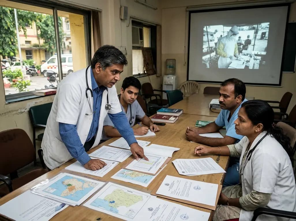
Following Nigeria's first recorded Ebola case linked to regional travel, the Africa CDC has convened an emergency ministerial summit. This alert underscores critical clinical priorities for healthcare professionals, including differentiating early viral infection from malaria, implementing barrier nursing, and triage.
Today

Leucine Rich Bio has introduced South Asia's first comprehensive vaginal microbiome test, BugSpeaks. By profiling microbial dysbiosis, pathogens, and antimicrobial resistance, this next-generation diagnostic platform provides clinicians with deeper insights into recurrent infections, fertility, and reproductive health.
Today

Review how tailored exercise programs benefit frail older patients with heart failure. Explore pathophysiological mechanisms, individualized training prescriptions, and strategies to overcome adherence barriers in clinical practice.
Today

This comparative study evaluates posterior labral tears with and without glenohumeral instability, highlighting distinct demographic patterns, MRI underestimation in stable shoulders, and successful arthroscopic repair outcomes.
Today

A clinical study demonstrates that early intervention with tocilizumab, either as monotherapy or bridged with corticosteroids, achieves significant proptosis reduction and resolves active inflammation in treatment-naive thyroid eye disease.
Today
