.webp&w=384&q=75)

Loading, please wait...
Loading, please wait...

Major depressive disorder represents a leading cause of global disability, yet its clinical manifestations remain remarkably diverse. Among its most challenging features, anhedonia in MDD presents a significant therapeutic obstacle for practicing physicians. Clinicians define anhedonia as the diminished ability to anticipate, experience, or pursue pleasure. While conventional antidepressants often stabilize negative affect, they frequently fail to resolve hedonic deficits. Consequently, affected individuals experience protracted functional impairment and an increased risk of treatment resistance. Understanding the biological underpinnings of this persistent deficit is therefore vital for developing more effective clinical strategies.
Recent advances in translational psychiatry emphasize that major depression is not a uniform neurobiological disorder. Instead, distinct clinical phenotypes arise from specific neuropathological mechanisms. To examine these differences, investigators deployed ultra-high-field 7 Tesla magnetic resonance imaging to assess cortical morphology in depressed cohorts. They stratified participants into nineteen patients with high anhedonia, nineteen patients with low anhedonia, and twenty matched healthy controls. The researchers utilized validated interest-activity psychometric metrics to differentiate reward processing deficits.
The neuroimaging results revealed widespread cortical thickness reductions across both patient groups compared with healthy individuals. However, patients presenting with high anhedonia exhibited significantly more pronounced cortical thinning than their low-anhedonia peers. Standard clinical scanners often miss these subtle structural variations due to limited spatial resolution. In contrast, 7 Tesla neuroimaging provided the resolution required to identify these fine morphological shifts across cortical boundaries. These findings demonstrate that anhedonia correlates with a distinct neuroanatomical profile characterized by accelerated gray matter loss. Thus, severe reward blunting serves as a marker for extensive structural degradation.
Determining how macroscale structural thinning connects to microscale molecular biology represents a central frontier in neuropsychiatry. To bridge this divide, the researchers correlated spatial neuroimaging patterns with whole-brain transcriptomic datasets from the Allen Human Brain Atlas. This spatial analysis demonstrated that regional cortical thinning significantly aligns with specific cortical gene expression profiles. In particular, transcriptional signatures linked to anhedonic atrophy were connected to mitochondrial dysfunction and energy failure.
Functional pathway enrichment analyses revealed that anhedonia-related genes participate prominently in oxidative phosphorylation, adipogenesis, and mTORC1 signaling pathways. Under physiological conditions, neuronal and synaptic survival demands continuous mitochondrial adenosine triphosphate generation. When mitochondrial function falters, oxidative stress escalates, leading to cellular exhaustion and progressive dendritic spine elimination. Furthermore, dysregulation of mTORC1 signaling disrupts local protein synthesis necessary for maintaining synaptic plasticity. Consequently, vulnerable brain regions cannot sustain normal structural complexity. These discoveries firmly link macroscopic cortical thinning in depressed individuals to fundamental defects in cellular respiration and metabolic signaling.
Psychiatric research historically emphasized neuronal abnormalities, but contemporary neurobiology highlights the indispensable contributions of non-neuronal cells. In this investigation, cell-type specific enrichment analyses revealed that anhedonia-related transcriptional signatures involve neurons, astrocytes, and oligodendrocytes. This multi-lineage involvement underscores that cortical thinning reflects widespread tissue remodeling rather than isolated neuronal loss. Therefore, addressing anhedonia requires a comprehensive cellular perspective.
Astrocytes fulfill critical homeostatic responsibilities, including glutamate clearance, blood-brain barrier maintenance, and metabolic fuel delivery to active neurons. When astrocyte gene expression falters, extracellular glutamate accumulates, promoting excitotoxic stress and synaptic loss. Similarly, oligodendrocytes provide metabolic sustenance through lactate shuttling and maintain myelin sheaths that facilitate rapid neuronal communication. The transcriptional vulnerability detected across oligodendrocytes suggests that impaired axonal insulation destabilizes cortical circuits. In addition, dysfunctional glia fail to buffer local oxidative stress generated by compromised mitochondria. As a result, non-neuronal cell failure directly compounds neuronal degeneration across vulnerable cortical territories. Recognizing this multi-cellular pathology helps clinicians appreciate the profound cellular damage accompanying severe anhedonia.
To contextualize these structural findings, the investigators analyzed cytoarchitectural variations and functional large-scale networks. By applying the classical von Economo-Koskinas atlas, they observed that high anhedonia manifested substantial cortical thinning in agranular cortical zones. Agranular cortices lack an internal granular layer and coordinate efferent motor projections and autonomic visceral responses. Consequently, degradation within these regions disrupts the physical translation of motivated impulses into purposeful actions.
Furthermore, network-level parcellations revealed significant cortical thickness reductions localized to the somatomotor and ventral attention networks. The ventral attention network directs cognitive focus toward novel and biologically meaningful environmental stimuli. Concurrently, the somatomotor network executes motivated behavioral outputs. When both networks sustain structural damage, patients display marked avolition, reduced environmental exploration, and physical apathy. In addition, functional meta-analytic decoding using Neurosynth demonstrated that these structural reductions correlate significantly with cognitive terms related to somatosensory processing and pain perception. This observation provides an intuitive neurobiological explanation for clinical presentations. Specifically, it clarifies why severely anhedonic individuals frequently report visceral emotional numbness alongside heightened physical pain sensitivity.
The discovery of distinct transcriptomic and structural substrates provides exciting opportunities for refining psychiatric practice. Traditional monoaminergic antidepressants, such as selective serotonin reuptake inhibitors, often demonstrate minimal efficacy against profound anhedonia. Because this research links hedonic deficits to mitochondrial dysfunction and disrupted mTORC1 signaling, clinicians can explore targeted metabolic and neuroplastic interventions. Therapeutic candidates that enhance mitochondrial bioenergetics or modulate rapid glutamatergic neurotransmission offer promising avenues to rescue faltering cortical circuits.
Additionally, combining high-resolution neuroimaging with transcriptomic profiling establishes a foundation for objective biomarker development. Using these quantitative parameters, clinicians could eventually stratify depressed patients at initial presentation, identifying individuals at high risk for persistent anhedonia. Although cross-sectional study designs and behavioral scoring metrics present unavoidable limitations, these findings offer a solid mechanistic blueprint. Future longitudinal investigations should evaluate whether novel neuroprotective agents can arrest or reverse cortical thinning over time. Ultimately, integrating molecular medicine into psychiatric care represents a vital transition toward personalized therapy, helping patients achieve meaningful functional recovery.
Ultra-high-field 7 Tesla MRI provides superior signal-to-noise ratio and exquisite spatial resolution compared to conventional imaging systems. Consequently, this technology enables clinicians and researchers to detect subtle, layer-specific cortical thickness reductions across distinct cortical regions. These fine structural alterations often escape detection when using standard clinical scanners.
Transcriptomic mapping links anhedonia-associated cortical thinning to severe mitochondrial dysfunction and impaired bioenergetic cascades. Specifically, gene enrichment analyses highlight dysregulated oxidative phosphorylation, adipogenesis pathways, and altered mTORC1 signaling. These cellular disruptions compromise metabolic homeostasis, which subsequently triggers synaptic degradation and localized morphological atrophy in vulnerable cortical regions.
Uncovering transcriptomic substrates allows clinicians to move beyond traditional monoaminergic antidepressant strategies. Because standard medications frequently fail to resolve severe hedonic deficits, identifying mitochondrial and mTORC1 involvement highlights promising targets for metabolic and neuroprotective pharmacotherapies. Furthermore, these molecular markers facilitate objective patient stratification and support personalized therapeutic interventions.
Disclaimer: This content is for informational and educational purposes only. It should not be used as a substitute for professional medical advice, diagnosis, or treatment. Always consult your physician or another qualified healthcare provider with any questions you may have regarding a medical condition. Refer to the latest local and national guidelines for clinical practice.
References

Read summarized clinical updates, watch expert medical content, and earn CME certifications right from your smartphone.
Open your smartphone camera to scan and install.





A landmark 7 Tesla imaging and transcriptomic study reveals that anhedonia in MDD drives pronounced cortical thinning linked to mitochondrial dysfunction, altered mTORC1 signaling, and glial pathology, offering novel biological targets for treatment-resistant depression.
Today

An Australian cohort of 34,449 adults in an unsubsidized tirzepatide digital weight loss service showed that peer-referred patients achieved significantly higher 6-month adherence (48% vs 42%) and greater weight loss (16.16% vs 14.06%) than matched non-referred peers, highlighting social support in obesity care.
Today
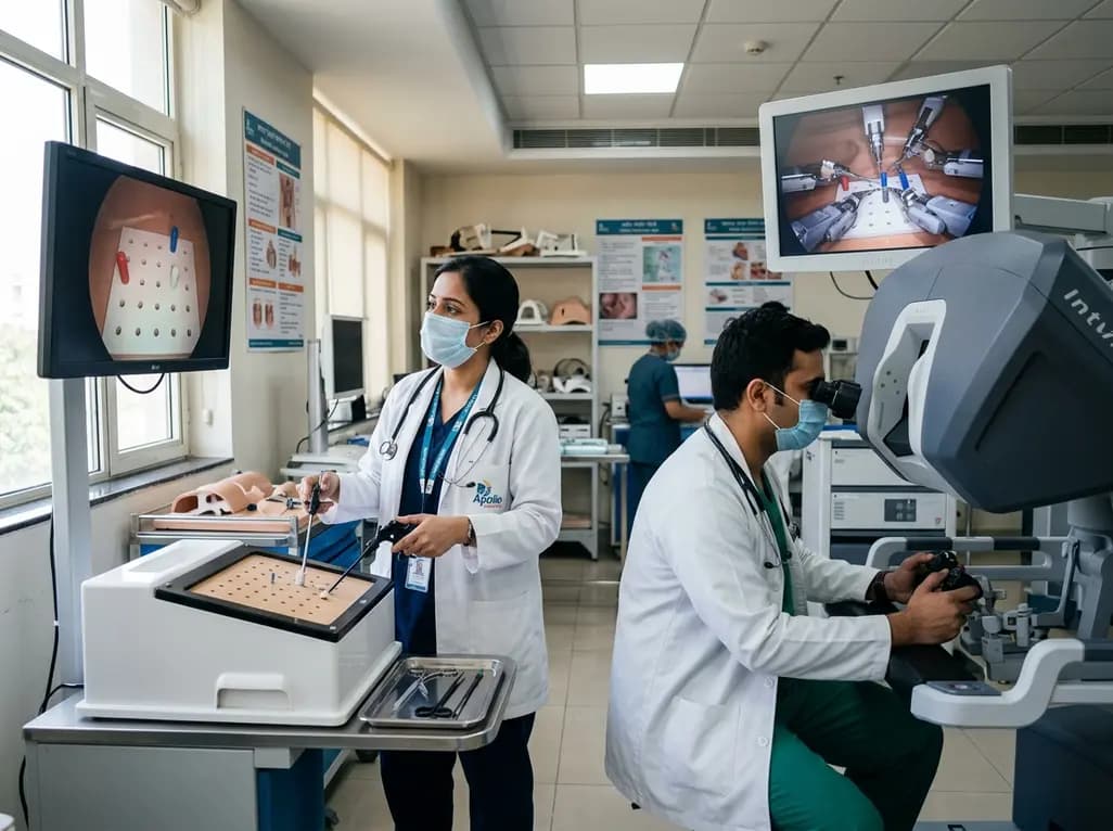
A randomized crossover study demonstrates that conventional and robotic laparoscopy require distinct neuropsychological and psychomotor skills. While trainees master conventional laparoscopy faster, robotic proficiency demands individualized, adaptive curricula tailored to specific learner profiles.
Today

A retrospective study of 109 patients undergoing elective carotid artery stenting evaluated radiation exposure on biplane flat-panel systems. Standardized workflows and dedicated low-dose protocols provide crucial baseline data to establish neurointerventional diagnostic reference levels and optimize patient safety.
Today

A scoping review of 51 studies reveals that spinal manual therapy on the lumbo-pelvic area is commonly used for isolated lower extremity pain, predominantly knee conditions. While thrust manipulation is frequent, reporting deficits highlight the need for standardized technique descriptions to guide practice.
Today

The Supreme Court has reserved its judgment on FSSAI's proposed front-of-pack nutritional warning labels for packaged foods. With debates over compliance timelines, added versus total sugars, and ultra-processed food definitions, this landmark decision carries profound public health implications for India.
Today
