

Loading, please wait...
Loading, please wait...

Aneurysmal subarachnoid hemorrhage often triggers severe delayed neurological deterioration. Clinicians frequently turn to cerebral vasospasm endovascular treatment when medical rescue strategies fail to restore adequate microvascular flow. However, emerging clinical evidence indicates a critical divergence between visual vascular restoration on digital subtraction angiography and ultimate patient recovery. Interventional neuroradiologists regularly achieve robust mechanical or pharmacological vasodilation in narrowed intracranial conduits. Nevertheless, patient recovery remains constrained by complex parenchymal ischemic insults.
Medical teams encounter post-hemorrhagic cerebral vasospasm in up to seventy percent of patients surviving initial aneurysm rupture. Historically, neurointensivists initiated hyperdynamic hemodynamic therapy to boost cerebral perfusion pressure. When neurological deficits persist or escalate, neurointerventional specialists deploy rescue endovascular procedures. These maneuvers include intra-arterial vasodilator infusions and percutaneous transluminal balloon angioplasty.
Clinicians historically assumed that reversing visible arterial caliber reduction directly protects brain parenchyma from permanent ischemic injury. Consequently, angiographic success metrics guided operative decision-making and bedside prognostic estimates. However, contemporary investigations indicate that mechanical expansion of conduct vessels fails to guarantee improved microcirculatory replenishment. A seminal retrospective analysis evaluated eighty-three patients undergoing endovascular rescue across sixteen years. The research team examined two hundred forty-six affected intracranial vessels. Strikingly, despite successful visual restoration of lumen dimensions, sixty-five percent of treated individuals demonstrated unfavorable functional outcomes at six months.
Therefore, interventionalists cannot rely purely on immediate angiographic enlargement as a surrogate marker for neuroprotection. The primary vascular spasm represents only a single component of delayed neurological deterioration. Microvascular thrombosis, cortical spreading depolarization, and neuroinflammation also play decisive roles. Consequently, clinicians must recognize the limitations of catheter-based interventions.
The physiological disconnect between macrovascular diameter and clinical recovery demands rigorous biomechanical scrutiny. In the study cohort, interventionalists delivered balloon angioplasty combined with intra-arterial vasodilators to twenty-six vessels. Meanwhile, they administered intra-arterial vasodilator monotherapy to two hundred twenty vessels. Angioplasty combined with pharmacotherapy achieved significantly greater caliber expansion in the middle cerebral artery trunk compared to pharmacotherapy alone.
However, quantitative hemodynamic assessment revealed an intriguing phenomenon. Cerebral circulation time, measured from arterial inflow to venous clearance, decreased identically across both therapeutic groups. Moreover, subsequent statistical modeling revealed that structural caliber gains did not influence neurological scores at six months. Therefore, widening a spastic conduct vessel does not systematically restore downstream capillary transit.
Furthermore, severe vasospasms impair downstream autoregulatory mechanisms within the cerebral microcirculation. Subarachnoid blood degradation products elicit pervasive endothelial dysfunction, mural edema, and perivascular constriction of precapillary arterioles. Even when an interventional catheter widens proximal arteries, microvascular flow often remains stagnant. In addition, delayed cerebral ischemia frequently stems from microscopic platelet microthrombi rather than isolated proximal stenosis. Consequently, clinicians must evaluate parenchymal transit times and capillary blush rather than relying strictly on proximal luminal profiles.
To enhance clinical prognostic precision, researchers investigated demographic, radiological, and physiological determinants of patient recovery. Multivariate regression models identified several independent variables that predicted unfavorable functional status on the modified Rankin Scale at six months. Notably, male sex, Black race, and preexisting chronic hypertension conferred elevated odds of enduring neurological disability.
In addition, the chronological tempo of vascular dysfunction provided crucial prognostic insight. Earlier onset of post-hemorrhagic spasm strongly correlated with worse six-month functional morbidity. Similarly, patients presenting with prolonged baseline pre-treatment cerebral circulation times experienced poor clinical outcomes. A sluggish baseline transit time reflects profound microcirculatory collapse and elevated intracranial resistance that mechanical vasodilators cannot easily reverse.
Conversely, isolated cephalalgia without accompanying focal neurological deficits exhibited an inverse association with poor functional recovery. Patients presenting exclusively with headache likely experienced mild focal spasm without widespread tissue hypoperfusion. Therefore, early symptomatology assists clinicians in distinguishing benign arterial irritation from malignant tissue ischemia. Recognizing these independent variables allows critical care teams to risk-stratify high-risk patients early in their hospital course.
Interventional neuroradiologists frequently debate the optimal pharmacological regimen for intra-arterial spasmolysis. Verapamil represents the most prevalent calcium channel blocker utilized for direct intra-arterial infusion due to its favorable safety profile. Clinicians frequently question whether combining verapamil with secondary agents, such as nicardipine or milrinone, confers additive clinical efficacy.
The study scrutinized angiographic and clinical outcomes between verapamil monotherapy and combination vasodilator infusions. Interestingly, verapamil monotherapy demonstrated comparable angiographic expansion and retreatment frequencies to multi-agent regimens. Furthermore, patient functional recovery did not vary between the two pharmacological approaches. Therefore, escalating vasodilator combinations provides no measurable therapeutic superiority over standardized verapamil infusions alone.
Additionally, researchers analyzed procedural retreatment requirements across procedural cohorts. Percutaneous balloon angioplasty establishes durable mechanical disruption of vascular smooth muscle tone. Consequently, balloon angioplasty reduced the necessity for repetitive endovascular sessions compared to transient intra-arterial vasodilators. However, mechanical manipulation carries distinct procedural hazards, including vessel rupture, intimal dissection, and thromboembolic dislodgement. Because balloon angioplasty failed to translate its structural durability into superior clinical survival or independence, operators must exercise discretion before escalating beyond gentle pharmacotherapy.
Given the evident decoupling of angiographic success from neurological recovery, researchers developed a six-variable predictive scoring paradigm. Incorporating baseline circulation delay, symptom onset timing, race, sex, hypertension, and focal symptom burden generated an area under the curve of 0.746. This predictive capacity underscores that holistic patient and microvascular parameters dictate functional trajectories far more than luminal caliber.
Therefore, neurocritical care paradigms must evolve beyond purely macrovascular endpoints. Modern bedside monitoring tools, including multimodal neuromonitoring, transcranial Doppler ultrasonography, and continuous electroencephalography, provide superior functional insights into tissue preservation. Furthermore, critical care specialists must aggressively counter secondary physiological insults. Maintaining euglycemia, strictly regulating core body temperature, preserving euvolemia, and preventing cortical spreading depolarizations are essential strategies.
In summary, catheter-directed therapies remain valuable rescue tools for alleviating acute, hemodynamically significant conduct vessel collapse. However, interventionalists cannot equate an aesthetic angiographic result with clinical success. Comprehensive neurointensive care must target microcirculatory flow, capillary thrombosis, and cellular metabolic survival to achieve meaningful functional recovery.
Angiographic widening of large proximal conduct arteries does not guarantee restored microvascular perfusion or tissue survival. Delayed cerebral ischemia involves microvascular constriction, capillary thrombosis, cortical spreading depolarizations, and neuroinflammation. Consequently, structural arterial expansion on digital subtraction angiography often fails to reverse the downstream parenchymal injuries that dictate long-term functional recovery.
Prolonged baseline cerebral circulation time reflects elevated intracranial resistance, microcirculatory stagnation, and severe capillary hypoperfusion prior to intervention. Studies identify prolonged circulation time as an independent predictor of poor six-month functional recovery. Even if interventions dilate major proximal vessels, elevated transit times highlight persistent downstream tissue hypoxia and ongoing microvascular distress.
No, combining verapamil with additional vasodilators does not improve angiographic caliber, procedural retreatment rates, or six-month clinical recovery compared to verapamil monotherapy. Verapamil alone provides comparable therapeutic benefit while minimizing the systemic hemodynamic risks, such as hypotension and reduced cerebral perfusion pressure, that often accompany aggressive multi-agent vasodilator protocols.
Disclaimer: This content is for informational and educational purposes only... Refer to the latest local and national guidelines for clinical practice.
References

Read summarized clinical updates, watch expert medical content, and earn CME certifications right from your smartphone.
Open your smartphone camera to scan and install.





A retrospective cohort study reveals that angiographic improvements following endovascular rescue therapy for post-hemorrhage cerebral vasospasm do not translate to superior 6-month functional outcomes, highlighting the divergence between arterial diameter and clinical recovery.
Today

A complex clinical case reveals how adipsic hypernatremia after traumatic brain injury, combined with hyperosmolar hyperglycemic state, triggers life-threatening dehydration and extensive venous thromboembolism, emphasizing the vital role of meticulous fluid management and vigilance.
Today

Large genomic databases like gnomAD and All of Us offer crucial insights into the population prevalence of partial RHD genotypes. Identifying these variants helps clinicians mitigate anti-D alloimmunization risks in transfusion medicine and obstetric care across diverse ancestral populations.
Yesterday
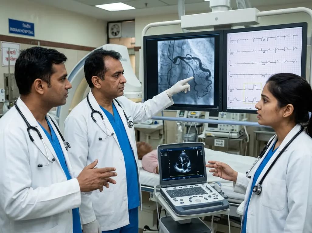
A hyperdominant left circumflex artery supplies extensive myocardial regions, creating a perilous diagnostic blind spot during acute occlusion. Standard ECGs often conceal posterior infarction, requiring clinicians to employ posterior leads and rapid echocardiography for early invasive catheterization.
Today
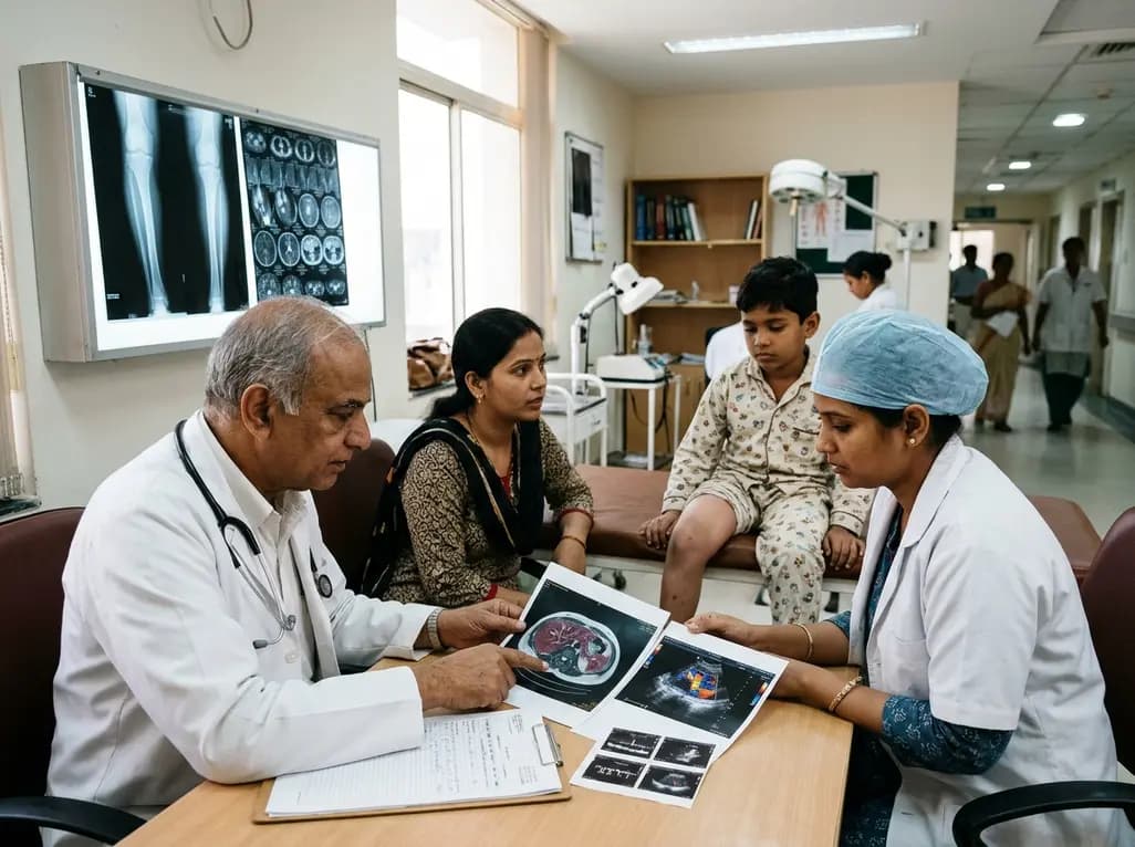
Deep intramuscular vascular malformations in pediatric Klippel-Trenaunay syndrome can mimic Maffucci syndrome. Multimodality imaging with Doppler ultrasound and MRI, along with histopathology, ensures prompt differential diagnosis, targeted surgical debulking, and optimized conservative surveillance.
Today

Discover essential clinical insights into AGLP1-type amyloidosis, a rare localized complication of glucagon-like peptide-1 analog injections. Learn how histopathology and mass spectrometry differentiate benign iatrogenic amyloidomas from systemic disease, preventing misdiagnosis and unwarranted therapies.
Yesterday
