.webp&w=384&q=75)

Loading, please wait...
Loading, please wait...
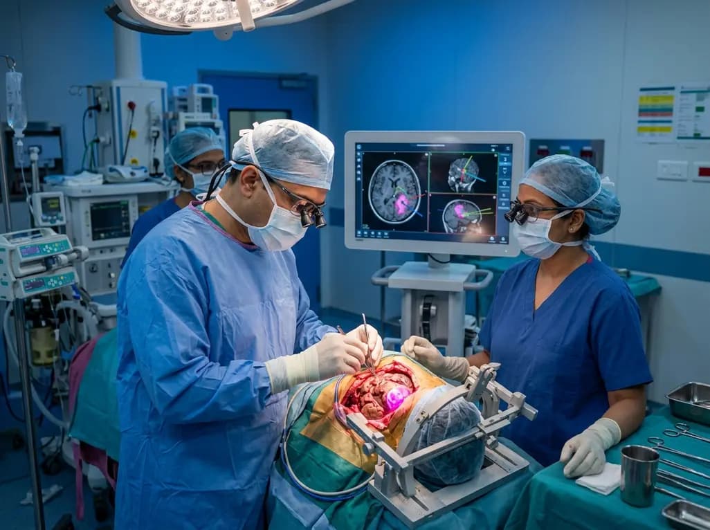
Central nervous system tumors involving bilateral or biventricular regions present intricate surgical challenges for neurosurgeons worldwide. Gangliogliomas are generally indolent, low-grade glioneuronal neoplasms defined by slow growth and favorable prognoses. However, these tumors occasionally undergo malignant transformation into anaplastic variants, which exhibit aggressive histological patterns and high recurrence rates. Achieving an optimal outcome during anaplastic ganglioglioma surgical resection requires careful operative planning, advanced intraoperative guidance, and coordinated multidisciplinary care. When lesions cross deep midline brain structures such as the corpus callosum, traditional surgical corridors carry substantial risk of neurological injury. Therefore, surgical teams must adopt innovative operative pathways that balance maximal tumor cytoreduction with functional preservation. Furthermore, understanding the transition from WHO Grade 1 lesions to malignant Grade 3 features remains critical for surveillance protocols. Consequently, modern neuro-oncological management emphasizes tailored surgical techniques combined with timely adjuvant therapies. Surgeons must constantly evaluate functional corridors to minimize postoperative cognitive, sensory, and motor deficits in these complex neurosurgical cases.
A detailed clinical case report illustrates the diagnostic and therapeutic complexities associated with bilateral intraventricular neoplasia in an adult patient. Initially, a 63-year-old female presented with a progressive intraventricular lesion infiltrating her left frontal lobe. Preliminary imaging and biopsy results confirmed a well-differentiated WHO Grade 1 ganglioglioma, prompting initial surgical intervention. Subsequent clinical monitoring, however, revealed the emergence of a new contralateral lesion expanding into the corpus callosum and adjacent ventricular spaces. As a result, the clinical team faced a major challenge regarding surgical accessibility and neurological preservation. Additionally, the rapid progression of contralateral disease raised strong suspicions of malignant transformation within the glial tissue. Furthermore, the lesion's location adjacent to vital deep structures restricted standard transcortical or transcallosal trajectories. Therefore, neurosurgeons designed a novel surgical strategy capable of reaching both sides of the tumor pathway safely. Consequently, thorough pre-operative mapping was conducted to evaluate vascular anatomy, fiber tracts, and ventricular corridors. This comprehensive evaluation allowed surgeons to formulate a customized operative plan.
To execute a precise intervention, the surgical team utilized advanced neuronavigation alongside intraoperative fluorescence imaging. Neuronavigation systems allow real-time localization of deep intraventricular boundaries, reducing reliance on static anatomical estimation. Meanwhile, intraoperative fluorescence imaging enhances the visualization of tumor margins, helping surgeons distinguish neoplastic tissue from healthy brain parenchyma. Therefore, integrating these modalities allows for safer dissection along the surgical corridor without compromising adjacent eloquent structures. Additionally, neurosurgeons extended the surgical corridor contralaterally along the established tumor route, utilizing a specialized transtumoral approach. Consequently, this method allowed direct access across the corpus callosum without necessitating additional extensive cortical incisions or excessive brain retraction. Furthermore, intraoperative neurophysiological monitoring verified functional neural integrity throughout the tumor extirpation process. As a result, the surgical team successfully navigated difficult ventricular anatomical landmarks with high precision. Thus, combining technological tools with an adaptive operative trajectory significantly improved intraoperative control, minimized tissue trauma, and facilitated maximum tumor cytoreduction.
Through the implementation of the specialized transtumoral corridor, the neurosurgical team achieved an extensive cytoreduction. Postoperative magnetic resonance imaging confirmed complete resection across most intraventricular sites, except for a small residual infiltration in the right posterior lateral ventricle. Subsequent histological analysis of the resected tissue revealed severe nuclear atypia, elevated mitotic activity, and microvascular proliferation. Consequently, neuropathologists updated the definitive diagnosis to anaplastic ganglioglioma, corresponding to a high-grade central nervous system neoplasm. Therefore, this aggressive histological transformation emphasized the necessity of immediate, comprehensive adjuvant therapy. Furthermore, the clinical team promptly scheduled postoperative radiotherapy to control residual disease and prevent further local recurrence. Meanwhile, the patient demonstrated remarkably favorable functional preservation postoperatively, avoiding severe motor, speech, or cognitive deficits. Consequently, this case demonstrates that anaplastic ganglioglioma surgical resection can be accomplished safely even when lesions infiltrate deep, bilateral ventricular structures. Ultimate success depends on combining meticulous microsurgical technique with accurate intraoperative visualization.
Operating within the ventricular system requires careful selection of surgical trajectories to minimize brain retraction and preserve functional neural pathways. Standard interhemispheric transcallosal approaches provide excellent access to midline lesions, yet they may increase the risk of disconnection syndromes when extended bilaterally. In contrast, extending a surgical corridor directly along the tumor pathway allows surgeons to exploit existing anatomical disruption caused by the lesion. Consequently, this transtumoral method creates a natural operative corridor that reduces the need for fresh parenchymal dissection. Furthermore, intraoperative image guidance continuously updates structural boundaries, accounting for brain shift as tumor volume is removed. Therefore, neurosurgeons can safely operate near critical vascular structures, such as the internal cerebral veins and anterior cerebral artery branches. Additionally, combining fluorescence technology with neuronavigation enhances surgical clarity in poorly demarcated tumor margins. As a result, utilizing adaptive, tumor-guided corridors represents a valuable strategy for managing deep-seated intraventricular masses while maintaining maximum surgical efficacy.
The management of aggressive glioneuronal tumors extends beyond surgical extirpation, requiring sustained multidisciplinary collaboration. Anaplastic gangliogliomas possess unpredictable biological behavior, necessitating close cooperation among neurosurgeons, neuropathologists, radiation oncologists, and medical oncologists. Furthermore, adjuvant radiation therapy plays a central role in limiting progression after subtotal or near-total resections. Consequently, serial postoperative neuroimaging is mandatory to detect early tumor recurrence or malignant progression in remaining brain regions. In addition, molecular profiling, including testing for BRAF V600E mutations, offers essential prognostic insights and potential targeted therapeutic options. Therefore, future treatment algorithms for recurrent or high-grade gangliogliomas will increasingly integrate targeted molecular agents alongside traditional surgery and radiation. Meanwhile, sharing novel surgical techniques through detailed clinical case reports expands the therapeutic armamentarium for managing complex, biventricular lesions. Overall, individualized operative trajectories combined with rigorous oncological follow-up offer the best opportunity for optimizing patient outcomes in challenging neuro-oncological scenarios.
Anaplastic ganglioglioma is a rare, aggressive World Health Organization Grade 3 glioneuronal tumor. Unlike low-grade WHO Grade 1 gangliogliomas, which grow slowly and exhibit benign features, anaplastic variants show high mitotic activity, nuclear atypia, cellular pleomorphism, and necrosis. Consequently, these malignant tumors require comprehensive treatment strategies including maximal surgical resection and adjuvant radiation therapy.
Fluorescence-guided surgery utilizes contrast agents that selectively accumulate in neoplastic tissue, causing tumor cells to glow under specific light wavelengths. Consequently, this technique helps neurosurgeons clearly delineate neoplastic boundaries from adjacent healthy brain tissue, facilitating maximal safe tumor resection while minimizing accidental damage to critical intraventricular vascular and neural structures during complex operations.
Adjuvant radiotherapy serves a critical role in controlling residual tumor cells following subtotal or complete surgical resection of anaplastic gangliogliomas. Because these high-grade tumors exhibit aggressive biological behavior and high local recurrence rates, postoperative radiation therapy helps suppress cellular proliferation, reduce local disease recurrence, and improve overall progression-free survival in affected patients.
Disclaimer: This content is for informational and educational purposes only and does not constitute formal medical advice. Healthcare providers should rely on clinical judgment and refer to the latest local and national guidelines for clinical practice.
References
Umana GE et al. [object Object] Br J Neurosurg. 2025 Dec. doi: 10.1080/02688697.2024.2400146. PMID: 39246067.

Read summarized clinical updates, watch expert medical content, and earn CME certifications right from your smartphone.
Open your smartphone camera to scan and install.





Managing bilateral intraventricular brain lesions infiltrating the corpus callosum presents surgical challenges. This case highlights a novel transtumoral approach using neuronavigation and fluorescence imaging to achieve maximal safe resection of an anaplastic ganglioglioma while preserving function.
3 months ago

A landmark 12-year longitudinal study highlights significant reductions in opioid prescribing for nonspecific low back pain following clinical guidelines and prescription monitoring systems, emphasizing non-opioid multimodal pain care.
Today

A study in Ultrasound in Obstetrics & Gynecology shows that quantitative vascularization of the anterior fornix/cervix with transvaginal ultrasound reliably identifies placenta accreta spectrum and predicts severe intraoperative hemorrhage, offering vital objective risk stratification.
Today
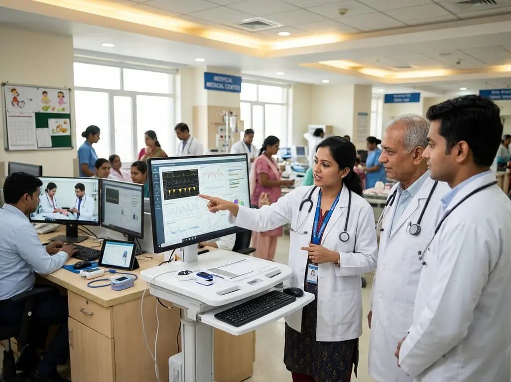
A landmark study details how an enterprise pediatric remote patient management model expanded from a single cardiology pilot to 16 diverse specialty programs by combining centralized digital operations with specialty clinical leadership.
Yesterday
A breakthrough study shows that 3D-planned corrective osteotomy successfully resolves distal radioulnar joint instability in pediatric diaphyseal forearm malunions. The precise osseous realignment eliminates chronic wrist pain and restores joint function without requiring secondary soft-tissue reconstruction.
Today

Protein O-GlcNAcylation couples cellular nutrient status to vital neuronal functions. This review explores how disrupted O-GlcNAc cycling drives both neurodevelopmental delays and neurodegenerative cascades, positioning metabolic post-translational modifications as therapeutic targets in clinical neurology.
Today
