.webp&w=384&q=75)

Loading, please wait...
Loading, please wait...
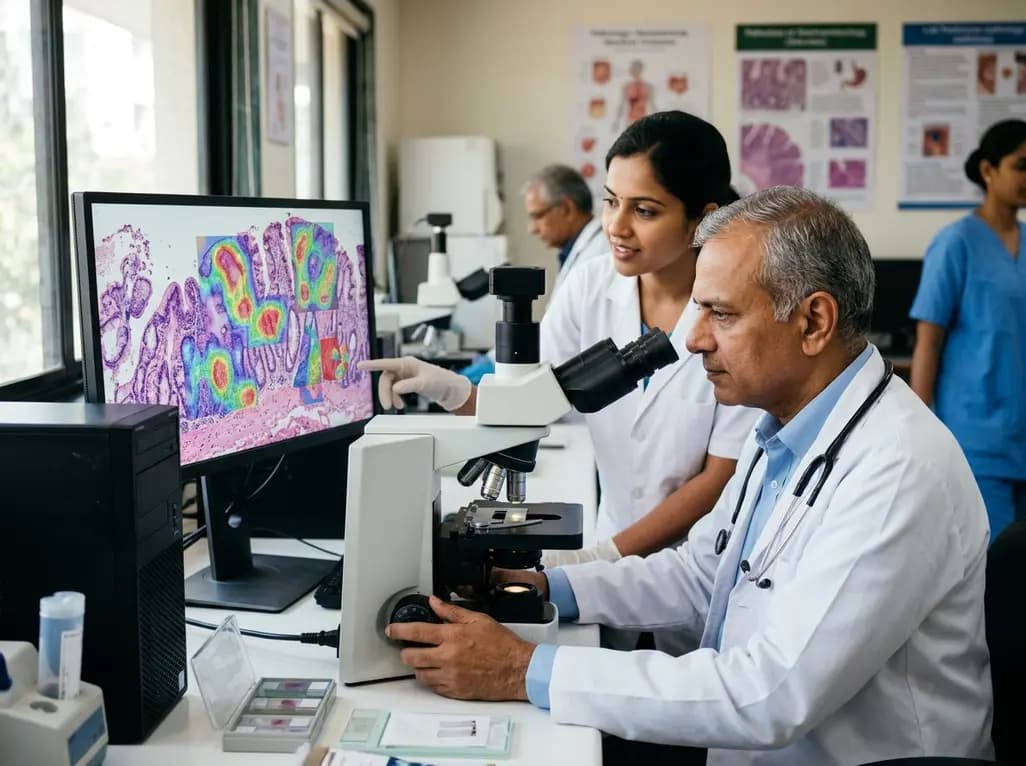
Gastric cancer remains a leading cause of oncological morbidity and mortality across the globe, especially in Asian populations. Histological examination of mucosal tissue plays an indispensable role in detecting premalignant lesions and preventing invasive disease. Standardized Updated Sydney System grading provides clinicians with critical diagnostic criteria to evaluate chronic gastritis, identify bacterial colonization, and stratify malignant transformation risks. However, executing thorough microscopic evaluations on high-volume biopsy samples creates substantial cognitive burdens for practicing pathologists. Recent advancements in deep learning offer viable solutions to resolve these diagnostic bottlenecks. A groundbreaking multi-task computational framework now demonstrates the feasibility of achieving automated, consistent, and highly reliable histopathological assessments in routine clinical workflows.
The Updated Sydney System serves as the universal gold standard for classifying and grading chronic gastritis in modern gastroenterology. Pathologists evaluate five core microscopic features across gastric antral and oxyntic mucosa: chronic mononuclear cell infiltration, neutrophilic activity, glandular atrophy, intestinal metaplasia, and Helicobacter pylori density. Each attribute receives a semi-quantitative score ranging from absent to marked severity. Consequently, this detailed scoring provides essential prognostic data that directly guides patient management.
Furthermore, accurate classification allows clinicians to calculate operative link for gastritis assessment (OLGA) and operative link on gastric intestinal metaplasia (OLGIM) stages. High-stage scores signal extensive mucosal damage and require structured endoscopic surveillance protocols. However, complete manual grading across all five variables remains time-consuming in high-volume pathology laboratories. Therefore, pathologists frequently provide abbreviated descriptive summaries rather than exhaustive numerical scoring. This practice inadvertently reduces the prognostic clarity required for personalized patient risk stratification.
Routine microscopic analysis of gastric mucosa presents several diagnostic hurdles that affect diagnostic precision. First, significant interobserver variability persists among pathologists when assigning subjective visual grades to inflammatory infiltrates. Differentiating between mild and moderate neutrophilic activity or assessing subtle mononuclear densities often leads to discordant interpretations. Consequently, treatment plans and surveillance intervals may vary across different clinical institutions.
Additionally, identifying mucosal atrophy poses distinct technical challenges. Pathologists cannot accurately grade glandular atrophy without visualizing the muscularis mucosae layer beneath the gastric glands. Tangential biopsy cuts or superficial tissue fragments often lack this crucial anatomical landmark. When pathologists attempt to score inadequate samples, false-positive atrophy diagnoses frequently occur. Therefore, diagnostic workflows require explicit mechanisms to categorize non-assessable specimens appropriately. Addressing these structural inconsistencies is essential for building trustworthy computational pathology tools.
To overcome these longstanding operational barriers, researchers developed SydneyMTL, a weakly supervised multi-task multiple instance learning framework. The neural network was trained on a massive repository containing 50,765 whole-slide images gathered from routine diagnostic practice. Unlike single-task algorithms that process histological markers independently, SydneyMTL jointly predicts all five Updated Sydney attributes simultaneously. This multi-task configuration mimics human cognitive reasoning by capturing intricate biological interrelationships among inflammatory patterns, metaplastic changes, and microbial presence.
Moreover, the engineering team designed an innovative algorithmic module dedicated to anatomical adequacy. The model incorporates a separate non-applicable classification category for glandular atrophy whenever the muscularis mucosae is absent. As a result, the artificial intelligence avoids forced misclassifications on superficial mucosal fragments. By embedding robust morphological safeguards into whole-slide image analysis, the platform achieves clinical realism that reflects actual laboratory requirements.
The investigators conducted rigorous validation studies to benchmark the artificial intelligence framework against human clinical performance. First, they assessed the model against routine diagnostic labels generated by 24 board-certified pathologists. SydneyMTL achieved an outstanding mean lenient accuracy of 89.1% across this diverse cohort of human readers. Furthermore, the algorithm attained at least 80% agreement for 21 out of the 24 pathologists on the retrospective dataset.
Subsequently, the researchers evaluated the platform using an independent, consensus-adjudicated golden dataset established by senior gastrointestinal subspecialists. SydneyMTL demonstrated exceptional concordance with expert consensus across all five individual histological features. The deep learning system reliably distinguished between varying densities of Helicobacter pylori and accurately quantified mucosal infiltration. Consequently, these robust validation metrics confirm that multi-task computational models can match the diagnostic reliability of experienced gastrointestinal pathologists.
Beyond standalone analytical accuracy, the clinical utility of artificial intelligence depends on its practical impact within real-time diagnostic environments. The researchers executed a two-reader randomized crossover trial to measure changes in physician performance and workflow speed during biopsy evaluation. Pathologists evaluated gastric whole-slide images with and without the interactive assistance of the SydneyMTL decision-support interface.
Importantly, the integration of artificial intelligence markedly improved interobserver agreement across all graded Sydney parameters. The software highlighted suspicious regions of active inflammation, metaplasia, and bacterial clusters, allowing pathologists to make confident determinations rapidly. Additionally, artificial intelligence assistance reduced slide review time by 34.2% for complete histological grading. Thus, automated assistance effectively eliminates diagnostic ambiguities while accelerating laboratory turnaround times without compromising diagnostic rigor.
The successful clinical validation of SydneyMTL represents a major milestone in computational gastroenterology and digital pathology. By transforming exhaustive manual scoring into an intuitive, AI-assisted review, laboratories can reliably deliver full Updated Sydney profiles for every gastric biopsy. Consequently, treating gastroenterologists gain comprehensive histological datasets that enhance post-endoscopy management decisions and long-term cancer surveillance protocols.
Furthermore, integrating multi-task artificial intelligence models into laboratory information systems establishes standard diagnostic baselines across regional health centers. As healthcare systems transition toward fully digital pathology pipelines, tools like SydneyMTL will help alleviate workforce shortages and minimize diagnostic discrepancies. Therefore, artificial intelligence decision-support platforms will play a pivotal role in delivering standardized, high-quality histopathological evaluations worldwide.
The Updated Sydney System evaluates five essential microscopic attributes in gastric biopsies: chronic mononuclear cell infiltration, active neutrophilic inflammation, glandular atrophy, intestinal metaplasia, and Helicobacter pylori colonization density. Each feature is semi-quantitatively categorized from normal or absent to marked severity. Together, these scores provide critical information regarding mucosal inflammation, structural injury, and patient risk for developing gastric malignancies.
SydneyMTL incorporates a specialized anatomical feature detection module that identifies the absence of muscularis mucosae. When tissue samples are superficial or cut tangentially, the artificial intelligence assigns a separate non-applicable status to glandular atrophy. This explicit handling prevents erroneous atrophy scoring on inadequate biopsies, ensuring reliable and clinically realistic diagnostic decision-support for practicing pathologists.
In a controlled randomized crossover study, artificial intelligence assistance reduced the review time required for complete Updated Sydney System grading by 34.2%. By automatically highlighting key areas of bacterial colonization, active neutrophilic inflammation, and intestinal metaplasia, the computational model streamlines microscopic slide interpretation, significantly enhances interobserver concordance among pathologists, and accelerates routine laboratory diagnostic workflows.
Disclaimer: This content is for informational and educational purposes only and should not be considered medical advice. Always consult a qualified healthcare professional regarding any medical condition or treatment. Refer to the latest local and national guidelines for clinical practice.
References
1. Kim HH et al. Comprehensive and reproducible grading of the Updated Sydney System in gastric biopsies using artificial intelligence: multi-pathologist validation and a reader study. Histopathology. 2026 Aug 15. doi: 10.1111/his.70258. PMID: 42601852.
2. Dixon MF, Genta RM, Yardley JH, Correa P. Classification and grading of gastritis. The updated Sydney System. International Workshop on the Histopathology of Gastritis, Houston 1994. Am J Surg Pathol. 1996;20(10):1161-1181.
3. Rugge M, Meggio A, Pennelli G, et al. Gastritis staging in clinical practice: the OLGA staging system. Gut. 2007;56(5):631-636.

Read summarized clinical updates, watch expert medical content, and earn CME certifications right from your smartphone.
Open your smartphone camera to scan and install.





SydneyMTL, an AI model trained on over 50,000 whole-slide images, enables comprehensive Updated Sydney System grading in gastric biopsies, achieving 89.1% accuracy, improving pathologist agreement, and reducing review time by 34.2%.
last month

A clinical report details delayed metal hypersensitivity following laparoscopic surgery, manifesting as progressive neuropathy and autonomic failure. Surgical clip removal and kink correction stabilized symptoms, underscoring the necessity of diagnostic vigilance when standard workups remain negative.
Today

A multiparametric MRI approach combining quantitative T2 shading, apparent diffusion coefficient (ADC) measurements, and morphological signs provides superior diagnostic accuracy over subjective evaluation to differentiate ovarian endometriomas from benign hemorrhagic cysts.
Today

A landmark case report reveals a donor heart beating for a century in a 63-year-old recipient, challenging conventional age limits in cardiac transplantation and offering critical lessons on organ longevity, donor selection, and long-term graft survival.
Today
A randomized trial compared myofascial release and dry needling for chondromalacia patellae in adult females. While both reduced knee pain, myofascial release combined with exercise produced superior quadriceps strength and improved the hamstring-to-quadriceps torque ratio.
Today

New research reveals that M1 macrophage-derived exosomes aggravate diabetic nephropathy by transferring WTAP to stabilize S1PR2 mRNA. Silencing WTAP in these vesicles attenuates endothelial injury and renal fibrosis, pointing toward innovative nanomedicine therapies.
Yesterday
