.webp&w=384&q=75)

Loading, please wait...
Loading, please wait...
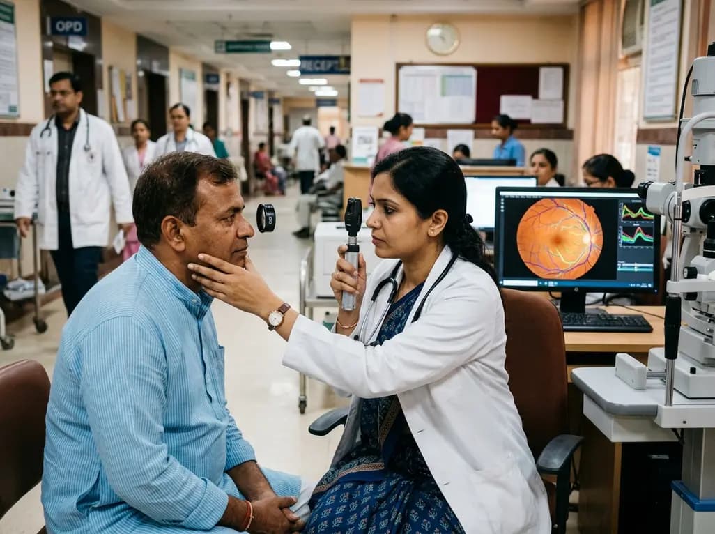
Diabetic nephropathy remains the leading contributor to end-stage renal disease worldwide, placing immense pressure on healthcare infrastructure. Clinicians frequently encounter diagnostic dilemmas when differentiating diabetic kidney damage from non-diabetic renal diseases in diabetic patients. While renal biopsy remains the diagnostic gold standard, its invasive nature and procedural risks restrict routine adoption. Consequently, non-invasive biomarkers have become vital for modern clinical nephrology. Recent advances now show that retinal microvasculature reflects microvascular alterations occurring simultaneously within the renal parenchyma. By harnessing advanced machine learning algorithms, non-invasive diabetic nephropathy diagnosis is rapidly becoming an accurate, accessible reality for global clinical practice.
The microvascular systems in the human eye and kidney share striking structural and developmental similarities. Both organs rely on extensive microvascular networks that remain highly susceptible to chronic hyperglycemia and systemic hypertension. Consequently, pathological processes affecting renal glomeruli often mirror changes within the retinal capillary bed. The retinal microvasculature provides the only directly observable microvascular bed in the living human body. Therefore, fundus photography enables clinicians to inspect microvascular alterations without performing invasive procedures.
Pathologically, long-standing diabetes triggers basement membrane thickening, pericyte loss, and capillary occlusion in both anatomical sites. In addition, hemodynamic shear stress damages the delicate endothelial lining in both glomerular and retinal capillaries. Researchers have historically leveraged qualitative diabetic retinopathy grading to estimate systemic microvascular risk. However, conventional qualitative assessments often fail to capture subtle morphological shifts. By integrating deep learning, clinicians can now quantify precise vascular geometric characteristics. These precise geometric metrics offer deep pathophysiological insight into ongoing subclinical renal microvascular damage.
To establish a robust diagnostic framework, investigators built a comprehensive multimodal database combining retinal imaging and granular clinical metrics. The multicentre trial incorporated 397 patients presenting with type 2 diabetes mellitus alongside chronic kidney disease. Specifically, the researchers developed unsupervised learning and ResNet neural network architectures to segment delicate retinal vessel branches. The artificial intelligence platform extracted intricate retinal vascular geometric parameters from standard fundus photographs with exceptional precision.
Furthermore, investigators applied weighted quantile regression and Lasso regularization to evaluate the mixed effects of complex retinal geometric parameters. They combined these image metrics with seven routine clinical parameters, including blood pressure, glycemic markers, and serum creatinine. This unified diagnostic model achieved an outstanding area under the receiver operating characteristic curve of 0.98. Moreover, the model demonstrated an overall diagnostic accuracy of 0.92 during test set evaluations. These quantitative results substantially outperformed traditional models that relied solely on conventional clinical predictors. Thus, combining multimodal data streams provides superior diagnostic power over isolated laboratory evaluations.
Generalizability remains a major barrier when deploying artificial intelligence systems into diverse clinical environments. To address this critical hurdle, the investigators evaluated the novel model against an external validation cohort spanning five distinct hospital centres. The multimodal diagnostic algorithm sustained remarkable diagnostic performance on the external dataset, yielding an accuracy of 0.91 and an AUC of 0.95. Consequently, these multi-centre results confirm the model's reliability across varying demographic profiles and photographic imaging equipment.
In addition to diagnostic screening, predicting disease trajectory is paramount for preventing end-stage renal disease. The researchers developed a dedicated prognostic index by deriving a mixed-effects parameter termed WQS-prognosis. They subsequently constructed a predictive model utilizing Cox regression combined with random forest algorithms. This prognostic model achieved an impressive AUC of 0.88 in predicting renal functional decline over longitudinal follow-up. Therefore, the artificial intelligence tool serves a dual role in modern clinical practice. It delivers immediate non-invasive diagnostic classification while simultaneously forecasting long-term renal outcomes for high-risk diabetic individuals.
India currently faces an unprecedented epidemic of type 2 diabetes, leading to an overwhelming burden of diabetic kidney disease. Unfortunately, access to specialized nephrologists and renal biopsy facilities remains severely constrained in rural and semi-urban Indian healthcare settings. Performing routine percutaneous kidney biopsies is impractical and carries risks of severe bleeding, pain, and hospitalisation. Therefore, implementing an automated, non-invasive screening platform based on digital fundus photography offers transformative potential for Indian primary healthcare centres.
Moreover, routine diabetic eye screening programs are already active across numerous district hospitals and vision centres in India. Integrating automated microvascular geometric analysis into these existing ophthalmic workflows creates a seamless, opportunistic screening pipeline. General physicians and endocrinologists can identify early renal microvascular involvement well before overt proteinuria or serum creatinine elevation occurs. Consequently, clinicians can initiate timely organ-protective interventions, including sodium-glucose cotransporter-2 inhibitors and renin-angiotensin system blockade. This proactive clinical approach can significantly reduce downstream dialysis dependency and renal replacement therapy demands nationwide.
Translating artificial intelligence models from academic research into daily clinical practice requires strategic operational implementation. First, medical device manufacturers must integrate algorithmic software directly into portable, non-mydriatic fundus cameras. This technological integration will empower community health workers and family physicians to generate instant risk profiles at the point of care. Furthermore, interoperability with existing electronic medical records will allow automated clinical decision support to guide primary care doctors.
However, prospective randomized validation trials must evaluate how AI-guided decision-making influences hard renal outcomes in diverse real-world populations. Clinicians must also recognize that artificial intelligence tools serve as clinical decision support systems rather than outright replacements for clinical acumen. In addition, ongoing research must explore serial retinal geometric tracking to assess whether therapeutic interventions actively reverse microvascular remodeling. As computational ophthalmology and nephrology continue to converge, multimodal non-invasive screening will undoubtedly establish a new paradigm for chronic diabetic complication surveillance.
Retinal imaging provides direct visualization of the human body's microvasculature, which shares developmental and physiological characteristics with renal glomeruli. By analyzing retinal vascular geometric parameters through artificial intelligence, clinicians can detect microvascular remodeling that mirrors renal microvascular damage. This non-invasive modality allows accurate differentiation between diabetic nephropathy and other renal pathologies without the complications and costs associated with invasive renal biopsies.
Traditional diagnosis relies primarily on serum creatinine, proteinuria, and clinical history, which often exhibit limited sensitivity in early stages. In recent multicentre trials, combining AI-derived retinal vascular geometric metrics with routine clinical markers achieved an AUC of 0.98 and 92% accuracy. This multimodal strategy significantly outperformed conventional clinical models, demonstrating that quantitative microvascular features add crucial diagnostic and prognostic value beyond standard biochemical testing alone.
Yes, the integrated artificial intelligence system effectively predicts longitudinal kidney disease progression. Researchers established a mixed-effects prognostic index using retinal vascular geometric parameters combined with random forest Cox regression models. This prognostic system achieved an AUC of 0.88 in predicting renal function deterioration over follow-up. Consequently, clinicians can accurately identify rapid progressors and optimize renoprotective medical therapies early in disease development.
Disclaimer: This content is for informational and educational purposes only and does not constitute formal medical advice. Healthcare professionals should make clinical decisions based on individual patient assessment and local clinical guidelines. Refer to the latest local and national guidelines for clinical practice.
References

Read summarized clinical updates, watch expert medical content, and earn CME certifications right from your smartphone.
Open your smartphone camera to scan and install.





A breakthrough multimodal AI framework combines retinal vascular geometric parameters and clinical data to achieve high diagnostic and prognostic accuracy in diabetic nephropathy, offering a powerful non-invasive alternative to traditional renal biopsy.
last month

UK Research and Innovation is deploying £80 million across the NHS to trial nine cutting-edge technologies. The initiative features seven-minute brain MRIs, blood biomarker assays, and predictive AI tools, aiming to evaluate 92% of suspected cases within 18 weeks and overcome lengthy diagnostic backlogs.
Today

A mixed-methods study identifies key drivers of PMTCT service utilization among adolescent girls and young women aged 15-24. Highlighting age disparities, PrEP uptake, and systemic barriers, the evidence guides clinicians toward adolescent-responsive, integrated maternal HIV prevention protocols.
Today

PepsiCo and Monster Beverage have challenged the FSSAI ban on the energy drink label in Indian courts. While companies highlight supply disruptions, regulatory actions spotlight severe cardiovascular and metabolic risks associated with excessive caffeine, sugar, and taurine consumption, especially in adolescents.
Today

A narrative review investigates whether the articularis genus muscle functions as an independent anatomical entity or blends with the vastus intermedius. We evaluate its morphology, role in retracting the suprapatellar bursa, and direct clinical significance in anterior knee pain and arthroplasty.
Today

A Bayesian multilevel meta-analysis reveals that aerobic training combined with moderate carbohydrate restriction modestly lowers HbA1c in type 2 diabetes. However, sparse data and very low certainty leave incremental benefits over exercise or diet alone unproven, highlighting the need for individualized care.
Yesterday
