.webp&w=384&q=75)

Loading, please wait...
Loading, please wait...
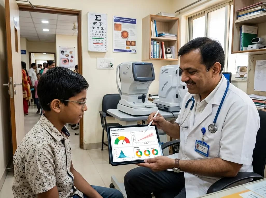
Childhood myopia has rapidly escalated into a major public health challenge across the globe. Consequently, pediatric ophthalmologists and optometrists encounter increasing numbers of school-aged children at risk of rapid visual deterioration. Early detection and timely clinical intervention remain the most reliable strategies to prevent high myopia and decelerate axial elongation. In recent years, researchers have developed novel computational models to address these diagnostic demands. Specifically, AI in myopia prediction offers clinicians transformative tools to identify vulnerable eyes before substantial refractive errors develop. Modern algorithms leverage complex mathematical architectures to analyze ocular biometry, environmental exposures, and familial patterns. By integrating these disparate data streams, clinicians can forecast refractive trajectories with unprecedented precision. This comprehensive review examines the current landscape of algorithmic innovation, highlighting how computational platforms enhance screening accuracy, personalize therapy, and reshape pediatric eye care paradigms worldwide.
Traditional vision screening programs in schools frequently face operational bottlenecks, including limited specialist availability and subjective measurement variability. However, machine learning algorithms and deep neural networks are transforming these mass screening initiatives into efficient, automated workflows. These systems evaluate non-cycloplegic autorefraction findings, corneal topography, and visual acuity metrics to classify myopic risk accurately. Consequently, primary care screeners can triage high-risk children rapidly and route them to tertiary eye centers for comprehensive cycloplegic examinations.
Furthermore, automated screening platforms substantially reduce false referral rates, conserving valuable clinical resources in high-volume public health settings. Convolutional neural networks can process non-mydriatic fundus photographs captured by portable cameras, detecting subtle peripapillary changes and early tessellated fundus patterns. As a result, community health workers can conduct high-throughput assessments in underserved urban and rural regions.
In addition, digital platforms facilitate seamless data transmission from community screening vans directly to centralized ophthalmic databases. Clinicians can then review automated risk scores and prioritize urgent pediatric consultations. Thus, integrating artificial intelligence into community screening bridges geographic gaps and establishes timely pathways for preventative pediatric eye care.
Accurate determination of future refractive error and ocular biometric changes is essential for effective pediatric myopia management. Advanced machine learning models, such as random forest classifiers, gradient boosting machines, and recurrent neural networks, now accurately project spherical equivalent trajectories across multi-year horizons. Rather than relying on simple linear regression, these algorithms capture complex nonlinear relationships between corneal curvature, anterior chamber depth, lens thickness, and axial length.
Moreover, longitudinal studies show that deep learning models can predict axial elongation rates with remarkable accuracy over three-to-five-year intervals. By evaluating past biometric data alongside baseline refractive measurements, algorithms identify fast progressors who might otherwise escape clinical detection during routine annual checkups. Therefore, practitioners can shift from retrospective management to prospective therapeutic planning.
Additionally, algorithmic forecasting helps clinicians distinguish between normal physiologic emmetropization and accelerated pathological elongation. Because excessive axial elongation directly correlates with future retinal detachment and choroidal neovascularization, early prediction provides a critical window for intervention. Consequently, predictive modeling allows clinicians to initiate targeted visual therapies before irreversible anatomical changes occur in young eyes.
The etiology of pediatric myopia involves an intricate interplay between genetic predisposition, environmental stressors, and behavioral habits. Fortunately, modern artificial intelligence architectures excel at integrating these heterogeneous data types into cohesive predictive frameworks. Neural networks combine polygenic risk scores with clinical parameters and parental refractive history to generate individualized risk profiles.
Furthermore, modern predictive algorithms increasingly incorporate behavioral metrics captured through smart wearable devices and mobile applications. These digital tools objectively quantify continuous outdoor light exposure, working distance during near tasks, and cumulative screen time. Therefore, the algorithm does not rely on subjective, recall-biased parental questionnaires. Instead, it evaluates real-time environmental data to assess how daily habits influence refractive progression.
In addition, machine learning models reveal subtle synergistic interactions among distinct risk factors. For example, an algorithm can identify how low ambient light exposure disproportionately accelerates axial lengthening in children with myopic parents. By illuminating these nuanced relationships, computational tools provide clinicians with actionable insights to deliver tailored lifestyle recommendations. Consequently, families receive evidence-based behavioral guidance designed specifically for their child's unique risk profile.
High myopia significantly elevates the lifetime risk of sight-threatening complications, including posterior staphyloma, myopic maculopathy, retinal detachment, and choroidal neovascularization. Deep learning algorithms trained on large datasets of ultra-widefield fundus photography and optical coherence tomography images demonstrate exceptional diagnostic accuracy in detecting these early structural alterations.
Specifically, convolutional neural networks can automatically segment retinal layers, quantify choroidal thinning, and detect subtle peripapillary atrophy before these changes become clinically evident on standard ophthalmoscopy. Moreover, automated image processing identifies early tractional maculopathy and microvascular abnormalities in juvenile patients with progressive high myopia. As a result, clinicians can monitor delicate microstructural changes with high objective precision over time.
Additionally, deep learning platforms facilitate automated risk stratification for secondary glaucoma and optic nerve head deformation associated with axial elongation. Because progressive scleral stretching alters optic disc biomechanics, AI systems evaluate lamina cribrosa morphology and retinal nerve fiber layer thickness to detect early subclinical neuropathy. Consequently, predictive image analysis empowers pediatric ophthalmologists to safeguard visual function and implement timely anatomical preservation strategies.
Selecting the optimal therapeutic regimen for childhood myopia requires balancing expected efficacy, patient compliance, and potential adverse effects. Currently, clinicians choose between low-concentration atropine eyedrops, orthokeratology lenses, peripheral defocus spectacle lenses, and multifocal soft contact lenses. Machine learning decision-support systems analyze baseline ocular metrics to recommend the most effective intervention for each individual child.
Moreover, predictive algorithms forecast individual treatment response trajectories under specific therapeutic modalities. For instance, decision tree models can identify whether a young patient will achieve superior axial elongation control with optical defocus lenses or pharmacological receptor blockade. Consequently, practitioners can avoid the lengthy trial-and-error periods that frequently lead to uncontrolled refractive progression.
Furthermore, artificial intelligence platforms monitor real-time therapeutic adherence and physiological responses during follow-up visits. If an algorithm detects suboptimal axial slowing under monotherapy, it prompts clinicians to consider combination protocols, such as combining low-dose atropine with orthokeratology. Therefore, data-driven treatment customization maximizes clinical efficacy, stabilizes refractive error, and enhances long-term visual outcomes for growing children.
Despite substantial computational advancements, several technical and clinical hurdles must be overcome before widespread deployment occurs in routine ophthalmic practice. A primary challenge involves algorithmic generalizability across ethnically diverse pediatric populations. Many existing models rely predominantly on East Asian training cohorts, which may limit predictive accuracy when applied to children of South Asian, Caucasian, or African descent.
Additionally, the black-box nature of complex deep learning architectures poses interpretability challenges for prescribing clinicians. To foster clinical trust, developers must integrate explainable AI techniques that clearly illustrate which biometric and environmental features drive specific predictions. Furthermore, ensuring stringent data privacy and establishing standardized data collection protocols remain essential prerequisites for multi-center clinical adoption.
Looking forward, the integration of federated learning and foundation vision models promises to address these regulatory and data-sharing constraints. Federated networks enable collaborative multi-institutional model training without sharing sensitive pediatric patient health information across borders. Therefore, ongoing global research collaborations will refine predictive precision, overcome regional disparities, and successfully embed intelligent algorithms into everyday pediatric eye care workflows.
Artificial intelligence models predict myopia progression by analyzing baseline refractive measurements, axial length elongation rates, corneal curvature, and demographic factors. Deep learning algorithms and machine learning classifiers evaluate these multidimensional clinical data points alongside lifestyle metrics, such as near-work duration and outdoor exposure time. Consequently, the software forecasts future spherical equivalent refraction and identifies children at high risk of rapid progression, allowing timely clinical intervention.
Yes, advanced machine learning architectures differentiate physiological emmetropization from progressive myopia by evaluating longitudinal biometric trajectories against normative ocular growth curves. Algorithms assess the rate of axial length elongation relative to corneal power and anterior chamber depth changes over time. Therefore, computational systems accurately pinpoint accelerated axial elongation that exceeds normal age-matched developmental thresholds, alerting clinicians to initiate targeted preventive treatments before significant refractive deterioration occurs.
AI models utilize diverse multimodal inputs to achieve high predictive accuracy in pediatric cohorts. These include non-cycloplegic and cycloplegic autorefraction, optical biometry measurements like axial length and corneal radius, fundus photographs, and optical coherence tomography scans. Additionally, models incorporate behavioral parameters from wearable sensors, such as ambient light levels and screen distance, alongside parental refractive history and polygenic risk scores to generate comprehensive prognostic assessments.
Disclaimer: This content is for informational and educational purposes only. It is not intended to provide medical advice, diagnosis, or treatment. Healthcare professionals must rely on their clinical judgment and verify information independently. Refer to the latest local and national guidelines for clinical practice.
References

Read summarized clinical updates, watch expert medical content, and earn CME certifications right from your smartphone.
Open your smartphone camera to scan and install.





Artificial intelligence is transforming pediatric myopia care. This review outlines AI-driven refractive power prediction, axial length monitoring, risk factor profiling, and customized treatment planning for children and adolescents.
4 weeks back
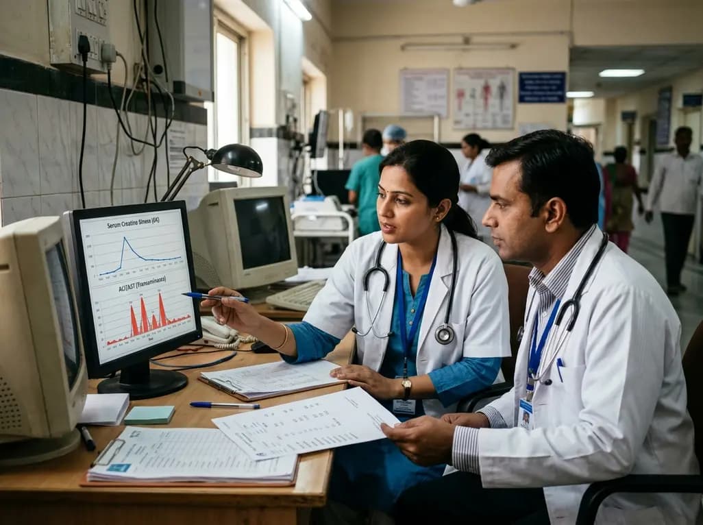
A recent case shows that exertional rhabdomyolysis can trigger marked transaminase spikes despite moderate creatine kinase elevation. Preserved hepatic synthetic function confirms muscle injury rather than liver failure, guiding conservative hydration and preventing unnecessary invasive diagnostics.
Today

A pharmacometric and machine learning study reveals that specific vaginal bacteria alter cervical antiretroviral exposure, highlighting the need for precision HIV PrEP strategies.
Today

Abdominal aortic aneurysm rupture remains a catastrophic vascular emergency. Groundbreaking research reveals that USP53 accelerates aneurysm progression by reprogramming smooth muscle cell metabolism toward aerobic glycolysis, highlighting a promising target beyond conventional diameter-based surveillance.
Today

A breakthrough study leverages natural language processing to accurately identify incident medication-related osteonecrosis of the jaw and track antiresorptive drug holidays from clinical narratives in electronic health records, advancing osteoporosis pharmacovigilance and dental safety.
Today

Recent research defines ferro-aging as a conserved iron-lipid axis driving organ decline across primates. Age-related iron dyshomeostasis stimulates ACSL4-mediated lipid peroxidation, promoting cellular senescence. Notably, vitamin C directly inhibits ACSL4, suppressing phospholipid oxidation and multi-organ decay.
Yesterday
