.webp&w=384&q=75)

Loading, please wait...
Loading, please wait...
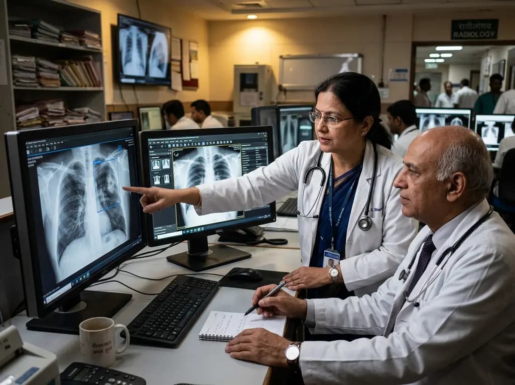
Evaluating the real-world utility of AI in chest radiography has become crucial as healthcare institutions adopt automated imaging tools. While artificial intelligence algorithms show immense promise in simulated benchmarks, their actual clinical performance often diverges when clinicians integrate them into daily workflows. A landmark prospective crossover study investigated four commercial deep learning models across 1,861 chest radiographs interpreted by multiple readers. The researchers sought to determine whether automated diagnostic assistance genuinely enhances diagnostic accuracy, optimizes interpretation speed, or alters escalation decisions. Their findings deliver critical insights for clinicians navigating modern technological integration in clinical practice.
The prospective crossover study evaluated five radiology residents with varying clinical experience ranging from one to six years. Over several structured interpretation phases, the readers reviewed 1,200 consecutive patients undergoing routine chest radiography. Each radiologist evaluated the 1,861 radiographs under five distinct conditions: an unassisted baseline reading and four separate interpretations using four distinct commercially available algorithmic platforms. To minimize recall bias, investigators implemented a strict 14-day washout period between reading sessions and randomized the case presentation order.
Furthermore, the investigative team established a robust reference standard based on finalized clinical reports, supplemented by confirmatory computed tomography scans whenever available. The readers evaluated five core pathological entities: pulmonary infiltrates, pleural effusions, mediastinal masses, pneumothorax, and pulmonary nodules. In addition to primary diagnostic accuracy, the researchers tracked critical secondary outcomes, including total interpretation time, diagnostic confidence scores, senior radiologist escalation, and computed tomography recommendations. Consequently, this rigorous multi-reader multi-case design provided an exceptionally transparent picture of algorithmic impact within real clinical environments. As a result, the trial established a practical foundation for analyzing whether deep learning software truly assists front-line clinicians during high-volume diagnostic shifts.
Contrary to common expectations, the study revealed that algorithmic assistance did not improve overall diagnostic accuracy across reader cohorts. In fact, diagnostic precision actually decreased for specific radiographic findings, particularly pleural effusions and pulmonary nodules. This decline occurred primarily because the automated tools generated a substantial volume of false-positive detections. As a result, readers frequently overcalled benign or artifactual densities as genuine pathology when prompted by algorithmic bounding boxes.
Moreover, these results highlight how sensitive detection thresholds can distort clinical discernment. When algorithms flag subtle opacities without sufficient specificity, clinicians often experience diagnostic confusion. Readers in the trial accepted automated suggestions that contradicted normal baseline interpretations. Therefore, while software developers design high sensitivity to prevent missed pathology, excessive false alarms compromise overall diagnostic integrity. Consequently, clinicians must balance algorithmic prompts against careful independent visual assessment to prevent unnecessary downstream workups. Ultimately, unchecked reliance on false alerts risks driving unneeded interventions, patient anxiety, and increased healthcare costs across radiology departments. In addition, junior residents appeared particularly vulnerable to misleading markers when evaluating subtle pulmonary nodules. Because early-career practitioners naturally seek diagnostic confirmation, ambiguous algorithmic flags can easily sway their reporting choices. Maintaining sharp clinical skepticism is therefore paramount.
Although diagnostic accuracy did not improve, the integration of algorithmic software demonstrated measurable workflow advantages. Specifically, three out of the five participating residents achieved statistically significant reductions in total interpretation time per case. Median reading times decreased by approximately 6 to 17 seconds per radiograph across various tool pairings. Thus, algorithmic assistance helped clinicians navigate routine studies more rapidly by offering immediate automated focal points during image triage.
Additionally, four out of five readers documented noticeably higher subjective diagnostic confidence when reviewing examinations alongside automated overlays. This elevated confidence likely facilitated quicker report finalization for ambiguous findings. However, researchers emphasize that speed gains must remain secondary to diagnostic precision. If shorter reading times stem from passive agreement with algorithmic prompts, clinicians risk perpetuating diagnostic errors. Therefore, departments must track turnaround metrics alongside rigorous quality assurance protocols to ensure that workflow acceleration does not compromise patient safety. Furthermore, time savings must translate into improved diagnostic thoroughness rather than rushed image sign-offs. When applied judiciously, minor per-case time savings accumulate into substantial efficiency gains across high-volume emergency and outpatient services. Nevertheless, imaging leaders must ensure that accelerated throughput never eclipses rigorous radiological evaluation.
The study also evaluated how automated software alters downstream clinical decision-making and consultation behavior. Notably, algorithmic support reduced the frequency of senior radiologist escalations in selected reader-software combinations. By providing secondary validation, the tools allowed junior residents to finalize complex studies independently without requiring immediate supervisor oversight. In one specific reader-algorithm pairing, automated assistance even lowered the rate of unnecessary computed tomography recommendations.
Furthermore, this reduction in escalation demonstrates that automated assistance can influence institutional resource allocation and staff utilization. When residents feel supported by consistent software predictions, they may reserve senior consultations for truly indeterminate presentations. Nevertheless, department leadership must monitor this trend closely. While decreasing escalation lightens supervisory workloads, uncritical reliance on software could inadvertently bypass essential senior guidance in borderline cases. Thus, structured supervisory safety nets remain vital during initial technology deployment. In addition, preventing inappropriate cross-sectional imaging orders conserves substantial financial resources and spares patients from unnecessary ionizing radiation exposure. Consequently, healthcare facilities can optimize resident autonomy while preserving senior oversight for genuinely precarious cases. Achieving this balance ensures that lower escalation rates reflect genuine diagnostic competence rather than overconfident diagnostic shortcuts.
The discrepancy between elevated reader confidence and stagnant diagnostic accuracy underscores the omnipresent danger of automation bias. Automation bias occurs when clinicians disproportionately trust automated recommendations, overriding their own sound clinical judgment. Because deep learning models often flag minor visual variations, inexperienced clinicians can easily misinterpret normal anatomy as true pathology. Consequently, institutions must establish comprehensive educational curricula that train medical staff to critique, rather than passively accept, algorithmic outputs.
Moreover, healthcare systems must recognize that commercial software requires careful local adaptation before routine implementation. Radiologists practice across diverse patient demographics, varying disease prevalences, and heterogeneous imaging equipment. Therefore, hospital departments should conduct rigorous on-site validation trials to fine-tune sensitivity thresholds and verify algorithmic generalizability. In addition, future prospective clinical trials must evaluate direct patient outcomes rather than relying solely on reader simulation metrics. Only through meticulous calibration and continuous practitioner oversight can healthcare systems harness the genuine benefits of automated image analysis while protecting clinical precision and patient safety across daily practice. Specifically, clinicians must learn to recognize recurring algorithmic artifacts and common false-positive patterns. By combining seasoned human perception with computational power, medical centers can cultivate a safe, synergistic diagnostic environment.
Recent multi-reader prospective evidence demonstrates that commercial algorithmic tools do not automatically improve diagnostic accuracy for chest radiographs. In fact, diagnostic accuracy for pulmonary nodules and pleural effusions occasionally declined due to increased false-positive detections. Because deep learning models prioritize high sensitivity, they often highlight subtle normal variants. Consequently, clinicians who rely excessively on automated alerts may misclassify benign findings as active pathology without improving overall clinical detection rates.
Algorithmic assistance provides tangible operational benefits, including faster interpretation times and enhanced diagnostic confidence. In prospective reader evaluations, radiology residents achieved statistically significant median time savings between 6 and 17 seconds per radiograph. Furthermore, automated prompts reduced unnecessary senior radiologist consultations and computed tomography escalation in specific pairings. Thus, the software helps streamline routine reporting workflows, provided clinicians maintain active oversight to ensure interpretation quality.
Hospitals must implement structured training programs that teach clinicians to evaluate automated prompts critically rather than accepting them passively. Furthermore, departments should conduct local validation studies to calibrate algorithm sensitivity thresholds to their specific patient demographics and imaging hardware. Establishing continuous quality assurance audits and maintaining mandatory senior review protocols for ambiguous cases will ensure that workflow efficiency gains never compromise diagnostic accuracy or patient safety.
Disclaimer: This content is for informational and educational purposes only and should not be considered medical advice. Always consult a qualified healthcare provider for diagnosis and treatment decisions. Refer to the latest local and national guidelines for clinical practice.
References

Read summarized clinical updates, watch expert medical content, and earn CME certifications right from your smartphone.
Open your smartphone camera to scan and install.





A prospective multi-reader crossover study evaluated four commercial AI tools for chest radiography, finding faster reading times and higher confidence but no accuracy gain due to increased false positives.
last month

A systematic review of 41 preclinical studies reveals that baicalin dampens systemic inflammation, inhibits NF-κB and NLRP3 pathways, and improves organ preservation in sepsis models. However, substantial study heterogeneity and a complete lack of human clinical trials highlight the need for further translational research.
Today
Discover the crucial distinctions between mesonephric and mesonephric-like proliferations of the female genital tract. Learn the key histomorphologic, immunohistochemical, and molecular differences essential for accurate diagnosis and clinical management.
Today

A large UK Biobank study reveals that plasma proteomic signatures capture preclinical organ damage and improve multiorgan risk prediction across early CKM syndrome stages 0 to 2 beyond traditional PREVENT clinical models.
Today
A narrative review investigates whether the articularis genus muscle functions as an independent anatomical entity or blends with the vastus intermedius. We evaluate its morphology, role in retracting the suprapatellar bursa, and direct clinical significance in anterior knee pain and arthroplasty.
Today

A Bayesian multilevel meta-analysis reveals that aerobic training combined with moderate carbohydrate restriction modestly lowers HbA1c in type 2 diabetes. However, sparse data and very low certainty leave incremental benefits over exercise or diet alone unproven, highlighting the need for individualized care.
Today
