.webp&w=384&q=75)

Loading, please wait...
Loading, please wait...

Accurate estimation of rectal cancer prognosis remains a vital clinical priority for multidisciplinary oncology teams worldwide. Although standard tumor-node-metastasis staging provides essential baseline guidance, it often fails to capture individual biological heterogeneity. Consequently, clinicians frequently encounter unexpected distant metastasis in patients who initially present with localized disease. Magnetic resonance imaging serves as the cornerstone for local staging and surgical planning. However, conventional qualitative evaluations cannot reliably detect subtle microscopic invasion or aggressive molecular phenotypes. Distant metastasis continues to represent the primary cause of treatment failure and disease-related mortality in rectal cancer. Therefore, researchers actively seek robust multimodal biomarkers to refine risk stratification before systemic progression occurs. Integrating macroscopic radiological features with microscopic immunological determinants offers an innovative avenue to overcome these challenges. Emerging predictive architectures blend computational artificial intelligence with host immune profiling. By combining these complementary modalities, oncologists can anticipate metastatic propensity much earlier in the disease course. Ultimately, improving prognostic precision enables clinicians to individualize systemic regimens and optimize surveillance intensity.
Preoperative high-resolution magnetic resonance imaging contains vast amounts of latent spatial data beyond conventional visual assessment. In recent investigations, artificial intelligence algorithms have demonstrated remarkable capability in extracting subvisual texture patterns from T2-weighted sequences. Specifically, deep learning architectures automatically learn hierarchical feature representations directly from raw imaging volumes. Researchers recently trained a specialized convolutional neural network on preoperative pelvic scans from a cohort of 306 rectal cancer patients. This computational pipeline generated a patient-level metric termed the DL_score. Because deep learning models capture complex spatial interactions, the score effectively reflects intratumoral heterogeneity and invasive tumor margins. Furthermore, this automated process eliminates inter-observer subjectivity, thereby delivering standardized risk assessment across diverse hospital environments. When applied before definitive surgery, the imaging signature provides early insight into the biological behavior of the primary lesion. Therefore, clinicians can identify aggressive tumor biology prior to histopathological confirmation. As a result, incorporating deep learning into routine imaging workflows significantly enhances clinical capabilities, providing oncologists with objective decision support.
While imaging quantifies macroscopic tumor behavior, host immune interactions dictate metastatic survival and disease trajectory. The Immunoscore provides a validated quantitative measure of the host antitumoral immune response within the tumor microenvironment. Pathologists quantify the density of CD3-positive total T lymphocytes and CD8-positive cytotoxic T cells using standardized immunohistochemical staining. Crucially, these measurements assess both the tumor core and the invasive margin on resected surgical specimens. In the studied cohort, patients exhibiting a lower Immunoscore showed significantly elevated risk for distant metastasis and substantially poorer metastasis-free survival. An immune-depleted microenvironment facilitates tumor cell escape, intravasation, and colonization in distant organs like the liver or lungs. Conversely, brisk infiltration of cytotoxic lymphocytes suppresses micrometastatic dissemination and reinforces durable systemic control. Thus, immune profiling delivers indispensable biological insight that complements radiological findings. Moreover, evaluating local host immunity helps explain why tumors with identical anatomical stages manifest vastly different clinical outcomes. Accordingly, integrating immune density metrics transforms qualitative pathology into highly reproducible, quantitative prognostic information.
To establish a practical clinical instrument, investigators synthesized computational imaging, digital pathology, and clinicopathological variables into a comprehensive nomogram. They employed multivariable Cox proportional hazards regression to identify independent predictors of distant metastasis. The final predictive model successfully integrated the magnetic resonance DL_score, the tissue Immunoscore, and key clinical risk factors. Model discrimination was rigorously assessed using the concordance index and time-dependent receiver operating characteristic curves. Furthermore, calibration curves demonstrated strong agreement between predicted metastasis probabilities and actual longitudinal clinical observations. The investigators also utilized SHapley Additive exPlanations to interpret feature contributions transparently. This explainable artificial intelligence approach confirmed that both deep learning features and immune scores contributed substantial prognostic weight. Additionally, decision curve analysis highlighted significant net clinical benefit across relevant decision thresholds. Consequently, the combined nomogram outperformed conventional clinical staging systems in predicting distant metastasis and overall patient prognosis.
Implementing reliable risk prediction tools carries profound clinical implications for colorectal cancer management in India. In recent years, Indian tertiary oncology centers have observed a concerning rise in young-onset rectal cancer cases presenting with aggressive histologies. Many patients present with locally advanced tumors that carry high intrinsic risks of distant dissemination. Therefore, precise stratification at the time of definitive surgery or restaging can transform therapeutic decision-making. Kaplan-Meier survival analyses from the study confirmed that the integrated nomogram robustly differentiates high-risk individuals from low-risk counterparts. For high-risk patients, oncologists can proactively intensify adjuvant systemic chemotherapy or implement novel consolidation strategies. Conversely, low-risk patients may safely avoid toxic overtreatment and prolonged hospitalizations. Furthermore, resource-conscious healthcare systems can utilize this risk model to tailor postoperative surveillance schedules. High-risk individuals benefit from frequent surveillance imaging, allowing rapid detection and timely salvage of oligometastatic lesions. Ultimately, integrating artificial intelligence and immunological scoring fosters equitable, data-driven personalized oncology across India.
The successful combination of deep learning and immune profiling heralds a new era in multimodal precision oncology. Moving forward, prospective multicenter trials must validate this integrated nomogram across heterogeneous imaging hardware and diverse patient demographics. Researchers are currently exploring whether biopsy-adapted immune scoring can bring this predictive capability into the true baseline, pre-neoadjuvant setting. In addition, incorporating circulating tumor DNA and next-generation genomic sequencing could further elevate model discrimination. Medical software developers are also working to automate the pipeline, embedding deep learning scores directly into picture archiving and communication systems. Such integration ensures seamless access for multidisciplinary tumor boards without disrupting clinical workflows. Moreover, expanding these techniques to assess therapy response will allow clinicians to adapt treatment regimens in real time. As computational infrastructure and molecular diagnostics become more cost-effective, immuno-radiomic models will establish a new benchmark for rectal cancer care.
Standard MRI interpretation relies on visual evaluation of gross anatomical features, which can miss microscopic tumor invasion and cellular heterogeneity. In contrast, deep learning models analyze vast arrays of subvisual pixel patterns within T2-weighted images. These algorithms calculate an objective numerical score that directly reflects intratumoral heterogeneity and aggressive metastatic behavior. Consequently, deep learning delivers consistent, reproducible risk estimation while eliminating observer variability during prognostic assessment.
The Immunoscore quantifies host antitumoral immune defenses by measuring CD3-positive and CD8-positive T lymphocyte densities at the tumor core and invasive margin. Patients with low immune infiltration exhibit higher rates of vascular intravasation and distant dissemination. Therefore, assessing the immune microenvironment reveals crucial biological vulnerability that conventional anatomical staging overlooks. This localized immune quantification provides an independent, robust predictor of long-term distant metastasis-free survival.
Multidisciplinary tumor boards can utilize the nomogram to stratify patients into distinct recurrence risk categories following surgical resection. Oncologists can confidently recommend intensified adjuvant chemotherapy regimens and tighter imaging surveillance for individuals identified as high-risk. Conversely, patients stratified into low-risk categories can be spared overtreatment and unnecessary toxicities. Thus, the model provides actionable, data-driven guidance that optimizes clinical resources and personalizes systemic therapeutic strategies.
Disclaimer: This content is for informational and educational purposes only... Refer to the latest local and national guidelines for clinical practice.
References

Read summarized clinical updates, watch expert medical content, and earn CME certifications right from your smartphone.
Open your smartphone camera to scan and install.





A novel nomogram integrating MRI deep learning and Immunoscore accurately predicts distant metastasis and refines rectal cancer prognosis, offering a powerful tool for personalized oncology.
Today

Nanoscale graphene oxide demonstrates remarkable neuroprotective potential in Parkinson's disease models by remodeling toxic alpha-synuclein fibrils, rescuing dopaminergic neurons, and curbing neuroinflammation, marking a new milestone for translational nanomedicine.
Today

The FEBE study investigates the prevalence of post-traumatic stress disorder symptoms in women receiving high-dose-rate brachytherapy for cervical cancer, highlighting the critical role of procedural sedation and trauma-informed clinical care.
Today

A landmark study reveals that intraoperative goal-directed perfusion targeting oxygen extraction ratio significantly reduces renal replacement therapy and two-year mortality in LVAD explant heart transplantation, offering a vital metabolic target for cardiac surgery teams.
Today

A Level I trauma center study shows a brief bedside mental health intervention boosts 30-day follow-up screening adherence by 84% in nonviolent injury patients. However, outcomes for violence survivors reveal critical care gaps, underscoring the urgent need for specialized community violence intervention programs.
Today
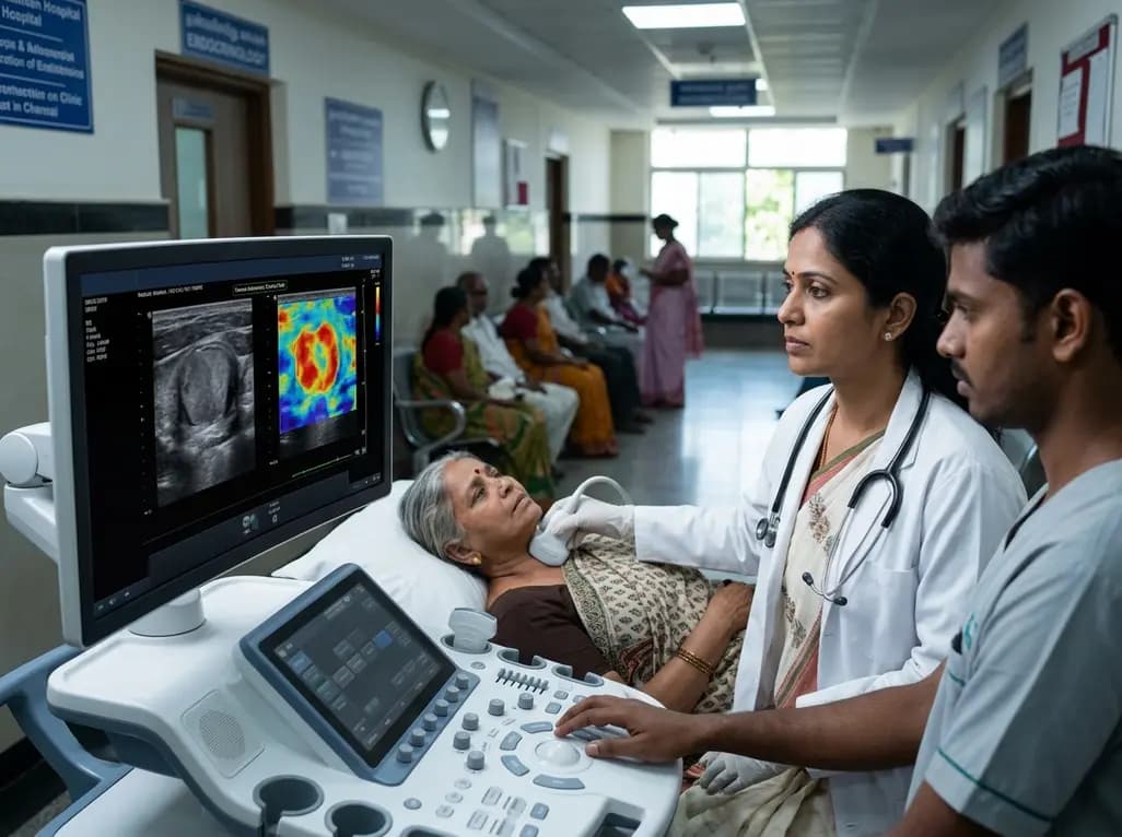
A study introduces a dual-discriminator generative adversarial network (DD-GAN) capable of generating virtual elastography ultrasound from standard B-mode images. The tool matches real elastography performance and improves thyroid nodule risk stratification when combined with TI-RADS.
Today
