.webp&w=384&q=75)

Loading, please wait...
Loading, please wait...
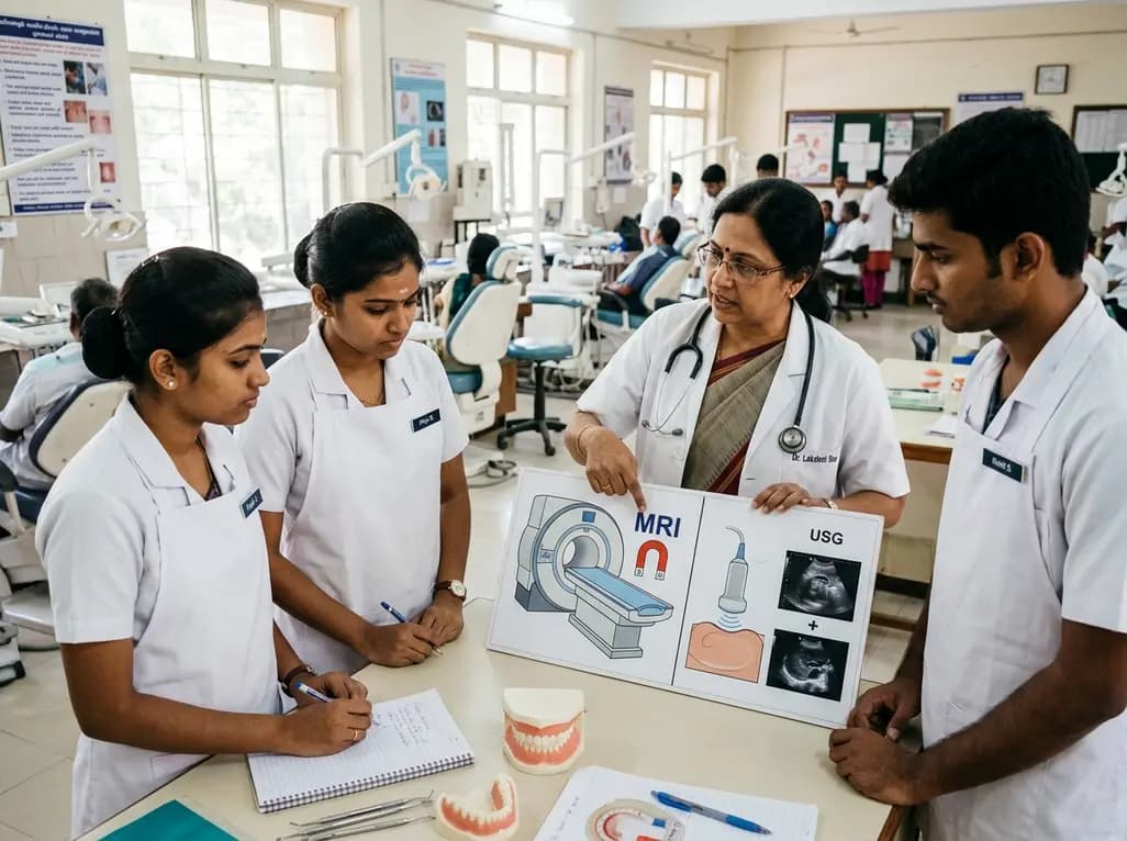
Contemporary dental diagnostics has historically relied on ionizing radiation, yet modern diagnostic pathways increasingly demand proficiency in advanced modalities. Integrating non-ionising imaging in dentistry represents a vital pedagogical shift that protects patients from cumulative radiation hazards while improving diagnostic resolution. However, dental graduates often transition into clinical practice with uneven competencies regarding advanced magnetic resonance imaging (MRI) and point-of-care ultrasound. A cross-sectional investigation evaluated academic-year differences among clinical dental undergraduates to understand how knowledge of these techniques evolves during formal clinical training.
Diagnostic imaging in oral healthcare is evolving beyond standard intraoral and panoramic radiographs. Consequently, clinicians must appreciate alternative imaging techniques that eliminate radiation exposure entirely. Non-ionising modalities provide superior soft-tissue differentiation, which is indispensable for evaluating temporomandibular joint internal derangements, deep space infections, salivary pathologies, and vascular lesions. Furthermore, these modalities mitigate biological risks for vulnerable patient demographics, including pediatric cases and pregnant women. Therefore, modern dental accreditation standards increasingly demand that undergraduate programs incorporate foundational instruction in diagnostic physics, clinical indications, and cross-sectional interpretation. Despite these evolving guidelines, structured didactic curricula frequently overlook the operational aspects of non-ionising technologies. As a result, many senior dental students understand ionizing protection principles well, yet they struggle to leverage advanced soft-tissue diagnostic modalities effectively during comprehensive treatment planning.
Magnetic resonance imaging utilizes static magnetic fields, rapid gradient shifts, and radiofrequency pulses that actively interact with metallic biomaterials. In clinical dentistry, patients routinely present with stainless steel orthodontic brackets, cobalt-chromium prostheses, amalgam restorations, and titanium dental implants. Clinicians must recognize that these biomaterials can create three distinct hazards: magnetic displacement forces, radiofrequency-induced thermal heating, and severe susceptibility artefacts that obscure vital anatomical landmarks. Stainless steel components and ferromagnetic retainers can experience significant torque, which risks mucosal dislodgement. In addition, closed conducting metallic loops may induce localized tissue burns during extended scanning sequences. Ferromagnetic items also distort local magnetic fields, generating signal voids that compromise adjacent neurovascular or oncologic diagnoses. Dental practitioners therefore bear a direct medico-legal responsibility to identify, record, and safely manage all restorative appliances before clearing patients for medical MRI examinations.
Recent research investigated how clinical dental students advance in their comprehension of non-ionising modalities across consecutive academic years. Investigators examined 188 clinical-year dental students across third, fourth, and fifth academic years using validated, anonymous questionnaires. Statistical analysis revealed that overall MRI knowledge scores improved significantly across academic cohorts. Specifically, total scores rose from 16.98 in the third year to 21.61 in the fourth year and 21.72 in the final year. However, post hoc analyses demonstrated that this advancement stemmed predominantly from theoretical physics questions rather than material-specific clinical safety evaluations. Students consistently scored lower when required to judge the dislodgement potential, heating propensity, and artifact severity of distinct restorative alloys. This disparity indicates that senior students acquire theoretical definitions as they progress through clinical training, yet they fail to cultivate operational decision-making skills.
Ultrasonography provides a dynamic, non-invasive, radiation-free diagnostic option that operates efficiently within point-of-care dental practice. High-frequency transducers reliably evaluate superficial head and neck structures, offering rapid chairside differentiation between cystic and solid swellings, identifying sialoliths, and assessing masseteric muscle hypertrophy. Moreover, Doppler ultrasound provides real-time functional hemodynamics, which aids clinicians in characterizing vascular malformations and diagnosing cervical lymphadenopathy. Despite these obvious advantages, clinical evaluation demonstrates that student knowledge regarding ultrasonographic limitations and indications remains stagnant across academic years. Many senior students erroneously believe that ultrasound can reliably penetrate intact cortical bone to evaluate intraosseous pathology. Furthermore, students consistently underestimate operator dependence and acoustic shadowing limitations. These pervasive misconceptions illustrate that without supervised ultrasound rotations, trainees cannot accurately integrate sonographic data into surgical or diagnostic protocols.
Addressing persistent knowledge gaps in advanced imaging requires comprehensive educational redesign. Academic institutions must move beyond purely theoretical lectures by embedding hands-on clinical simulations and collaborative interdisciplinary clerkships into dental training. Dental students benefit greatly from dedicated rotations through hospital radiology departments, where they witness real-time MRI screening and sonographic evaluations. Additionally, clinical departments should introduce practical case-based scenarios focusing on material selection, implant clearances, and orthodontic appliance management prior to cranial imaging. Dental faculties must also establish standardized algorithmic checklists that guide undergraduate students through metallic appliance risk stratification. By aligning didactic physics with practical chairside decision-making, dental colleges will graduate practitioners who confidently navigate complex non-ionising diagnostic scenarios, thereby safeguarding patient safety across diverse clinical settings.
Ultimately, enhancing competency in non-ionising imaging strengthens the interdisciplinary bridge connecting general dentistry with medical radiology. As maxillofacial interventions grow increasingly sophisticated, dental professionals routinely co-manage patients presenting with complex head and neck pathologies. When general dentists understand MRI interactions, they select non-ferromagnetic prosthetic components and draft precise referral communications for medical colleagues. Similarly, integrating point-of-care ultrasonography into dental operatories accelerates emergency triage, minimizes unnecessary computed tomography requests, and optimizes patient comfort. Therefore, advancing non-ionising imaging education elevates the standard of patient-centered care while eliminating unwarranted radiation exposure. Academic institutions must champion these instructional improvements to equip future clinicians with the diagnostic expertise required for modern clinical practice.
Dental materials cause artefacts because their magnetic susceptibility differs sharply from surrounding oral soft tissues and bone. When placed within a static magnetic field, ferromagnetic or paramagnetic components distort field uniformity. Consequently, the scanner misregisters spatial signals, which produces dark signal voids and spatial distortions. Clinicians must identify these materials beforehand because these artefacts can completely obscure anatomical structures in the brain, orbits, and maxillofacial region.
Titanium dental implants are considered safe during medical MRI procedures because commercially pure titanium is non-ferromagnetic. Osseointegrated fixtures do not experience significant displacement forces, nor do they generate hazardous radiofrequency heating in fields up to 3 Tesla. However, practitioners must remember that titanium abutments and metallic retaining screws can produce localized image distortion. Technicians can mitigate these minor distortions by modifying scan sequences or adjusting gradient angles.
Dentomaxillofacial ultrasonography is indicated primarily for superficial soft-tissue assessments across the head and neck. Clinicians utilize ultrasound to detect salivary gland sialolithiasis, evaluate superficial lymphadenopathy, assess temporomandibular joint disc displacement, and distinguish between solid neoplasms and fluid-filled cysts. Furthermore, duplex Doppler imaging enables chairside evaluation of vascular lesions. Conversely, ultrasound cannot penetrate intact cortical bone, meaning it cannot diagnose deeper intraosseous dental pathology.
Disclaimer: This content is for informational and educational purposes only and should not be considered professional medical advice. Healthcare professionals should make decisions based on individual clinical circumstances and patient needs. Guidelines, indications, drug dosages, and contraindications may change over time. Refer to the latest local and national guidelines for clinical practice.
References

Read summarized clinical updates, watch expert medical content, and earn CME certifications right from your smartphone.
Open your smartphone camera to scan and install.





A cross-sectional investigation highlights significant gaps in dental students' understanding of non-ionising imaging. While theoretical knowledge improves across academic years, clinical judgements regarding MRI material safety and ultrasonography applications require structured curricular enhancement.
Today
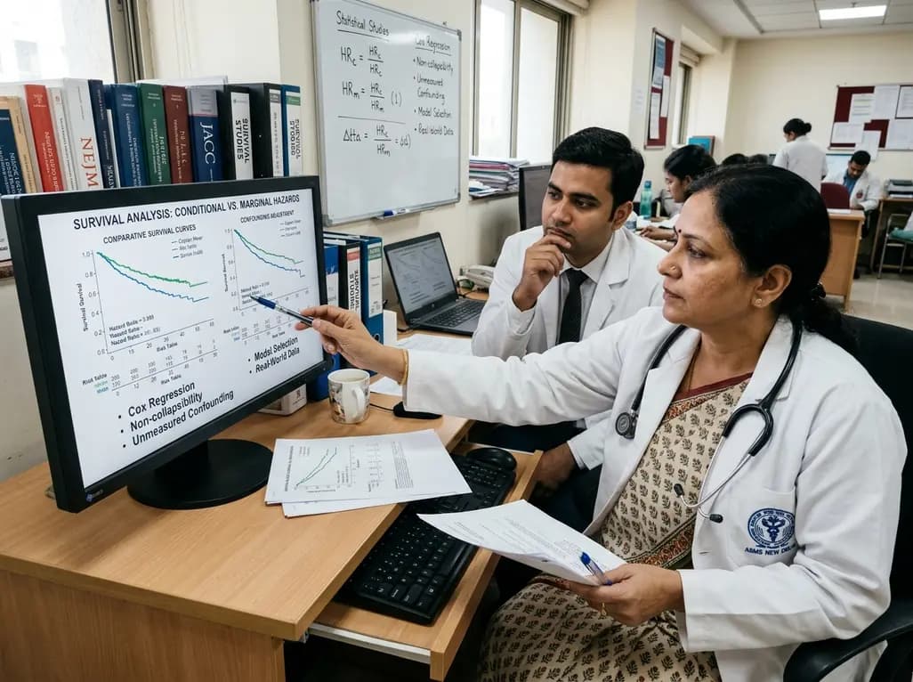
Survival analyses often report hazard ratios, but mathematical non-collapsibility creates crucial differences between conditional and marginal estimates. Clinicians must account for unmeasured confounding and model selection when interpreting trial endpoints and real-world cardiovascular outcomes.
Today

A cross-sectional study of 700 college students highlights significant contraceptive knowledge disparities between sexually active and inactive young adults. The findings demonstrate a critical need for proactive, comprehensive sexual health counseling before sexual debut.
Today
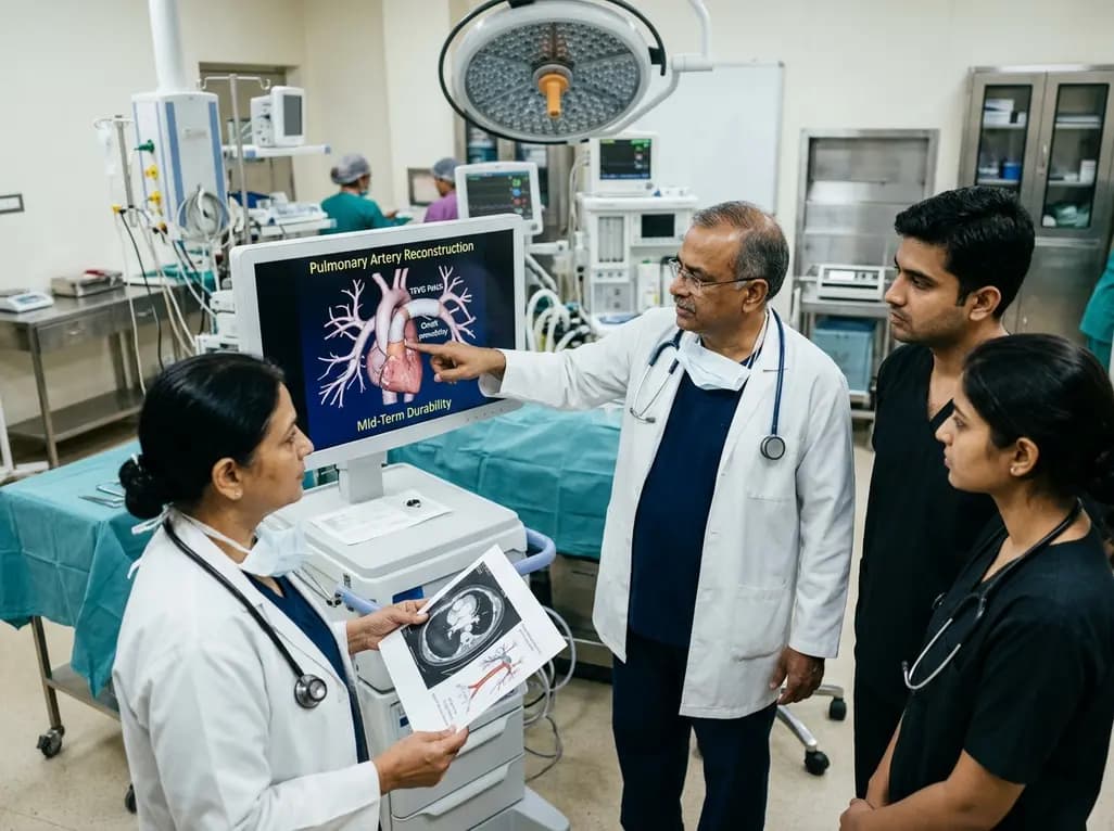
Autologous tissue-engineered vascular graft patches created via in-body tissue architecture show excellent mid-term durability and safety for pulmonary artery reconstruction in congenital heart surgery, particularly for peripheral branch augmentation.
Today
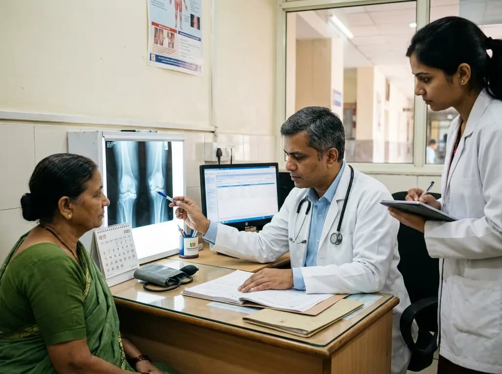
A novel cluster analysis of real-world datasets reveals four distinct CPPD disease phenotypes, validating existing EULAR categories while uncovering previously unrecognized monoarticular and axial presentations to refine patient management.
Today
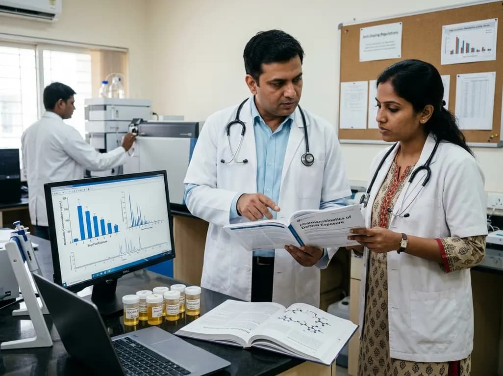
Recent pharmacokinetic research demonstrates that trace dermal exposure to microgram quantities of trenbolone and metenolone produces detectable urinary metabolites for days to over a week, providing critical objective data for evaluating unintentional contamination claims in sports drug testing.
Today
