.webp&w=384&q=75)

Loading, please wait...
Loading, please wait...
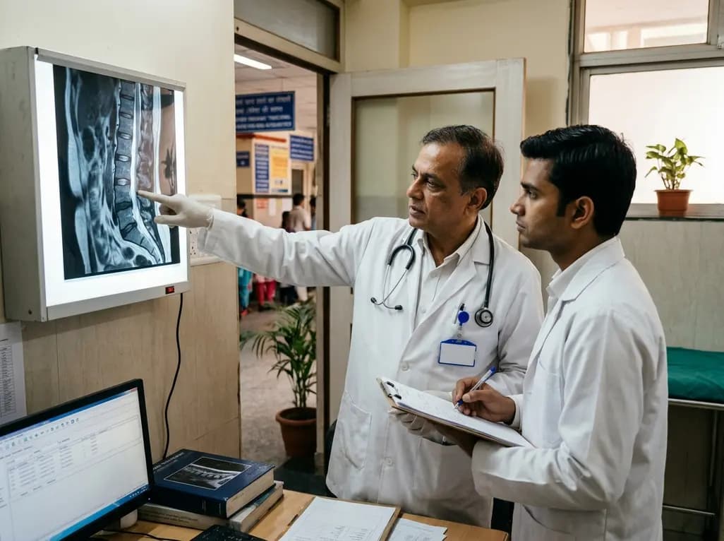
Spinal surgeons frequently utilize lateral retroperitoneal corridors to access the lumbar spine while preserving posterior musculoskeletal structures. However, these minimally invasive access techniques occasionally induce peripheral neurological complications that manifest unexpectedly. An abdominal wall pseudohernia represents one such rare yet distressing complication that challenges clinicians across multiple surgical specialties. Unlike true incisional hernias, this condition involves regional muscular flaccidity rather than a structural aponeurotic defect. Consequently, accurate identification prevents unnecessary prosthetic mesh repairs and directs appropriate neural intervention.
An abdominal wall pseudohernia produces visible unilateral or focal flank protrusion that mimics a mechanical abdominal defect. Patients typically notice asymmetrical abdominal bulging that worsens during standing or during maneuvers that increase intra-abdominal pressure. However, physical palpation reveals intact aponeurotic layers without a palpable fascial ring or hernial orifice. The underlying pathology stems directly from the functional denervation of local abdominal wall musculature. Specifically, interruption of motor conduction across the transverse abdominis, internal oblique, or external oblique muscles causes complete muscular flaccidity.
In addition, patients frequently describe sensory alterations across the corresponding cutaneous dermatome. These sensory changes range from localized paresthesia and numbness to severe neuropathic dysesthesia. Furthermore, patients often experience significant psychological distress due to the prominent cosmetic deformity and functional instability. Because the clinical appearance closely mimics an incisional hernia, misdiagnosis remains remarkably common in primary care and general surgery clinics. Consequently, clinicians must maintain high diagnostic suspicion when evaluating flank asymmetry following retroperitoneal or lateral spinal procedures. Early recognition prevents misdirected surgical interventions, such as unnecessary mesh placement, which fails to correct the underlying muscular denervation.
Lateral transpsoas and retroperitoneal approaches expose several peripheral nerve branches to potential intraoperative trauma. Among these neural structures, the subcostal nerve, which represents the twelfth thoracic ventral ramus, occupies a particularly vulnerable corridor. This nerve emerges beneath the twelfth rib and courses ventrally between the transversus abdominis and internal oblique muscles. Therefore, lateral incision placement, direct retractor blade pressure, or prolonged mechanical stretching can readily induce ischemic neuropraxia or structural axonotmesis.
Furthermore, dense scarring and postoperative fibrosis can envelop the neural trunk long after index surgery concludes. In some clinical cases, electrothermal injury from monopolar cautery or aggressive dissection incites extensive perineural inflammation. Consequently, fibroblasts deposit abundant collagen fibers around the nerve sheath, forming a constrictive cicatricial band. This chronic mechanical compression impedes axonal axoplasmic flow and promotes sustained neuromuscular decoupling. Moreover, the subcostal nerve provides pivotal motor innervation to the upper anterolateral abdominal wall. When chronic perineural entrapment halts motor transmission, the denervated muscle fibers rapidly undergo progressive atrophy. Thus, surgeons operating within retroperitoneal corridors must respect precise intercostal and subcostal trajectories to minimize peripheral mechanical traction.
Accurate differentiation between a true incisional hernia and an abdominal wall pseudohernia requires rigorous multimodality evaluation. First, dynamic physical examination provides immediate diagnostic clues. A pseudohernia becomes prominently visible during standing or Valsalva maneuvers, but it lacks a fascial defect upon manual palpation. Furthermore, gentle manual reduction does not push visceral contents through a distinct fibrous margin.
In addition, high-resolution computed tomography of the abdomen and pelvis serves as the imaging gold standard. Cross-sectional imaging clearly demonstrates intact musculoskeletal continuity without fascial disruption or visceral herniation. Instead, computed tomography typically illustrates marked thinning, loss of muscle volume, and fatty infiltration of the affected abdominal wall layers. Dynamic abdominal ultrasound also corroborates muscle laxity and distinguishes denervation from genuine wall defects. Moreover, electromyography provides essential neurophysiological confirmation by detecting active denervation potentials, such as fibrillations and positive sharp waves, within the affected muscular segments. Subsequently, clinicians can integrate these electrodiagnostic findings with magnetic resonance neurography to locate discrete entrapment points. Consequently, a comprehensive diagnostic workup protects patients from premature, ineffective prosthetic mesh repairs and guides targeted neural decompression.
A recent clinical report described a 75-year-old male with multiple prior spinal operations who developed a persistent lateral abdominal wall pseudohernia. Despite one full year of conservative management, the asymmetric flank bulge showed no spontaneous improvement. Therefore, the surgical team pursued targeted operative exploration to investigate possible peripheral nerve entrapment.
Intraoperatively, surgeons encountered dense perineural fibrosis completely encasing the subcostal nerve along its retroperitoneal course. Notably, direct intraoperative nerve stimulation demonstrated markedly diminished motor responses in the corresponding abdominal wall muscles. The surgical team meticulously performed external and internal neurolysis, freeing the encased nerve trunk from dense cicatricial bands. Following this meticulous decompression, repeat intraoperative nerve stimulation produced robust, visible muscular contractions across the flank. Consequently, the restored neuromuscular conduction facilitated rapid clinical recovery. In fact, the patient achieved complete resolution of abdominal wall laxity within just one month following surgery. This dramatic outcome underscores that chronic entrapment neuropathy can remain biologically reversible even after prolonged clinical duration. Thus, timely surgical neurolysis offers a reliable, restorative therapeutic option for refractory post-surgical pseudohernias.
This striking case provides vital clinical lessons for spinal surgeons, general surgeons, and rehabilitation physicians alike. First, clinicians must thoroughly consider neuropathic etiologies whenever patients present with post-surgical flank bulging. While conservative management represents the appropriate initial strategy, spontaneous axonal regeneration often fails when mechanical entrapment exists. Therefore, clinicians should not prolong non-operative observation indefinitely when electrodiagnostic or anatomical evidence indicates focal nerve entrapment.
Additionally, surgical planning must emphasize preventive measures during lateral lumbar and thoracolumbar approaches. Surgeons should implement neuromonitoring protocols and avoid excessive, prolonged retractor pressure against the lateral abdominal wall. Furthermore, gentle tissue handling and judicious use of electrocautery prevent extensive postoperative fibrosis around retroperitoneal nerves. When patients present with refractory pseudohernia, multidisciplinary collaboration between spine surgeons, peripheral nerve specialists, and plastic reconstructive surgeons ensures optimal patient selection. Ultimately, surgical neurolysis targets the root anatomical cause rather than merely masking structural laxity with synthetic meshes. By understanding subcostal nerve vulnerability and mastering decompressive techniques, modern surgical teams can successfully resolve this challenging post-surgical complication.
A true incisional hernia involves a distinct structural breach within the abdominal fascia, allowing intra-abdominal contents to protrude through the defect. In contrast, an abdominal wall pseudohernia features intact fascial layers without any mechanical gap. The noticeable bulge occurs entirely because localized denervation paralyzes the abdominal musculature, causing marked regional flaccidity. Therefore, imaging studies demonstrate muscle atrophy rather than visceral herniation, and standard mesh repair fails to restore lost neuromuscular function.
The subcostal nerve travels along the lower border of the twelfth rib before piercing the anterolateral abdominal wall musculature. During lateral retroperitoneal spine approaches, surgeons frequently place retractors across this precise anatomical pathway. Consequently, mechanical stretching, prolonged retractor blade pressure, or direct thermal injury can severely compromise the nerve. In addition, postoperative healing often induces dense perineural fibrosis. This cicatricial tissue gradually entraps the nerve trunk, disrupting motor signaling to the flank muscles.
Clinicians generally recommend an initial observation period of six to twelve months because neuropraxic injuries often recover spontaneously. However, when severe abdominal wall laxity persists without clinical or electrophysiological recovery, targeted surgical intervention becomes necessary. Surgical neurolysis is indicated when cross-sectional imaging or clinical findings demonstrate persistent nerve entrapment from dense scar tissue. Decompressing the encased nerve restores axonal continuity, facilitating remarkable muscular recovery even after prolonged post-surgical denervation.
Disclaimer: This content is for informational and educational purposes only... Refer to the latest local and national guidelines for clinical practice.
References

Read summarized clinical updates, watch expert medical content, and earn CME certifications right from your smartphone.
Open your smartphone camera to scan and install.





A recent case highlights abdominal wall pseudohernia secondary to subcostal nerve entrapment after lateral spinal surgery. Decompressive neurolysis achieved full functional recovery.
last month

Review how tailored exercise programs benefit frail older patients with heart failure. Explore pathophysiological mechanisms, individualized training prescriptions, and strategies to overcome adherence barriers in clinical practice.
Today

Leucine Rich Bio has introduced South Asia's first comprehensive vaginal microbiome test, BugSpeaks. By profiling microbial dysbiosis, pathogens, and antimicrobial resistance, this next-generation diagnostic platform provides clinicians with deeper insights into recurrent infections, fertility, and reproductive health.
Today

In high-risk pulmonary embolism complicated by severe anemia and active uterine bleeding, systemic thrombolysis is contraindicated. Large-bore aspiration thrombectomy provides rapid hemodynamic rescue without fibrinolysis, supported by inferior vena cava filter placement and multidisciplinary critical care.
Today

This comparative study evaluates posterior labral tears with and without glenohumeral instability, highlighting distinct demographic patterns, MRI underestimation in stable shoulders, and successful arthroscopic repair outcomes.
Today

A clinical study demonstrates that early intervention with tocilizumab, either as monotherapy or bridged with corticosteroids, achieves significant proptosis reduction and resolves active inflammation in treatment-naive thyroid eye disease.
Today
