.webp&w=384&q=75)

Loading, please wait...
Loading, please wait...
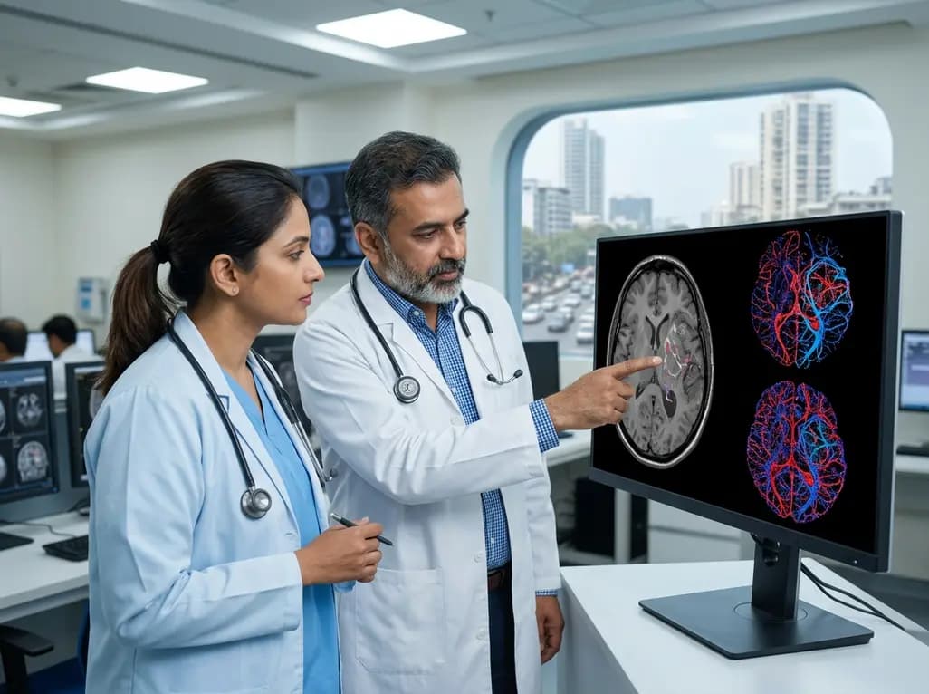
Accurate preoperative grading and molecular classification of gliomas are essential for tailoring neurosurgical resection, guiding adjuvant chemotherapy, and establishing prognosis. Historically, definitive identification of isocitrate dehydrogenase (IDH) mutation status and World Health Organization (WHO) tumor grade has required invasive stereotactic biopsy or surgical resection. However, noninvasive imaging biomarkers are rapidly evolving to provide preoperative insights into tumor biology. Utilizing 7T MRI glioma vascularity mapping presents a revolutionary noninvasive technique to profile microvascular patterns and decode angiogenic signatures. Ultra-high-field susceptibility-weighted imaging (SWI) at 7 Tesla offers unparalleled spatial resolution and contrast sensitivity, enabling detailed visualization of the tumor vascular microenvironment. Consequently, these radiological advancements help clinicians differentiate aggressive high-grade wildtype tumors from less aggressive mutant subtypes before performing invasive interventions.
The revised World Health Organization classification of central nervous system tumors places profound emphasis on molecular biomarkers, particularly IDH mutation status. Gliomas harboring IDH mutations typically demonstrate distinct biological behavior, improved treatment responsiveness, and prolonged overall survival compared to IDH-wildtype glioblastomas. In parallel, tumor progression and histological malignancy closely associate with microvascular proliferation. As tumors progress from lower grades to higher grades, intense neoangiogenesis leads to structurally disordered, tortuous, and hyperpermeable vessel networks.
Conventional magnetic resonance imaging frequently struggles to resolve these delicate capillary beds and microvascular alterations with sufficient fidelity. However, the vascular microenvironment acts as a key driver of tumor growth and local invasion. Therefore, obtaining detailed preoperative phenotypic maps of tumor angiogenesis provides valuable clinical intelligence. By analyzing vessel morphology, density, and spatial branching, clinicians can infer underlying genomic alterations and cellular differentiation. Ultra-high-field neuroimaging bridges the gap between macro-structural radiological observation and microscopic tissue architecture, establishing a new diagnostic paradigm in contemporary neuro-oncology.
Ultra-high-field 7T magnetic resonance imaging dramatically boosts signal-to-noise ratio and susceptibility contrast, making it exceptionally suited for susceptibility-weighted imaging. In recent clinical investigations, researchers acquired high-resolution T1-weighted MP2RAGE and 7T SWI scans across training and validation cohorts of glioma patients. Two independent neuroradiologists meticulously delineated three-dimensional regions of interest covering the full tumor volume. Subsequently, advanced computational image processing pipelines were deployed to characterize the microvascular bed.
Specifically, Frangi vesselness filtering was implemented to enhance tubular microvascular structures while suppressing background parenchymal noise. Next, three-dimensional skeletonization algorithms transformed complex vascular networks into quantifiable topological frameworks. This algorithmic workflow allowed researchers to extract granular quantitative features representing vascular topology, branching density, vessel length, and signal intensity attenuation. Because 7T SWI is extraordinarily sensitive to deoxygenated hemoglobin and microvascular magnetic susceptibility differences, it accurately captures microscopic intratumoral vessel architecture. Furthermore, quantitative feature extraction eliminates subjective reader variation, creating objective, reproducible metrics that reflect the true biological complexity of tumor neoangiogenesis.
Following automated feature extraction and rigorous dimensionality reduction, investigators evaluated multiple machine learning classifiers to predict molecular status and tumor grade. The evaluated architectures included logistic regression, support vector machines, naive Bayes, and random forest models. Feature selection was conducted using non-parametric statistical tests and Spearman rank correlation to eliminate redundant vascular parameters and avoid overfitting.
The resulting predictive models demonstrated robust diagnostic accuracy in both internal training and independent validation sets. When discriminating IDH-mutant from IDH-wildtype gliomas, the multi-parametric vascular classifiers achieved high sensitivity, specificity, and areas under the receiver operating characteristic curve. Similarly, the machine learning models differentiated WHO Grade 1–2 low-grade gliomas from Grade 3–4 high-grade malignancies with high precision. Intraclass correlation coefficients and Cohen kappa statistics confirmed excellent interobserver agreement between evaluating radiologists during tumor contouring. Thus, combining topological vascular metrics with machine learning algorithms establishes a reliable, noninvasive radiomics pipeline capable of decoding molecular and histological phenotypes directly from preoperative scan data.
The ability to profile glioma angiogenesis and predict IDH status preoperatively carries profound translational significance for neurosurgical planning and medical management. Knowing tumor grade and molecular subtype in advance allows neurosurgeons to optimize surgical margins, determine the feasibility of supratotal resection, and select intraoperative neuromonitoring techniques. For example, IDH-wildtype high-grade gliomas require aggressive cytoreduction combined with rapid postoperative chemoradiation protocols.
Conversely, accurately identifying lower-grade IDH-mutant tumors prevents overtreatment while guiding appropriate surgical timing and targeted therapeutic regimens. Furthermore, noninvasive vascular characterization provides critical baseline data for monitoring response to anti-angiogenic agents and novel IDH inhibitors. In patients where stereotactic biopsy carries high surgical risks due to eloquent or deep-seated tumor localization, 7T SWI radiomics offers a safe diagnostic alternative. As ultra-high-field MRI scanners become increasingly integrated into tertiary medical centers and specialized cancer institutions, these quantitative vascular algorithms can streamline preoperative multidisciplinary workflows and enhance personalized treatment planning for neuro-oncology patients worldwide.
Despite these encouraging diagnostic achievements, several technical and clinical challenges must be addressed before widespread clinical adoption occurs. First, 7T MRI systems remain limited in availability and incur substantial equipment and maintenance costs, restricting their present use primarily to academic research centers. Additionally, ultra-high-field scanning is vulnerable to magnetic susceptibility artifacts, particularly near skull base boundaries, tissue-air interfaces, and surgical bone windows.
Moreover, while machine learning models show superior discriminatory capacity in controlled cohorts, prospective validation across diverse, multi-institutional patient populations is essential. Future investigations must explore multi-modal integration by combining 7T SWI vascular metrics with chemical exchange saturation transfer, MR spectroscopy, and perfusion-weighted imaging. Incorporating deep learning architectures directly into clinical picture archiving and communication systems will also accelerate real-time workflow translation. Ultimately, standardizing image acquisition protocols and automating vessel segmentation pipelines will solidify the role of ultra-high-field vascular mapping as a staple tool in noninvasive neuro-oncological diagnostics.
Ultra-high-field 7T magnetic resonance imaging provides substantially higher signal-to-noise ratio and amplified magnetic susceptibility contrast compared to conventional 1.5T or 3T platforms. This enhanced sensitivity enables susceptibility-weighted imaging to detect minute differences in deoxygenated venous blood and microvascular iron content. Consequently, 7T SWI visualizes microscopic, tortuous neoangiogenic capillary beds and vessel branching patterns that remain largely invisible on lower-field scans.
Isocitrate dehydrogenase mutation status represents a foundational prognostic and predictive biomarker in modern neuro-oncology. IDH-mutant gliomas exhibit distinct metabolic profiles, slower proliferation rates, and significantly superior responses to adjuvant alkylating chemotherapy compared to IDH-wildtype tumors. Preoperative determination allows neurosurgical teams to plan optimal surgical margins, implement targeted pharmacological therapies earlier, and provide accurate prognostic counseling to patients and their families.
Frangi vesselness filtering enhances continuous tubular vascular geometries across MRI volumes while suppressing background soft tissue signal and scanner noise. Following enhancement, three-dimensional skeletonization algorithms compress the complex vessel network into single-voxel-width medial axes. This computational transformation enables automated extraction of objective quantitative parameters, such as vessel length, diameter, tortuosity, and branching density, which feed directly into predictive machine learning models.
Disclaimer: This content is for informational and educational purposes only. It is not intended to be a substitute for professional medical advice, diagnosis, or treatment. Always seek the advice of your physician or other qualified health provider with any questions you may have regarding a medical condition. Refer to the latest local and national guidelines for clinical practice.
References

Read summarized clinical updates, watch expert medical content, and earn CME certifications right from your smartphone.
Open your smartphone camera to scan and install.





A study evaluates 7T SWI for noninvasive profiling of the glioma vascular microenvironment. Using vessel filtering, 3D skeletonization, and machine learning, this ultra-high-field MRI technique accurately differentiates IDH mutation status and WHO tumor grade to guide neurosurgical and oncological care.
last month

The CARE study protocol introduces a multilingual, RAG-powered conversational interface designed by IIT Bombay to support tobacco cessation in India. Evaluating feasibility and behavioral outcomes across three cessation centers, this digital health initiative aims to enhance quitting support for diverse users.
Today
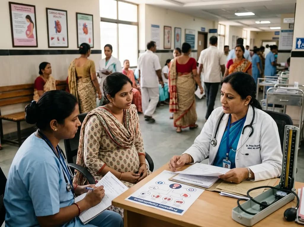
A large UK cohort study of 1.4 million women shows that pregnancy complications, including gestational diabetes, hypertension, and preterm birth, substantially raise long-term risks for hypertension, chronic kidney disease, type 2 diabetes, and cardiovascular events, highlighting early postpartum intervention needs.
Today
A cross-sectional study reveals that hypertension knowledge, illness perceptions, depression, and substance use significantly predict self-care behaviors. Addressing mental health and cognitive beliefs in primary care is crucial for optimal blood pressure control and cardiovascular disease prevention.
Today
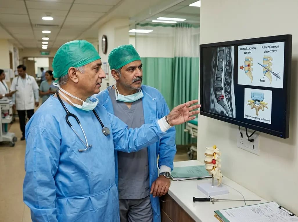
This clinical overview examines recent advancements in lumbar disc herniation surgery, comparing microscopic and full-endoscopic discectomy while highlighting biportal techniques and emerging motion-preserving innovations for spine specialists.
Today
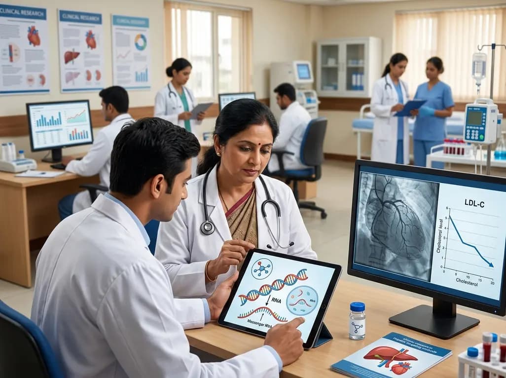
Small interfering RNA (siRNA) therapeutics harness RNA interference to degrade disease-causing messenger RNA. By delivering hepatocyte-targeted, durable gene silencing, agents targeting PCSK9, LPA, and angiotensinogen offer potent cardiovascular risk reduction with infrequent, biannual dosing schedules.
Today
