.webp&w=384&q=75)

Loading, please wait...
Loading, please wait...

Fluorescence-guided resection using 5-aminolevulinic acid has transformed neurosurgical oncology by delineating infiltrative tumor margins in high-grade gliomas. Historically, neurosurgeons relied on wide-field operating microscopes equipped with specialized optical filters to detect protoporphyrin IX fluorescence. However, the rapid clinical adoption of novel exoscopes in glioma surgery presents both technical opportunities and diagnostic dilemmas. Because digital exoscopes modify optical sensors and illumination dynamics, clinicians must ensure these devices match traditional diagnostic benchmarks to avoid inadvertent overresection or underresection.
Modern neurosurgery increasingly embraces digital visualization platforms to overcome physical constraints in operating rooms. Conventional operative microscopes require rigid ocular positioning, which often induces neck strain during lengthy tumor resections. In contrast, 3D digital exoscopes project high-definition stereoscopic imagery onto external monitors. Consequently, operating teams experience superior ergonomic posture and effortless collaborative viewing. Furthermore, high-resolution cameras provide magnification comparable to classical optics while maintaining an expansive working distance.
Nevertheless, optical transition requires thorough technical validation. Operating microscopes utilize long-pass filter systems such as the classical BLUE 400 platform. These standard optical filters selectively isolate the 635 nm red emission of protoporphyrin IX under violet-blue excitation light. Digital exoscopes, however, capture photons through electronic image sensors and digital post-processing algorithms. Therefore, neurosurgeons must confirm that digital filtration preserves tissue fluorescence characteristics. If a system artificially amplifies faint signals, a surgeon might resect functional brain parenchyma. Conversely, insufficient sensitivity might leave viable tumor tissue behind. Thus, verifying optical fidelity remains essential for surgical precision.
To evaluate diagnostic consistency across optical platforms, researchers conducted a rigorous technical evaluation. Specifically, the investigative team analyzed 73 resected glioma tissue specimens demonstrating heterogeneous fluorescence intensities. They examined these samples across three contemporary digital visualization systems: the Aeos, the ORBEYE, and the Kinevo exoscope. Additionally, they compared each system against the historical benchmark, the BLUE 400 filter. Six independent raters methodically assessed the demarcated fluorescent surface area across all captured images.
Interestingly, the evaluation revealed significant differences among the devices. The ORBEYE and Kinevo platforms demonstrated fluorescent surface area measurements comparable to the standard operating microscope. In contrast, the Aeos system delineated a significantly larger fluorescent surface area across identical tissue samples. Statistical evaluations using intraclass correlation coefficients confirmed excellent inter-rater reliability for both the Kinevo and Aeos exoscopes. Meanwhile, the ORBEYE platform demonstrated good reliability. Therefore, these findings confirm that while all three systems reproduce fluorescence signals, optical calibration varies between manufacturers. Neurosurgeons must understand these distinct operational profiles when navigating complex infiltrative margins.
The elevated fluorescence detection observed with the Aeos system raises pivotal clinical considerations. On one hand, higher optical sensitivity allows neurosurgeons to detect low-density infiltrative tumor boundaries that analog microscopes might miss. Consequently, enhanced visualization could facilitate more radical cytoreduction in non-eloquent brain regions. Furthermore, improved signal pickup might assist clinicians during the resection of lower-grade gliomas or recurrences where protoporphyrin IX accumulation remains modest.
On the other hand, heightened sensitivity creates substantial diagnostic ambiguity regarding tissue specificity. If an exoscope amplifies non-specific background scatter or autofluorescence, healthy brain tissue may appear malignant. Thus, surgeons risk resecting critical functional pathways, precipitating permanent neurological deficits. Additionally, the study noted that while the Aeos visualized larger fluorescent zones, researchers could not definitively confirm whether these expanded margins represented viable tumor cells. In contrast, the ORBEYE and Kinevo systems closely replicated established microscope thresholds. Therefore, they preserve historical specificity profiles validated over decades of clinical trials. Consequently, surgical teams must balance increased sensitivity against functional preservation.
In addition to static area measurements, the study explored the dynamic photophysical phenomenon of photobleaching. Continuous blue-light illumination rapidly degrades protoporphyrin IX molecules into non-fluorescent photoproducts. Consequently, fluorescence intensity naturally fades over time during prolonged surgical observation. In this comparative investigation, researchers quantified the decay rate of protoporphyrin IX under uninterrupted illumination across all evaluated systems.
Notably, the investigators discovered that modern digital exoscopes induce significantly faster photobleaching than traditional analog microscopes. Among the tested instruments, the Aeos platform exhibited the fastest photobleaching decay rate. This accelerated decay introduces practical challenges into neurosurgical workflows. For instance, if a surgeon leaves the blue illumination mode active while dissecting deep cavity margins, the vital signal may vanish prematurely. Consequently, residual neoplastic tissue could escape detection during subsequent cavity inspection. Therefore, neurosurgical teams must implement disciplined illumination habits. Surgeons should toggle excitation light intermittently and limit blue illumination to active margin assessment. Moreover, moderating illumination intensity preserves valuable fluorescence throughout tumor debulking.
Despite variations in photobleaching and sensitivity, novel digital exoscopes demonstrate high technical feasibility in brain tumor surgery. Furthermore, high intraclass correlation coefficients prove that different surgeons interpret exoscopic fluorescence signals with remarkable consistency. In particular, the ORBEYE and Kinevo systems function as dependable, safe alternatives to standard operating microscopes. They replicate familiar visualization paradigms while providing modern ergonomic comfort and crisp digital visualization.
Looking ahead, ongoing refinements in sensor engineering and software filtration will further enhance intraoperative precision. Digital platforms also create unprecedented opportunities to integrate artificial intelligence algorithms. For example, machine learning software can analyze pixel-level spectral decay in real time, automatically differentiating true tumor fluorescence from non-specific autofluorescence. Additionally, multi-spectral overlays could project vital navigation landmarks directly onto monitor screens without switching illumination modes. Thus, exoscopes bridge optical visualization and digital computer-assisted surgery. However, until multicenter trials validate long-term oncological outcomes, neurosurgeons should exercise prudence. Clinicians must combine digital visual cues with intraoperative neuromonitoring to achieve maximal safe resection.
Modern exoscopes provide equivalent or superior fluorescence visualization compared to conventional microscopes. While devices like the ORBEYE and Kinevo closely match standard BLUE 400 filter thresholds, platforms like the Aeos demonstrate higher sensitivity. Furthermore, exoscopes enhance team ergonomics and provide three-dimensional video display during complex cranial procedures.
Photobleaching degrades protoporphyrin IX molecules under continuous blue excitation light, causing tumor fluorescence to fade prematurely. Because novel exoscopes utilize intense digital illumination systems, photobleaching occurs more rapidly. Consequently, surgeons must toggle excitation lighting intermittently to preserve fluorophore visibility during meticulous tumor cavity inspection and radical resection.
Higher sensitivity detects faint fluorescence in infiltrative margins, potentially maximizing cytoreduction. However, excessive sensitivity may compromise diagnostic specificity by capturing background autofluorescence. Consequently, neurosurgeons risk inadvertently resecting non-tumorous functional brain parenchyma. Therefore, surgeons must combine visual inspection with intraoperative neuronavigation and neurophysiological monitoring to prevent neurological deficits.
Disclaimer: This content is for informational and educational purposes only and should not be construed as clinical or surgical advice. Healthcare professionals should evaluate device technical specifications and institutional protocols independently. Refer to the latest local and national guidelines for clinical practice.
References

Read summarized clinical updates, watch expert medical content, and earn CME certifications right from your smartphone.
Open your smartphone camera to scan and install.





A comparative technical evaluation assesses 5-ALA fluorescence across novel exoscopes (Aeos, ORBEYE, Kinevo) against the BLUE 400 standard. Findings highlight high reliability for ORBEYE and Kinevo, alongside increased sensitivity and photobleaching with Aeos, offering vital insights for glioma resection.
Today
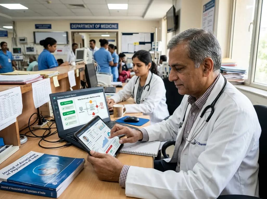
A multicenter study validates a hybrid clinical decision support system combining rule-based logic and machine learning to optimize anticoagulant prescription reviews, reducing alert fatigue and intercepting prescribing errors.
Today

The ClinGen Prenatal Gene Curation Expert Panel evaluated 63 disease relationships across 61 genes, establishing clinical validity for severe fetal phenotypes like hydrops and stillbirth to enhance prenatal genomic interpretation and clinical care.
Today
A metataxonomic study reveals distinct gut bacteriome biomarkers in type 2 diabetes, obesity, and cardiovascular complications, identifying specific bacterial shifts that pave the way for precision metabolic medicine.
Today

A psychometric validation study confirms that the Turkish Copenhagen Hip and Groin Outcome Score (HAGOS-T) offers excellent reliability, construct validity, and interpretability for patients with hip osteoarthritis, providing clinicians with a robust tool for functional assessment.
Today

Clinical guidelines rely heavily on isolated biomarkers like IGF-1 and HbA1c. However, portal insulin delivery fundamentally gates hepatic growth hormone sensitivity. This physiological continuum unites type 1 and type 2 diabetes, obesity, cirrhosis, and acromegaly, challenging conventional treatment strategies.
Today
