.webp&w=384&q=75)

Loading, please wait...
Loading, please wait...

The segmented Le Fort I osteotomy stands as a cornerstone in the surgical correction of complex dentofacial deformities. Unlike the single-piece procedure, this technique allows for the independent movement of maxillary segments, providing surgeons with the flexibility to address transverse discrepancies, vertical steps, and dental crowding simultaneously. Consequently, clinicians often favor this approach when patients present with a narrow maxilla or an irregular occlusal plane. However, despite its versatility, many medical professionals have traditionally perceived it as less predictable than its non-segmented counterpart. This skepticism often stems from the inherent complexity of stabilizing multiple bone segments against the pull of surrounding soft tissues and masticatory muscles. Therefore, understanding the precise three-dimensional accuracy of this procedure is essential for improving patient outcomes and surgical planning. As the dental field transitions toward digital workflows, the need for robust data on 3D stability has never been greater. Notably, recent systematic reviews have begun to provide a more nuanced view of how these segments behave post-surgery. Specifically, researchers are now looking beyond traditional 2D metrics to evaluate how well our surgical plans translate into real-world skeletal and dental positions.
Historically, surgeons evaluated the success of a segmented Le Fort I osteotomy using two-dimensional cephalometric radiographs and dental cast models. While these methods provided basic insights into sagittal and vertical changes, they lacked the depth required to capture the full complexity of transverse and rotational movements. Furthermore, 2D imaging often suffers from magnification errors and the overlapping of anatomical structures, which can obscure subtle discrepancies. To overcome these limitations, the field has increasingly adopted three-dimensional assessment methods, such as Cone Beam Computed Tomography (CBCT). Specifically, voxel-based registration has emerged as a highly reliable protocol for quantifying surgical accuracy. By aligning preoperative and postoperative 3D volumes at the voxel level, clinicians can achieve a much higher degree of precision in measuring bone movement. Consequently, this shift toward 3D analysis allows for a more comprehensive evaluation of how each maxillary segment moves in all six degrees of freedom. This detailed level of observation is crucial because even minor inaccuracies in the transverse or pitch dimensions can significantly impact the long-term functional and aesthetic success of the orthognathic intervention. Therefore, the adoption of standardized 3D protocols is now a primary recommendation for modern clinical practice.
Recent data indicates that the segmented Le Fort I osteotomy generally demonstrates clinically acceptable accuracy across most parameters. Nevertheless, specific movements appear to be more challenging to achieve precisely as planned. For instance, transverse widening often underachieves the intended goals, with discrepancies frequently ranging from 0.77 mm to 1.41 mm. Similarly, maxillary advancement and the control of the pitch (the forward or backward tilt) of the maxilla are prone to underachievement. Studies have shown that advancement discrepancies can range between 0.55 mm and 2.69 mm, while pitch errors can vary from 0.12 to over 5 degrees. These variances likely occur because the surgical site is subject to significant tension from the palatal soft tissues and the scarring process. In addition, the manual manipulation of segments during fixation can introduce small but cumulative errors that are only visible through detailed 3D mapping. Because these movements are central to correcting Class II and Class III malocclusions, surgeons must account for these potential undercorrections during the virtual surgical planning phase. By anticipating these discrepancies, clinicians can adjust their plans to ensure that the final postoperative outcome aligns more closely with the patient's functional needs and aesthetic goals.
One of the most critical aspects of any orthognathic surgery is the long-term stability of the results. In the case of a segmented Le Fort I osteotomy, stability is often evaluated in two distinct categories: skeletal stability and dental stability. Longitudinal follow-ups reveal that skeletal changes are relatively minimal, often staying within a range of 0.05 mm to 0.86 mm. This suggests that the bone segments themselves, once healed, remain fairly stationary. However, transverse relapse is markedly more pronounced at the dental level, with changes often measuring between 0.39 mm and 1.41 mm. This discrepancy between bone and tooth stability indicates that the dental arch tends to constrict more than the underlying skeletal foundation. Consequently, the teeth may shift back toward their original positions even if the bone segments remain properly aligned. This phenomenon highlights the importance of rigorous postoperative orthodontic management. Orthodontists play a vital role in maintaining the achieved width through the use of retainers or specific archwire protocols. Furthermore, the higher rate of dental relapse suggests that the soft tissue envelope exerts a continuous pressure that the dental components are less equipped to resist than the rigid skeletal segments.
Achieving perfect transverse and vertical control during a segmented Le Fort I osteotomy remains a significant clinical challenge. Specifically, the transverse expansion of the maxilla is often considered the most unstable of all orthognathic movements. This instability arises because the palatal tissues are naturally resistant to stretching, and they frequently pull the maxillary segments back toward the midline during the healing phase. Moreover, the vertical repositioning of segments, particularly when attempting to correct an open bite or a gummy smile, requires precise management of the posterior segments. If the pitch is not accurately controlled, it can lead to an unfavorable change in the occlusal plane or an inadequate display of the incisors. Therefore, surgeons must be particularly vigilant during the fixation process, ensuring that the plates and screws are positioned to resist the primary vectors of relapse. In addition, many experts recommend using auxiliary stabilization methods, such as palatal bars or specialized surgical splints, to provide extra support during the initial months of healing. By recognizing that certain movements are inherently less stable, the surgical team can implement proactive measures to minimize the risk of postoperative relapse and ensure a more predictable recovery for the patient.
To further improve the evidence base for segmented Le Fort I osteotomy, the medical community must move toward standardized assessment protocols. Currently, considerable methodological heterogeneity exists between studies, which makes it difficult to perform meta-analyses or compare outcomes across different surgical centers. For example, some researchers use surface-based registration, while others prefer voxel-based matching, leading to slight variations in reported accuracy. Consequently, establishing a universal 3D assessment framework would allow for a more robust comparison of surgical techniques and fixation methods. Furthermore, the integration of artificial intelligence in 3D planning software may soon help surgeons better predict individual relapse patterns based on a patient's unique soft tissue profile. As these digital tools become more accessible, the predictability of segmented maxillary movements is likely to increase significantly. Ultimately, the goal is to provide a highly personalized surgical experience where accuracy is maximized and the risk of relapse is minimized through data-driven planning and standardized postoperative monitoring. This evolution will ensure that the segmented Le Fort I osteotomy continues to be a reliable and effective option for patients with the most complex dental and skeletal requirements.
Three-dimensional assessment, particularly voxel-based registration, is superior because it captures the complex movements of individual bone segments in all spatial planes. Traditional 2D methods often hide transverse discrepancies and rotational errors due to image overlapping. In contrast, 3D imaging allows for precise measurements of skeletal stability and dental relapse, providing a more accurate reflection of the surgical outcome and helping clinicians refine their techniques for better predictability.
Research consistently shows that transverse widening, maxillary advancement, and pitch control are the movements most prone to underachievement during a segmented Le Fort I osteotomy. Transverse discrepancies often occur due to palatal soft tissue tension, while advancement and pitch can be affected by the mechanical limitations of surgical fixation and muscular pull. Recognizing these trends allows surgeons to adjust their planning to compensate for expected undercorrections during the actual procedure.
Dental relapse is typically more significant than skeletal relapse following this procedure. While the bone segments often remain stable with less than 1 mm of movement, the teeth can show significantly more constriction, sometimes exceeding 1.4 mm. This occurs because the dental arch is more susceptible to the pressures of the surrounding soft tissues. Therefore, long-term orthodontic retention is crucial to maintain the surgical expansion and ensure the stability of the final occlusion.
Disclaimer: This content is for informational and educational purposes only and does not constitute medical advice, diagnosis, or treatment. Always seek the advice of a qualified healthcare provider regarding any medical condition. The findings discussed are based on specific systematic reviews and may vary based on individual clinical cases and surgical techniques. Refer to the latest local and national guidelines for clinical practice.
References
da Costa Senior O et al. Three dimensional accuracy and stability of segmented Le Fort I osteotomy - a systematic review. Head Face Med. 2026 Jun 26. doi: 10.1186/s13005-026-00625-x. PMID: 42351191.
Kim YJ, Park YH, Koerich de Paula L, Conley RS. 3D Assessment of Orthognathic Surgical Outcomes. In: 3D Imaging in Medicine. Pocket Dentistry; 2015.
Blæhr TL, et al. Transverse Expansion and Stability after Segmental Le Fort I Osteotomy versus Surgically Assisted Rapid Maxillary Expansion: a Systematic Review. J Oral Maxillofac Res. 2016 Dec 28;7(4):e1. doi: 10.5037/jomr.2016.7401.

Read summarized clinical updates, watch expert medical content, and earn CME certifications right from your smartphone.
Open your smartphone camera to scan and install.





A systematic review highlights the 3D accuracy and stability of segmented Le Fort I osteotomy. While clinically acceptable, certain movements like transverse widening and pitch remain challenging to maintain and achieve.
3 months ago
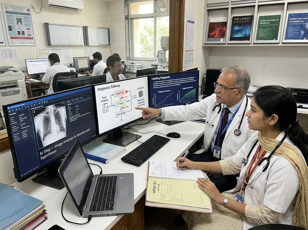
A breakthrough peer-reviewed study demonstrates that a sequential AI diagnosis system can mirror real-world clinical reasoning. By ordering stepwise tests and updating hypotheses dynamically, this multi-agent orchestrator achieved superior diagnostic accuracy while reducing investigative costs.
Today

A prenatal case highlights severe glutaric acidemia type II presenting as bilateral hyperechoic cystic kidneys at 18 weeks. Whole-exome sequencing revealed a novel homozygous ETFDH frameshift variant, demonstrating the power of rapid genetic testing in fetal renal anomalies.
Today

Review how tailored exercise programs benefit frail older patients with heart failure. Explore pathophysiological mechanisms, individualized training prescriptions, and strategies to overcome adherence barriers in clinical practice.
Yesterday

This comparative study evaluates posterior labral tears with and without glenohumeral instability, highlighting distinct demographic patterns, MRI underestimation in stable shoulders, and successful arthroscopic repair outcomes.
Yesterday

A clinical study demonstrates that early intervention with tocilizumab, either as monotherapy or bridged with corticosteroids, achieves significant proptosis reduction and resolves active inflammation in treatment-naive thyroid eye disease.
Yesterday
