.webp&w=384&q=75)

Loading, please wait...
Loading, please wait...

Femoroacetabular impingement syndrome frequently presents as a bilateral condition in young and athletic individuals. When conservative management fails, staged bilateral hip arthroscopy serves as an established intervention to restore joint biomechanics, alleviate pain, and repair damaged fibrocartilage. However, clinical debate continues regarding the morphological behavior of the unoperated contralateral hip during the interstage waiting period. Because several months or years often elapse between sequential interventions, clinicians must determine whether progressive structural degradation in the contralateral labrum compromises long-term post-surgical recovery and functional satisfaction.
Femoroacetabular impingement syndrome arises from morphological abnormalities of the femoral head-neck junction, the acetabular rim, or a combination of both. Consequently, abnormal contact occurs during terminal hip motion, generating repetitive shear stresses across the chondrolabral junction. Over time, these pathological abutments cause progressive labral tears, articular delamination, and premature osteoarthritis. Although many individuals first report unilateral groin discomfort, bilateral structural deformities exist in up to four out of five affected patients. Therefore, clinicians must carefully evaluate both hips during initial diagnostic consultations. While one hip may manifest debilitating mechanical pain, the opposite hip frequently harbors subclinical structural abnormalities. As patients modify their gait mechanics to protect the symptomatic joint, mechanical loads shift toward the contralateral side. Consequently, increased weight-bearing stress can accelerate tissue wear in the non-operated hip, creating an evolving clinical challenge for orthopaedic sports specialists.
Recent long-term imaging analyses provide critical clarity regarding contralateral joint health across extended surgical timelines. In a robust consecutive cohort evaluated with serial magnetic resonance imaging, patients undergoing staged bilateral hip arthroscopy demonstrated demonstrable tissue degradation between procedures. Specifically, researchers measured a statistically significant decrease in labral height at the 3:00 anterior-inferior position over an average interval of nearly fifty months. Furthermore, qualitative labral tear classifications revealed significant morphological worsening between the index operation and the second surgery. However, stable dimensions persisted at the superior and anterosuperior zones, indicating that mechanical attrition follows distinct regional patterns within the acetabulum. Because the anterior labrum experiences continuous shear forces during hip extension and external rotation, progressive thinning occurs reliably as the surgical interval lengthens. These findings prove that radiographic tear progression is a natural consequence of prolonged waiting times in patients with bilateral osseous impingement anatomy.
Despite progressive structural degradation within the contralateral labrum, clinical outcomes demonstrate reassuring durability after the second procedure. Patients achieved excellent functional improvements across comprehensive validated measurement tools, including the modified Harris Hip Score, the International Hip Outcome Tool, and the Hip Outcome Score subscales. Furthermore, patients achieved substantial reductions in visual analog pain scores and reported high satisfaction levels at a minimum two-year follow-up. Most importantly, objective rates of achieving the minimal clinically important difference and patient acceptable symptom state remained comparable to historical unilateral cohorts. Statistical correlation analyses revealed no negative association between the severity of interstage labral deterioration and final functional scores. Thus, modern arthroscopic restorative techniques, such as anatomical labral refixation and precise femoroplasty, successfully overcome morphological degeneration. Consequently, orthopaedic surgeons can reassure patients that delaying contralateral intervention until mechanical symptoms emerge will not adversely compromise their ultimate clinical prognosis.
Several interrelated biomechanical and biological factors explain why structural wear progresses without inevitably causing poor surgical results. First, compensatory mechanical loading occurs throughout the primary postoperative rehabilitation period. Because patients offload their surgical limb, the contralateral joint absorbs higher ground reaction forces during everyday ambulation. Furthermore, pre-existing cam or pincer deformities continue to create focal stress concentrations during standard daily tasks. Because the fibrocartilaginous labrum possesses limited intrinsic vascularity, spontaneous healing cannot occur once a microtear develops. Instead, repetitive mechanical impingement gradually extends the cleavage plane and diminishes tissue height. However, the acetabular labrum primarily functions as a hydraulic seal rather than a major direct load-bearing structure. Once arthroscopic intervention establishes anatomic clearance through bony resection and securely anchors the labral base, normal fluid dynamics and joint stability return. As a result, biological restoration overcomes preexisting structural degradation.
Determining the ideal staging interval requires balancing tissue preservation against physiological recovery from the primary surgery. Simultaneous bilateral procedures eliminate repeated hospitalizations, but they complicate immediate weight-bearing and functional rehabilitation. Conversely, staged bilateral hip arthroscopy permits full muscular conditioning and gait normalization between interventions. Because structural labral wear progresses gradually over several years, clinicians should base surgical indications primarily on patient symptoms and functional limitation rather than routine interval imaging alone. In addition, surgeons should counsel active individuals regarding activity modification, core strengthening, and hip abductor conditioning to shield the unoperated joint. When contralateral symptoms eventually intensify, scheduled repeat cross-sectional imaging accurately documents morphology and guides operative planning. In conclusion, careful symptom tracking, combined with contemporary arthroscopic repair techniques, ensures optimal clinical outcomes regardless of structural deterioration during the waiting window.
Approximately fifteen to twenty-five percent of individuals treated for symptomatic femoroacetabular impingement syndrome eventually undergo surgery on the opposite hip. Although bilateral morphological deformities exist in up to eighty percent of cases, bilateral mechanical symptoms do not always develop concurrently. Consequently, surgeons recommend addressing the contralateral joint only when persistent pain, functional disability, and failed conservative therapy establish a definitive clinical indication.
Clinical evidence demonstrates that delaying the second procedure does not compromise final functional recovery or patient satisfaction. Although serial magnetic resonance scans confirm progressive tissue thinning and tear propagation over prolonged interstage periods, post-surgical functional scores remain excellent. Specifically, patients achieve standard clinical milestones and pain relief comparable to unilateral cohorts once comprehensive arthroscopic debridement, osseous resection, and labral refixation are completed.
Clinicians should prioritize regular clinical evaluations and functional assessments rather than relying solely on frequent routine imaging. Providers should monitor contralateral groin pain, internal rotation discomfort, and subjective joint catching during regular postoperative checkups. However, if unoperated hip symptoms progress significantly or restrict rehabilitation milestones, repeating magnetic resonance imaging helps evaluate labral tear patterns and ensures timely surgical planning.
Disclaimer: This content is for informational and educational purposes only and should not be considered medical advice. Always seek the guidance of a qualified healthcare provider for any medical condition or treatment decision. Refer to the latest local and national guidelines for clinical practice.
References

Read summarized clinical updates, watch expert medical content, and earn CME certifications right from your smartphone.
Open your smartphone camera to scan and install.





A longitudinal study reveals that patients undergoing staged bilateral hip arthroscopy experience progressive contralateral labral deterioration during surgical intervals. However, structural degradation does not compromise 2-year postoperative clinical outcomes or patient-reported satisfaction scores.
Today

A landmark cluster randomized trial demonstrates that community-based mass dog vaccination achieves superior coverage and sustains protective herd immunity against rabies far better than conventional campaign models, offering vital lessons for global and Indian elimination targets.
Today
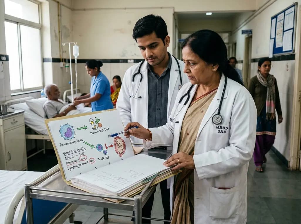
Explore the crucial link between dietary protein and immunocompetence. This review examines how low-protein nutrition shifts metabolic resources away from pathogen defense toward early reproduction, compromising host survival and immunity against severe bacterial infections.
Today

Differential oxygenation during peripheral TA-NRP can cause undetected coronary hypoxemia due to arch vessel clamping. A novel circuit with continuous aortic root monitoring detects desaturation early, facilitating left ventricular unloading and preventing allograft dysfunction and unnecessary heart discard.
Today

A post hoc analysis of the PARM-T2D study shows pemafibrate exerts baseline-dependent effects on LDL-C and particle size in type 2 diabetes and hypertriglyceridemia. It lowers triglycerides and remnants while favorably remodeling atherogenic LDL particles based on initial lipid control status.
Today
