.webp&w=384&q=75)

Loading, please wait...
Loading, please wait...
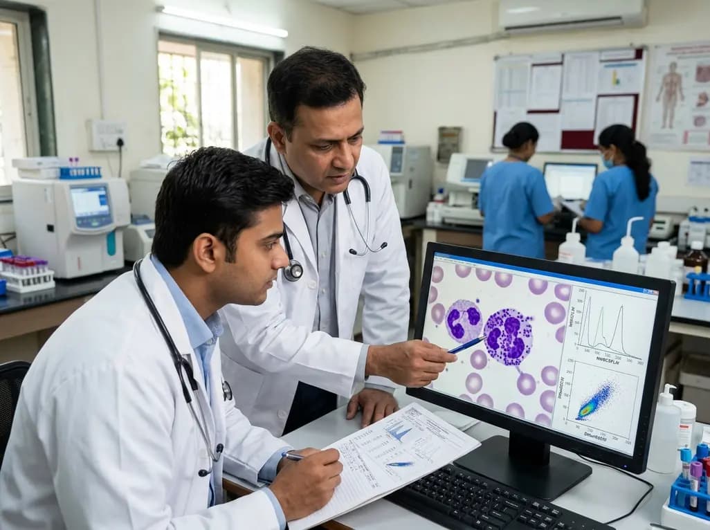
Sepsis remains one of the most significant challenges in modern clinical medicine, particularly within the intensive care units of India. Early and rapid identification of the systemic inflammatory response is critical because delayed treatment significantly increases mortality rates. While traditional markers like Procalcitonin and C-reactive protein provide value, they often lack the real-time speed required during the "golden hour" of treatment. Consequently, researchers have turned toward automated hematology analyzers to find more immediate solutions. Modern analyzers do more than just count cells; they generate complex data known as Cell Population Data (CPD). This data provides insights into the internal complexity and activation status of circulating white blood cells. Recent studies now suggest that sepsis morphological granularity biomarkers derived from these analyzers offer a breakthrough in early detection. By analyzing light scattering and fluorescence, clinicians can observe changes in leukocyte morphology long before traditional culture results return. This technological evolution transforms a standard complete blood count into a powerful diagnostic tool for critical care settings.
For years, hematologists observed that leukocytes became more granular during severe infections, yet the exact biological mechanism remained somewhat elusive. A landmark study has finally clarified this phenomenon, demonstrating that increased lysosomal content is the primary cause of heightened cytoplasmic granularity during sepsis. Specifically, the research utilized both in vitro human blood models and in vivo mouse models stimulated with lipopolysaccharide (LPS). The results showed that septic conditions trigger a significant expansion in the number and size of lysosomes within leukocytes and monocytes. Furthermore, this expansion directly correlates with specific scattergram changes on automated analyzers. The internal complexity of a cell, which the analyzer measures as "side scatter," increases as the lysosomal compartment grows to meet the demands of the inflammatory response. Therefore, the granularity we observe in a septic patient is not just a random morphological change; it is a direct hematological manifestation of aberrant lysosomal expansion. Understanding this biological link allows pathologists to interpret CPD parameters with much higher clinical confidence.
The study went beyond simple observation by investigating the intracellular signaling pathways that drive these morphological shifts. Researchers focused on the protein kinase C (PKC) and myeloid differentiation primary-response protein 88 (MyD88) pathways, which are pivotal in the innate immune response. By using specific inhibitors in mouse models, the team demonstrated that blocking these pathways significantly reduced the sepsis-associated increase in lysosomal content. Consequently, the characteristic changes in leukocyte granularity were also suppressed. This finding indicates that the sepsis morphological granularity biomarkers we track in the lab are direct products of these specific signaling cascades. Moreover, it suggests that the expansion of the lysosomal system is a highly regulated response to bacterial endotoxins. Because MyD88 acts as a crucial adapter for Toll-like receptors, its activation during sepsis essentially reconfigures the leukocyte's internal architecture. This molecular insight is vital for clinicians because it confirms that the scattergram shifts are reliable indicators of the patient's underlying physiological struggle against infection.
To implement these findings in a clinical setting, one must understand the specific parameters provided by the hematology analyzer. The study highlighted two critical parameters: NWBCSFLW and DMonSSCW. The parameter NWBCSFLW represents the fluorescence intensity and granularity of white blood cells, which reflects the increased lysosomal counts in leukocytes. Similarly, DMonSSCW measures the side scatter complexity specifically in monocytes, which also rises sharply under septic conditions. Notably, these parameters provide a quantitative look at cell activation that a manual peripheral smear cannot easily replicate. While a pathologist might describe "toxic granulation" qualitatively, these automated values offer a precise numerical scale for monitoring. Furthermore, because these parameters are calculated automatically during a routine white blood cell differential, they provide data in real-time without requiring additional reagents or specialized testing. Therefore, integrating NWBCSFLW and DMonSSCW into daily clinical rounds could significantly enhance the speed of sepsis recognition in emergency departments and ICUs across the country.
The practical application of these biomarkers extends beyond initial diagnosis into the realm of therapeutic monitoring. Since lysosomal expansion is a dynamic process, changes in NWBCSFLW and DMonSSCW can reflect the patient's response to antibiotic therapy or source control. If the treatment is successful, the intracellular granularity should theoretically stabilize or decrease as the systemic inflammatory response subsides. In addition, these biomarkers help differentiate between simple infection and true sepsis, which is a common diagnostic dilemma. Many traditional markers stay elevated for days, whereas morphological changes in leukocytes are often more sensitive to the immediate state of cellular activation. Moreover, the cost-effectiveness of using existing hematology infrastructure makes this an ideal solution for resource-constrained environments. By leveraging the data already being produced by automated counters, hospitals can implement a sophisticated screening system without significant capital investment. This approach prioritizes early intervention, which remains the single most effective strategy for improving survival outcomes in septic patients.
Despite the promising evidence, integrating these advanced parameters into routine clinical practice requires a shift in how we view the common hemogram. Clinicians must move beyond looking only at the total leukocyte count and start examining the morphological data hidden within the scattergram. Furthermore, laboratory informatics systems need to be updated to display these parameters clearly for the treating physician. Training for laboratory staff is also essential to ensure they can flag abnormal NWBCSFLW or DMonSSCW values for immediate review. In India, where the burden of infectious disease is high, such an objective and automated screening tool could save countless lives by reducing the time to first antibiotic dose. Additionally, future research should focus on establishing standardized cutoff values across different patient populations and analyzer platforms. Ultimately, the transition from research to bedside will depend on a multi-disciplinary effort between pathologists, intensivists, and emergency physicians to recognize the diagnostic power of the lysosome.
Lysosomes are dense cytoplasmic organelles that increase in size and number when a leukocyte becomes activated during sepsis. Automated hematology analyzers use laser light to measure internal cellular complexity, known as side scatter. Because lysosomes add density and structure to the cytoplasm, they cause more light to scatter and increase fluorescence intensity. These physical changes are captured as specific numerical parameters like NWBCSFLW, providing a real-time morphological map of the infection.
A standard white blood cell count only tells you the quantity of cells, which can be misleading in early sepsis or in immunocompromised patients. In contrast, morphological biomarkers like DMonSSCW measure the actual functional and structural state of the cells. They can detect activation and granularity changes even when the total count remains within normal limits. This allows for much earlier detection of the systemic inflammatory response compared to waiting for a significant rise or fall in cell numbers.
The MyD88 pathway is a central signaling hub that triggers the body's innate immune response to pathogens. The recent study proved that this pathway directly regulates the expansion of lysosomes in response to bacterial triggers like LPS. By identifying this link, researchers have confirmed that the changes seen on the hematology scattergram are not artifacts but are biologically significant markers of the patient's immune activation. This adds a layer of mechanistic validation to the diagnostic use of granularity parameters.
Disclaimer: This content is for informational and educational purposes only. It is not intended as a substitute for professional medical advice, diagnosis, or treatment. Always seek the advice of your physician or other qualified health provider with any questions you may have regarding a medical condition. Refer to the latest local and national guidelines for clinical practice.
References
Qi H et al. Lysosome-Induced Increase in Intracellular Granularity of Leukocytes: A Key Morphological Biomarker for Early Sepsis in Cell Population Data. J Leukoc Biol. 2026 Jul 16. doi: undefined. PMID: 42461689.
Urrechaga E et al. Role of leucocytes cell population data in the early detection of sepsis. J Clin Pathol. 2018;71(3):259-266. doi: 10.1136/jclinpath-2017-204524.
Park DH et al. Usefulness of Cell Population Data for the Early Diagnosis of Sepsis. Ann Lab Med. 2019;39(6):534-540. doi: 10.3343/alm.2019.39.6.534.

Read summarized clinical updates, watch expert medical content, and earn CME certifications right from your smartphone.
Open your smartphone camera to scan and install.





New research identifies lysosome expansion as the primary cause of increased leukocyte granularity in sepsis. Validating specific hematology parameters like NWBCSFLW and DMonSSCW provides clinicians with reliable, real-time morphological biomarkers for early sepsis detection and therapeutic monitoring.
Last week

Andhra Pradesh reported 10 new Covid-19 cases, taking the state tally to 49 while deaths remain at four. With 24 patients hospitalized and 16 under home isolation, the Health Department has intensified monitoring. Medical professionals should review regional distribution, diagnostic protocols, and management plans.
Today

An 11-year Swedish registry study of 618 uterine sarcoma patients found that minimally invasive surgery yielded survival comparable to open surgery in early stages. However, adjuvant chemotherapy conferred no survival benefit in localized or advanced disease, highlighting stage and histology as key outcomes.
3 days back

A cross-sectional study evaluates post-intensive care syndrome in cardiac patients 2-4 weeks post-ICU discharge, highlighting cognitive, psychological, and functional impairments and the need for structured multidisciplinary rehabilitation.
3 days back

Anterior cruciate ligament reconstruction failure lacks uniform definition. A narrative review proposes an integrative framework incorporating objective and subjective instability, persistent pain, restricted motion, graft rupture, and secondary meniscal injury to standardize clinical reporting.
3 days back

With World Obesity Atlas data warning that over 41 million Indian children are overweight or obese, ICMR and NIN have unveiled a 10-point policy roadmap. The initiative calls for mandatory front-of-pack labeling, HFSS taxes, strict marketing bans, and healthier school environments to curb non-communicable diseases.
Today
