.webp&w=384&q=75)

Loading, please wait...
Loading, please wait...
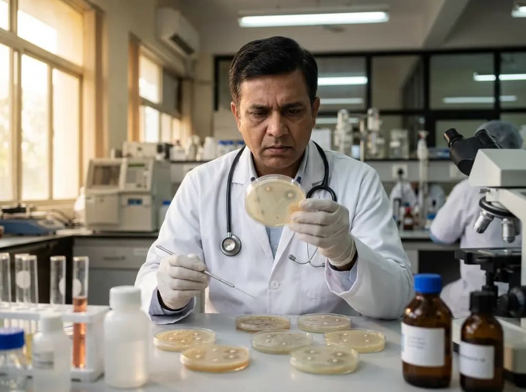
In the evolving landscape of oncology, immune checkpoint inhibitors (ICIs) have revolutionized the treatment of thoracic malignancies. While these therapies are primarily lauded for their survival benefits, they are also associated with a unique spectrum of immune-related adverse events. Among the more curious and visually striking phenomena observed in clinical practice is immunotherapy and hair repigmentation. This process involves the darkening of previously gray or white hair in patients undergoing cancer treatment. For years, clinicians have debated whether this effect was a byproduct of the chemotherapy often administered alongside ICIs or a specific reaction to the immunotherapy itself. Understanding this distinction is crucial for both oncologists and dermatologists, as it may serve as an external indicator of the body's internal immune response. Recent prospective studies have sought to provide definitive evidence by comparing patient cohorts receiving different therapeutic regimens. By utilizing advanced imaging techniques, researchers can now quantify these changes, moving beyond anecdotal case reports into the realm of evidence-based clinical observation. This exploration not only sheds light on hair biology but also highlights the systemic reach of modern cancer treatments.
The science behind hair color involves a complex interplay of melanocyte stem cells and mature melanocytes within the hair follicle bulge. As individuals age, these cells often enter a state of quiescence or are lost, leading to graying. However, immunotherapy agents, particularly those targeting the PD-1/PD-L1 pathway, appear to disrupt this status quo. Research suggests that certain melanocyte stem cells express PD-L1, which helps them maintain a dormant state. When a patient receives immunotherapy and hair repigmentation occurs, it is hypothesized that the inhibitor "releases the brake" on these dormant cells. This reactivation allows for the production of new pigment that is then deposited into the growing hair shaft. Furthermore, the inflammatory milieu created by immune activation may stimulate the production of melanocyte-stimulating hormones. Unlike chemotherapy, which typically targets rapidly dividing cells and often results in alopecia, immunotherapy can actually rejuvenate certain cellular functions. This phenomenon highlights the intricate connection between the immune system and the integumentary system. While the exact pathways are still being mapped, the presence of repigmentation suggests a robust reactivation of cellular processes that were previously considered permanently exhausted in the elderly or chronically ill.
A recent prospective study by Jiang S and colleagues has provided significant clarity regarding the drivers of hair darkening. The research team followed 29 patients with thoracic malignancies, dividing them into an immunotherapy group and a chemotherapy group to isolate the cause. Using standardized dermoscopic images, they applied grayscale analysis to quantify changes in hair pigmentation over time. The results were quite revealing. In the immunotherapy group, grayscale values decreased significantly, which indicates a transition toward darker pigmentation. In contrast, the chemotherapy group showed no statistically significant change in hair color. The magnitude of this change was notably greater in those receiving immune-targeted agents. Specifically, the immunotherapy group showed a large effect size, suggesting that the clinical observation is not just a fluke but a consistent biological trend. This controlled design effectively rules out chemotherapy as the primary cause of hair repigmentation in this patient population. By providing a quantitative framework, the study confirms that the immune system's modulation is the core factor at play. These findings allow clinicians to provide more accurate information to patients who may notice their hair changing during the course of their cancer journey.
Trichoscopy, or hair-specific dermoscopy, has emerged as an indispensable tool for documenting immunotherapy and hair repigmentation. Traditionally used to diagnose various forms of alopecia, this non-invasive technique allows for high-magnification visualization of the scalp and hair shafts. In the context of oncology, trichoscopy provides an objective method to track follicular health and pigment changes without the need for invasive biopsies. By using grayscale analysis on captured images, clinicians can measure the exact degree of darkening that occurs during treatment. This objective measurement is superior to simple visual inspection, which can be subjective and influenced by lighting conditions. In India, where patient diversity in hair texture and baseline color is high, such standardized tools are essential for accurate clinical recording. Trichoscopy also helps in differentiating between repigmentation and other scalp conditions that might arise during therapy, such as inflammatory dermatoses. As oncology moves toward more personalized monitoring, the integration of dermoscopic tools like trichoscopy ensures that even subtle side effects are documented accurately. This data can then be correlated with systemic treatment responses, providing a more holistic view of the patient's progress through their treatment cycles.
The observation of hair darkening in thoracic tumor patients carries potential prognostic significance. In several documented cases, the appearance of cutaneous immune-related adverse events has been linked to better overall survival and treatment response. If hair repigmentation is indeed a marker of a reactive immune system, it could serve as a visual surrogate for therapeutic efficacy. For oncologists, noticing these changes might provide a subtle clue that the immunotherapy is successfully modulating the patient's immune environment. However, it is important to note that the absence of hair darkening does not imply treatment failure. The phenomenon appears more frequently in certain demographics and may be influenced by genetic factors. Nevertheless, the link between immunotherapy and hair repigmentation offers a unique window into the biological activity of ICIs. Clinicians should be prepared to discuss these changes with patients, as darkening hair is often a welcome, though unexpected, side effect. While most discussions around oncology side effects focus on toxicity and discomfort, repigmentation represents a rare instance of a "positive" or at least neutral side effect. Documenting these occurrences helps build a more comprehensive profile of how various ICI agents affect the body beyond the primary tumor site.
Managing the expectations of patients undergoing treatment for thoracic malignancies involves clear communication about both common and rare side effects. When patients notice their hair darkening, they may feel confused or concerned about what it signifies. Healthcare providers can use the findings from recent studies to reassure patients that this is a documented response to immunotherapy. It is essential to integrate these dermatological observations into the overall care plan. For instance, if a patient experiences repigmentation alongside other immune-related symptoms, it may warrant a closer look at their inflammatory markers. Conversely, the psychological boost a patient might receive from seeing their hair return to a younger state should not be underestimated. In the clinical setting, maintaining a record of these changes through trichoscopy can be a part of routine dermatologic surveillance for cancer patients. This proactive approach ensures that any side effect, whether it be the darkening of hair or a more concerning rash, is managed within a standardized framework. As the use of immunotherapy continues to expand in India and globally, the medical community's understanding of these diverse manifestations will only grow, leading to better-informed patients and more precise clinical management.
Disclaimer: This content is for informational and educational purposes only. It is not a substitute for professional medical advice, diagnosis, or treatment. Always seek the advice of your physician or other qualified health provider with any questions you may have regarding a medical condition. Refer to the latest local and national guidelines for clinical practice.
References
Jiang S et al. The Impact of Immunotherapy on Hair Repigmentation in Patients With Thoracic Tumors: A Clinical Observation Based on Trichoscopy. Thorac Cancer. 2026 Jul undefined. doi: 10.1111/1759-7714.70338. PMID: 42387269.
Rivera N et al. Hair Repigmentation During Immunotherapy Treatment With an Anti-Programmed Cell Death 1 and Anti-Programmed Cell Death Ligand 1 Agent for Lung Cancer. JAMA Dermatol. 2017;153(11):1162-1165. doi:10.1001/jamadermatol.2017.2106.
Santoro C et al. Hair Repigmentation as a Sign of Response to Nivolumab in a Patient with Lung Adenocarcinoma. Case Rep Oncol. 2020;13(3):1214-1218. doi:10.1159/000510561.
Hair repigmentation is not a universal side effect. It is specifically associated with immune checkpoint inhibitors, such as PD-1 and PD-L1 blockers, and is rarely seen with standard chemotherapy. While it has been increasingly reported in patients with lung cancer and other thoracic tumors, it remains relatively uncommon. The frequency of this occurrence may vary based on the specific drug used, the patient's genetic background, and their baseline hair color before treatment began.
Trichoscopy is a non-invasive imaging tool that allows clinicians to examine the hair and scalp at high magnification. During cancer treatment, it provides an objective way to monitor changes in hair density, shaft diameter, and pigmentation. By using specialized software to perform grayscale analysis, doctors can calculate the exact change in hair color over time. This removes the subjectivity of human observation and creates a reliable record that can be compared against the patient's systemic response to therapy.
Generally, hair darkening during immunotherapy is considered a benign side effect and is not a cause for medical concern. In many cases, it is viewed as a sign that the immunotherapy is effectively stimulating the body's immune pathways. Some studies even suggest that cutaneous changes like repigmentation might correlate with a better clinical response to the cancer treatment. However, patients should always report any physical changes to their oncology team to ensure all aspects of their health are being monitored correctly.

Read summarized clinical updates, watch expert medical content, and earn CME certifications right from your smartphone.
Open your smartphone camera to scan and install.





A clinical study using trichoscopy reveals that immunotherapy is the primary driver of hair repigmentation in thoracic tumor patients, offering a quantitative framework to understand this unique immune-related phenomenon.
3 weeks back

Andhra Pradesh reported 10 new Covid-19 cases, taking the state tally to 49 while deaths remain at four. With 24 patients hospitalized and 16 under home isolation, the Health Department has intensified monitoring. Medical professionals should review regional distribution, diagnostic protocols, and management plans.
Today

An 11-year Swedish registry study of 618 uterine sarcoma patients found that minimally invasive surgery yielded survival comparable to open surgery in early stages. However, adjuvant chemotherapy conferred no survival benefit in localized or advanced disease, highlighting stage and histology as key outcomes.
3 days back

A cross-sectional study evaluates post-intensive care syndrome in cardiac patients 2-4 weeks post-ICU discharge, highlighting cognitive, psychological, and functional impairments and the need for structured multidisciplinary rehabilitation.
3 days back

Anterior cruciate ligament reconstruction failure lacks uniform definition. A narrative review proposes an integrative framework incorporating objective and subjective instability, persistent pain, restricted motion, graft rupture, and secondary meniscal injury to standardize clinical reporting.
3 days back

With World Obesity Atlas data warning that over 41 million Indian children are overweight or obese, ICMR and NIN have unveiled a 10-point policy roadmap. The initiative calls for mandatory front-of-pack labeling, HFSS taxes, strict marketing bans, and healthier school environments to curb non-communicable diseases.
Today
