.webp&w=384&q=75)

Loading, please wait...
Loading, please wait...
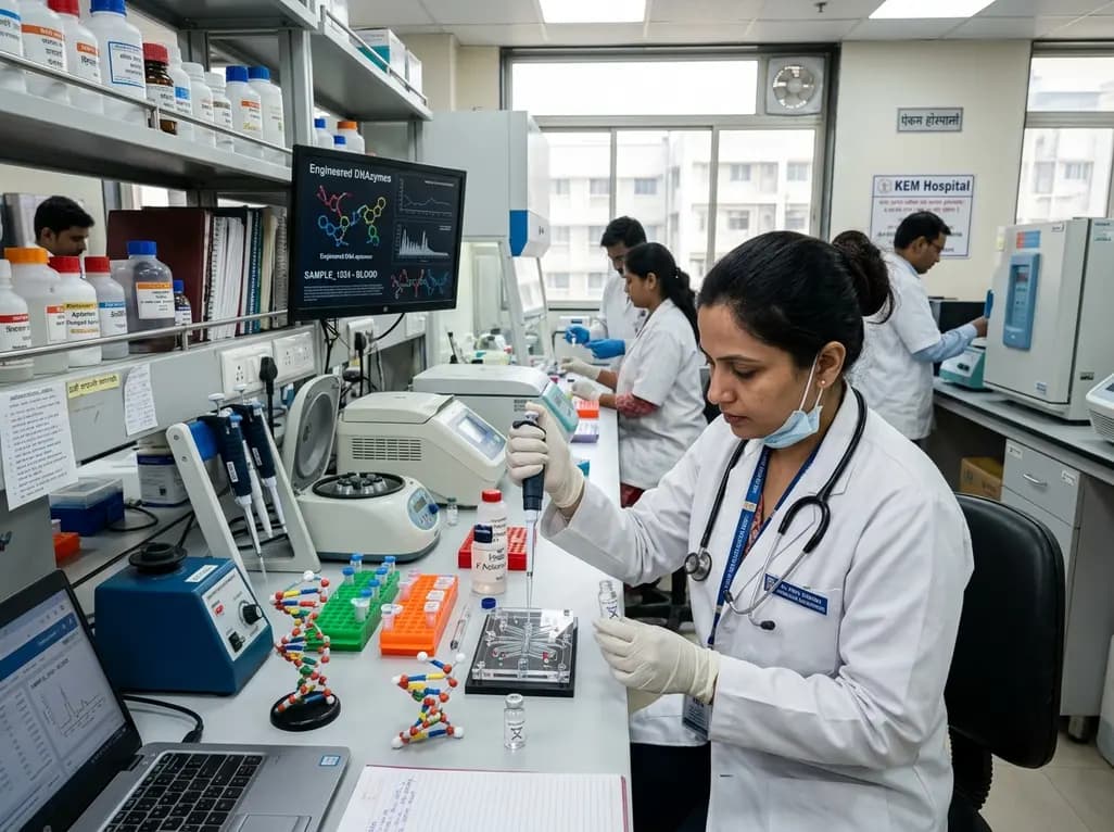
Clinical diagnostics increasingly demand rapid, highly selective molecular tools for point-of-care patient evaluation. Functional nucleic acids, comprising synthetic aptamers and catalytic DNAzymes, offer programmable recognition capabilities that rival conventional monoclonal antibodies. Furthermore, these synthetic oligonucleotides provide distinct manufacturing advantages, including negligible batch-to-batch variation and cost-effective chemical synthesis. Physicians rely on precise molecular assays to detect infectious pathogens, oncogenic drivers, and acute metabolic derangements. Consequently, researchers actively develop nucleic acid biosensors to address critical diagnostic gaps in outpatient and tertiary hospital settings. However, direct clinical implementation requires these synthetic sequences to withstand hostile physiological biofluids. Endogenous nucleases in unprocessed patient specimens rapidly hydrolyze unmodified phosphodiester backbones. Moreover, matrix proteins and fluctuating ionic strengths disrupt secondary folding structures necessary for ligand binding. Therefore, diagnostic translation requires a comprehensive definition of functional stability. True diagnostic durability encompasses far more than simple sequence persistence. Instead, molecular probes must retain target binding affinity, enzymatic cleavage, and reproducible signal transduction under assay conditions. Addressing these foundational degradation mechanisms establishes the pathway toward robust diagnostic devices in clinical pathology.
Chemical engineering serves as the primary defense against enzymatic destruction within biological matrices. Unmodified phosphodiester linkages remain highly vulnerable to serum phosphodiesterases and cellular exonucleases. To counteract this rapid degradation, bioengineers substitute native backbones with phosphorothioate or methylphosphonate linkages. These synthetic alterations prevent phosphodiester cleavage while preserving conformational flexibility. Furthermore, chemists modify ribose sugars at the 2'-position using 2'-O-methyl, 2'-fluoro, or locked nucleic acid chemistries. These sugar adjustments lock oligonucleotides into rigid, nuclease-resistant geometries that enhance hybridization affinity. In addition, synthetic biologists employ non-canonical bases and hydrophobic side chains to expand analyte recognition. For instance, modified nucleobases establish tight non-covalent contacts with uncharged small molecules and bulky protein biomarkers. Nevertheless, extensive chemical modifications can unintentionally compromise catalytic performance or diminish aptamer binding specificity. Each synthetic modification alters internal hydrogen bonding networks and spatial electron densities. Therefore, diagnostic developers must perform rigorous functional validation following chemical synthesis. Clinicians require diagnostic assays that maintain analytical sensitivity despite dense structural modification. Ultimately, rational chemical optimization guarantees that modified oligonucleotides exhibit superior stability without sacrificing target selectivity.
Topological engineering provides an effective physical strategy to protect synthetic probes without introducing disruptive internal modifications. Most clinical nucleases require accessible terminal ends to initiate enzymatic nucleic acid degradation. Consequently, researchers cap terminal endpoints with inverted thymidines, bulky fluorophores, or polyethylene glycol polymers to block exonuclease digestion. Similarly, chemical or enzymatic ligation circularizes linear aptamers into continuous, closed loops. Circular oligonucleotides lack vulnerable endpoints, which completely prevents exonuclease degradation in patient biofluids. Moreover, topological circularization constrains conformational entropy, stabilizing active binding geometries even under elevated physiological temperatures. Researchers also implement covalent crosslinking to lock fragile stem-loop assemblies into functional secondary states. In addition, advanced molecular designs exploit mechanically interlocked topologies, including catenanes and rotaxanes. These interlocked architectures restrict structural unfolding while maintaining responsive sensor signaling. Thus, topological engineering protects sensitive molecular recognition domains without impairing receptor-ligand engagement. Hospital laboratory teams benefit from these structurally locked designs because circularized assays resist degradation during ambient storage. As a result, topological protection transforms fragile oligonucleotides into stable analytical probes that withstand demanding field diagnostic challenges.
Architectural engineering organizes individual recognition elements into cooperative, multivalent nanostructures to enhance analytical sensitivity and physical resilience. Monovalent aptamers often demonstrate rapid dissociation kinetics in biological matrices, which impairs low-abundance biomarker detection. In contrast, multivalent displays significantly increase local probe density, triggering avidity effects that dramatically lower effective dissociation constants. Bioengineers frequently assemble DNA tetrahedrons, dynamic origami tiles, and dendritic scaffolds to present aptamers in uniform spatial orientations. Furthermore, rigid spatial scaffolding sterically crowds the surrounding microenvironment, which physically blocks nucleases from accessing vulnerable internal sequences. Beyond rigid polyhedra, diagnostic developers utilize rolling circle amplification to synthesize extensive concatemers bearing tandem catalytic and binding motifs. These concatemerized strands amplify analytical signals enzymatically, transforming minute target binding events into prominent colorimetric or fluorescent readouts. Therefore, clinicians can rapidly detect trace infectious antigens or early circulating cancer markers. Moreover, multivalent scaffolding buffers diagnostic probes against thermal fluctuations and mild surfactant exposure. Ultimately, scaffolded nanostructures bridge the gap between microscopic molecular recognition and reliable macroscopic diagnostic instrumentation, establishing dependable molecular detection across varied clinical settings.
Interfacial dynamics and local microenvironments dictate biosensor performance when direct clinical biofluids contact diagnostic substrates. Non-specific biofouling by abundant proteins frequently obscures sensor surfaces, generating background noise and causing signal attenuation. To prevent surface passivation, researchers functionalize biosensor interfaces with zwitterionic polymers and hydrophilic self-assembled monolayers. Furthermore, encapsulating functional oligonucleotides within hydrogel networks or porous microgels creates a selective, size-exclusion protective barrier. Hydrogel pores allow small analyte molecules to diffuse freely while excluding bulky nucleolytic enzymes and proteases. Consequently, microenvironment engineering stabilizes enzymatic activity and prevents non-specific binding events across diverse patient matrices. For instance, oral biosensors combine terminal capping with antifouling interfaces to quantify salivary cortisol and viral antigens without mucosal interference. Similarly, urine displays fluctuating ionic strengths, extreme pH variability, and urea concentrations that disrupt nucleic acid folding. Conformationally locked aptamers preserve their binding pockets despite these harsh urinary fluctuations, enabling reliable microalbuminuria detection. In addition, hydrogel confinement isolates enteric pathogens from crude fecal homogenates containing heavy particulate matter. Therefore, combining multiple stabilization levers overcomes matrix interference across every major human biofluid, enabling decentralized pathology testing.
Functional nucleic acids offer distinct manufacturing and logistical advantages over monoclonal antibodies. Researchers produce synthetic aptamers and DNAzymes through chemical oligonucleotide synthesis, eliminating biological batch variability and animal cell culture requirements. Furthermore, these synthetic probes demonstrate superior thermal stability, refolding reversibly after heat denaturation. This resilience simplifies room-temperature transport across tropical clinical settings. However, clinical implementation requires specialized chemical and topological stabilization to match the natural serum persistence that immunoglobulin structures exhibit.
Sequence persistence merely reflects whether the phosphodiester backbone remains intact following nuclease exposure. However, diagnostic assays require functional stability, which demands that the oligonucleotide maintains its tertiary folded conformation, binding affinity, and catalytic activity. Biofluids contain high ionic fluctuations and matrix proteins that can denature probe secondary structures without cleaving covalent bonds. Consequently, diagnostic developers must verify that stabilized sequences generate reproducible detection signals under real clinical assay conditions rather than monitoring physical strand survival alone.
Hydrogels and microgels provide protective physical microenvironments through size-exclusion molecular sieving. Their crosslinked three-dimensional polymer networks possess controlled nanoscale pore dimensions. These nanoscopic pores permit rapid diffusion of small clinical analytes, such as electrolytes, glucose, and bacterial toxins, toward the embedded nucleic acid probes. Conversely, the dense matrix sterically excludes bulky endogenous nucleases and proteolytic enzymes present in patient samples. Therefore, hydrogel encapsulation preserves probe integrity without requiring extensive internal chemical modification of the recognition sequence.
Disclaimer: This content is for informational and educational purposes only and does not constitute medical, diagnostic, or treatment advice. Clinical decisions should always rely on comprehensive patient assessments, standard diagnostic protocols, and multidisciplinary evaluation. Refer to the latest local and national guidelines for clinical practice.
References

Read summarized clinical updates, watch expert medical content, and earn CME certifications right from your smartphone.
Open your smartphone camera to scan and install.





Functional nucleic acids such as aptamers and DNAzymes enable programmable biosensing for clinical diagnostics. Stabilizing these synthetic probes through chemical, topological, architectural, and microenvironmental engineering ensures robust biomarker detection in complex samples like blood, saliva, and urine.
Yesterday
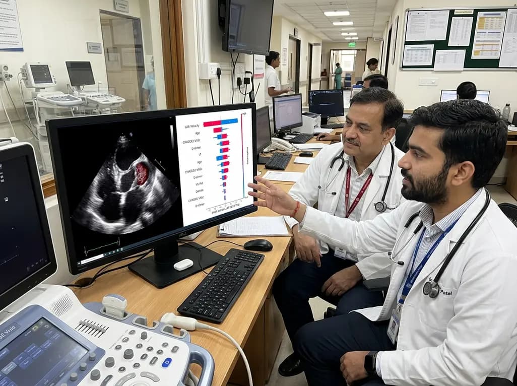
A study demonstrates an explainable machine learning framework integrating clinical and echocardiographic metrics to predict left atrial appendage thrombus in non-valvular atrial fibrillation, utilizing SHAP analysis for clinical interpretability.
Yesterday
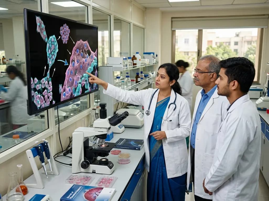
Recent research reveals that downregulating YAP1/TAZ-TEAD via Hippo signaling triggers spontaneous human trophoblast syncytialization in 3D cultures, illuminating placental biology and preeclampsia.
4 weeks back

Abdominal aortic aneurysm rupture remains a catastrophic vascular emergency. Groundbreaking research reveals that USP53 accelerates aneurysm progression by reprogramming smooth muscle cell metabolism toward aerobic glycolysis, highlighting a promising target beyond conventional diameter-based surveillance.
Last week

A breakthrough study leverages natural language processing to accurately identify incident medication-related osteonecrosis of the jaw and track antiresorptive drug holidays from clinical narratives in electronic health records, advancing osteoporosis pharmacovigilance and dental safety.
Last week

Recent research defines ferro-aging as a conserved iron-lipid axis driving organ decline across primates. Age-related iron dyshomeostasis stimulates ACSL4-mediated lipid peroxidation, promoting cellular senescence. Notably, vitamin C directly inhibits ACSL4, suppressing phospholipid oxidation and multi-organ decay.
Last week
