

Loading, please wait...
Loading, please wait...
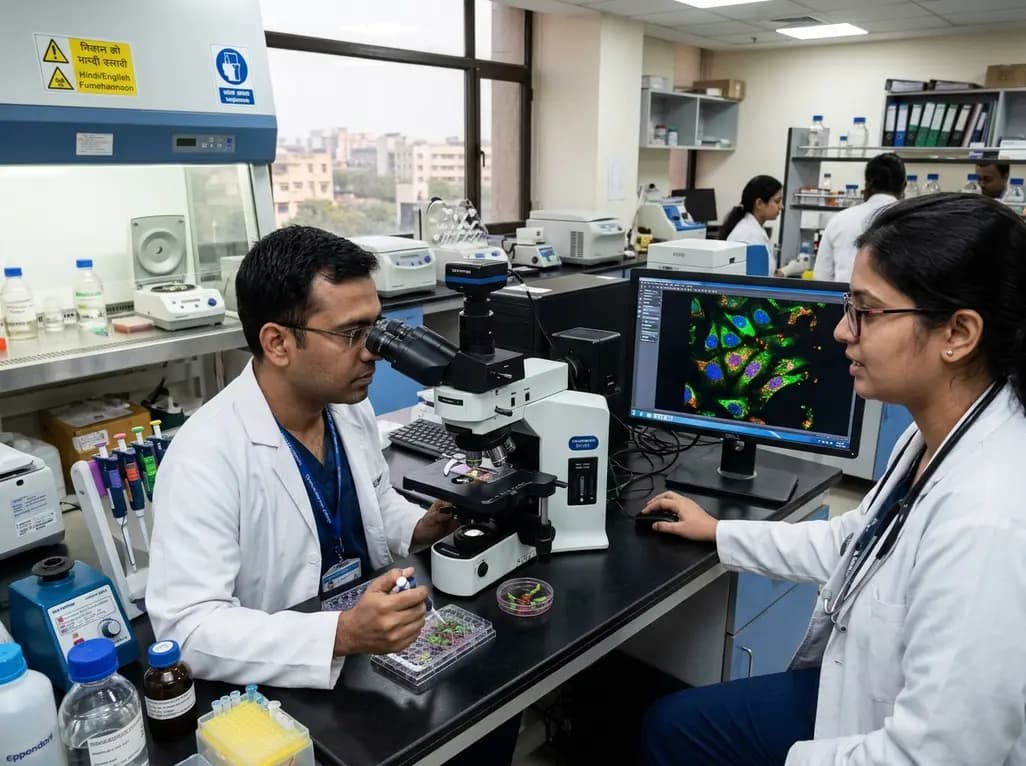
Modern cancer management demands unprecedented spatial precision to eradicate resistant malignant cells. Preclinical studies show that intracellular compartmentalization dictates drug efficacy. Consequently, organelle-directed DNA nanodevices have emerged as transformative tools in molecular oncology. Additionally, precise localization prevents off-target toxicity in healthy tissues. Conventional chemotherapeutics diffuse broadly throughout the intracellular space, often missing primary targets and provoking systemic toxicity. In contrast, programmable nucleic acid architectures can deliver potent therapeutic compounds directly to vital subcellular structures. DNA serves as an exceptional engineering material because investigators can manipulate its sequence, geometry, and valency with molecular exactness. Furthermore, researchers categorize these systems into static nanoframeworks, reconfigurable nanodevices, and dynamic nanomachines. Static nanoframeworks provide rigid structural support and spatial orientation for multiple therapeutic ligands. Meanwhile, dynamic nanodevices sense specific molecular triggers within the cellular microenvironment and execute physical reconfigurations. Ultimately, sophisticated nanomachines perform sequential, mechanical actions to disrupt internal cellular processes. By shifting the paradigm from broad intracellular delivery toward compartmentalized intervention, these platforms offer profound clinical benefits. They lower required drug dosages, diminish adverse events, and overcome multifaceted chemotherapy resistance. Thus, nanoscale nucleic acid architecture introduces a promising horizon for personalized oncology.
Effective organelle targeting requires a rigorous cascade of distinct biological milestones. Historically, scientists treated subcellular delivery simply as a localization problem. However, successful therapy requires safe passage through complex physiological and intracellular checkpoints. First, the engineered construct must maintain structural stability in systemic circulation against degrading nucleases. Second, surface-engineered ligands must recognize specific tumor-cell receptors to initiate receptor-mediated endocytosis. After cellular uptake, the nanodevice encounters severe entrapment within acidic endosomal vesicles. Therefore, researchers incorporate stimuli-responsive motifs to facilitate rapid endosomal escape before hydrolytic enzymes destroy the cargo. Once in the cytoplasm, the platform must navigate intricate cytoskeletal highways to locate its target organelle. Intracellular molecular crowding severely impedes passive diffusion of bulky nanostructures across the cytoplasm. Consequently, active transport mechanisms or specific organelle-homing peptide sequences must guide the nanostructure toward destination membranes. Each barrier represents a potential point of therapeutic failure if engineering designs lack comprehensive optimization. Therefore, modern DNA nanodevices integrate multi-stage responsiveness to systematically overcome each successive barrier. This sequential precision ensures that therapeutic payloads reach their designated target intact. Moreover, quantitative imaging tracks these trafficking kinetics with high temporal accuracy.
Different subcellular compartments serve distinct metabolic and signaling functions within malignant cells. Consequently, investigators design specialized DNA architectures to exploit the distinct vulnerabilities of each organelle. For instance, mitochondria regulate cellular energy synthesis and intrinsic apoptotic pathways. By conjugating triphenylphosphonium cations or mitochondrial-targeting peptides to DNA origami, researchers disrupt mitochondrial transmembrane potential. This localized disruption unleashes cytochrome c and triggers swift cell death without requiring massive systemic doses. Similarly, the cell nucleus remains a premier target for genotoxic drugs and gene-editing complexes. DNA nanodevices display nuclear localization signals that engage importin proteins to traverse the nuclear pore complex. In contrast, lysosome-targeted nanodevices exploit the acidic lumen and rich hydrolase environment to trigger payload release. Alternatively, nanomachines can mechanically perturb lysosomal membrane integrity to induce cathepsin leakage and necroptosis. Furthermore, recent innovations focus on the endoplasmic reticulum and Golgi apparatus. Targeting these biosynthetic hubs disrupts proteostasis and induces fatal unfolded protein stress. In addition, researchers incorporate fluorescent reporters to confirm precise organellar accumulation. Such multi-organelle targeting strategies may prevent the emergence of drug-resistant cancer phenotypes. Thus, tailored DNA nanoplatforms can dismantle vital cellular operations across multiple subcellular domains.
Precision medicine requires intelligent nanodevices that activate payloads exclusively within designated subcellular microenvironments. Fortunately, DNA nanotechnology offers unparalleled conformational flexibility through programmable base pairing. Engineers build responsive aptamer gates, toehold-mediated strand displacement circuits, and proton-sensitive non-canonical motifs. For example, i-motif DNA sequences undergo reversible conformational folding under slightly acidic pH conditions. When an engineered device enters the acidic lysosome, this structural change immediately opens molecular locks to release cytotoxic payloads. In addition, scientists utilize the elevated glutathione levels inside cancer cells to cleave disulfide-linked nucleic acid frameworks. Other advanced platforms respond dynamically to organelle-specific enzymes such as mitochondrial reactive oxygen species or lysosomal proteases. Moreover, dynamic DNA nanomachines can execute logical computation by processing multiple inputs before initiating actuation. A device might require both an acidic pH and a tumor-specific microRNA sequence before deploying its drug payload. Consequently, this multi-input Boolean logic eliminates premature drug leakage in normal tissues and prevents off-target toxicity. Furthermore, external physical triggers like near-infrared light can actuate photo-responsive DNA complexes remotely. As a result, clinicians gain temporal and spatial control over drug delivery mechanisms. Such smart actuation ensures maximum cytotoxic impact directly inside malignant compartments.
Translating complex DNA nanodevices from academic discovery to bedside clinical utility presents substantial challenges. Specifically, researchers must overcome physiological degradation, reticuloendothelial clearance, immunogenicity, and costly large-scale manufacturing hurdles. However, emerging technological integration provides remarkable solutions to expedite clinical progress. Artificial intelligence and self-driving laboratories now accelerate sequence design, predict tertiary folding patterns, and simulate intracellular interactions. Machine learning algorithms analyze high-content microscopy images to track organelle docking kinetics with sub-micron resolution. Furthermore, automated robotic synthesis platforms rapidly generate and evaluate structural variations through closed-loop experimentation. Moreover, surface passivation with protective polyethylene glycol coatings significantly extends systemic circulation time. Consequently, protected nanostructures evade hepatic sequestration and achieve superior tumor biodistribution. In addition to computational design, establishing rigorous regulatory evidence standards remains critical for clinical validation. Investigators must clearly differentiate true organelle actuation from passive subcellular accumulation. Standardized toxicological assays and pharmacokinetics studies must rigorously confirm biocompatibility and clearance mechanisms in preclinical models. Therefore, collaborative frameworks uniting oncologists, materials scientists, and regulatory agencies will shape translational success. Ultimately, these advanced engineering standards will transform theoretical nucleic acid architecture into reproducible, life-saving oncology therapeutics.
Conventional synthetic nanocarriers like liposomes and polymeric micelles offer variable cargo distribution and heterogeneous structural architectures. In contrast, DNA nanodevices provide atomic-level programmability with defined geometry, sequence, and valency. Furthermore, DNA scaffolds enable researchers to position targeting ligands, imaging fluorophores, and therapeutic payloads with nanometer precision. Consequently, these smart structures accurately sense biochemical cues and execute computations to release payloads exclusively within target organelles.
After cellular internalization, nanodevices face rapid entrapment within acidic endosomal vesicles. To overcome this critical hurdle, bioengineers incorporate stimuli-responsive chemical components into the DNA frameworks. For instance, researchers conjugate proton-sponge polymers or pH-responsive peptides directly to the DNA structure. Furthermore, protonation of these motifs inside the acidic endosomal lumen triggers osmotic swelling and membrane destabilization. Consequently, the nanodevices escape into the cytosol before hydrolytic lysosomal enzymes can degrade their therapeutic cargo.
Artificial intelligence significantly accelerates the design and experimental optimization of complex nucleic acid nanostructures. Specifically, machine learning algorithms predict thermodynamic folding pathways and optimize nucleotide sequences to prevent unwanted kinetic traps. Furthermore, deep learning models analyze high-throughput confocal microscopy data to evaluate subcellular delivery kinetics with exceptional accuracy. Therefore, combining computational intelligence with automated robotic platforms enables closed-loop experimentation. This integrated methodology rapidly identifies top-performing nanodevice configurations for precision cancer therapy.
Disclaimer: This content is for informational and educational purposes only... Refer to the latest local and national guidelines for clinical practice.
References

Read summarized clinical updates, watch expert medical content, and earn CME certifications right from your smartphone.
Open your smartphone camera to scan and install.





Organelle-directed DNA nanodevices offer spatial precision in cancer therapy by navigating intracellular barriers, selectively targeting subcellular compartments, and actuating payload release via stimuli-responsive mechanisms, ushering in a new era of programmable nanomedicine.
Yesterday

A multicenter study validates a machine learning-guided recalibrated SOFA score, demonstrating significantly improved ICU mortality discrimination while preserving the simple, additive, point-based structure critical for bedside decision-making.
2 days back
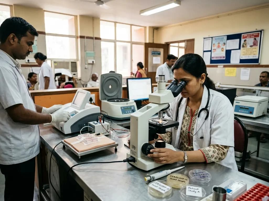
A pilot study evaluated WearDOXXN, a capillary microfluidic sweat patch, for noninvasive breast cancer monitoring by comparing sweat and serum CEA and PIP/GCDFP-15 levels.
Yesterday

Researchers have engineered novel light-on photoswitchable adrenoceptor agonists targeting β1-receptors. By remaining inactive until illuminated, these precision molecules offer reversible optical control, tailored relaxation kinetics, and distinct efficacy profiles, paving the way for targeted cardiovascular therapies.
3 days back

A single-cell transcriptomic atlas reveals how peripheral nerve injuries cause dorsal root ganglion remodeling. By evaluating proximal nerve crush and resection, researchers identify distinct glial and immune programs that resolve neuropathic pain and promote regeneration.
3 weeks back

A breakthrough 3D-printed piezoelectric collagen hydrogel activated by ultrasound accelerates diabetic wound repair, demonstrating 98.5% wound closure, robust antibacterial action, and enhanced vascularization without toxic chemical crosslinkers.
Yesterday
