

Loading, please wait...
Loading, please wait...
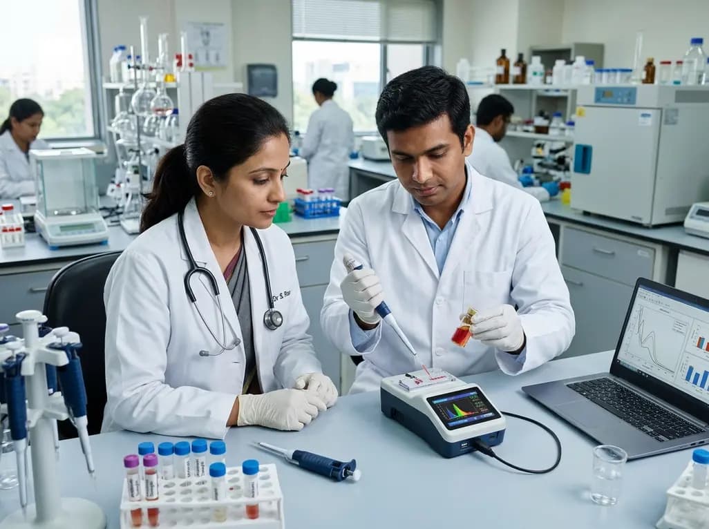
The widespread overuse of antimicrobial agents across healthcare, veterinary medicine, and agriculture has accelerated the worldwide crisis of antimicrobial resistance. Developing optical sensors for antibiotic detection represents an urgent priority to preserve drug efficacy and prevent toxic accumulation. Gold nanoparticles have emerged as premier nanostructured materials for optical sensing due to their unique optical and physicochemical properties. Specifically, gold nanoparticles exhibit localized surface plasmon resonance, generating intense extinction bands within visible wavelengths that respond sensitively to local refractive index fluctuations. When target antibiotic molecules interact with surface-engineered gold nanoparticles, the system produces a distinct, concentration-dependent optical signal. Investigators tailor the size, geometry, and surface capping agents of gold nanoparticles to optimize selective recognition. Consequently, researchers functionalize gold surfaces with aptamers, monoclonal antibodies, or molecularly imprinted polymers targeting specific antibiotic classes like beta-lactams, aminoglycosides, and fluoroquinolones. Furthermore, these nanostructures provide large surface-to-volume ratios, enhancing reaction kinetics and molecular capture efficiency. Routine chromatographic methods remain slow, expensive, and centralized. Therefore, plasmonic gold nanoplatforms establish a highly adaptable foundation for rapid, decentralized, and sensitive antibiotic screening in clinical and environmental settings.
Optical biosensors utilize diverse photochemical and photophysical signaling pathways to quantify antibiotic residues with extreme precision. The most widely employed modality involves aggregation-induced plasmonic coupling of colloidal gold nanoparticles. For example, specific binding between target antibiotic molecules and nanoparticle ligands triggers interparticle cross-linking. This aggregation shifts the localized plasmon band toward longer wavelengths, creating an immediate visual color transition from wine-red to purple or blue. Additionally, researchers employ fluorescence resonance energy transfer architectures, wherein gold nanoparticles serve as super-quenching acceptors. Upon binding the analyte, fluorescence is quantitatively restored or quenched relative to the antibiotic concentration. Surface-enhanced Raman scattering offers another ultra-sensitive pathway, achieving detection limits in the picomolar range. Gold nanostructures create localized electromagnetic hot spots that amplify the unique Raman vibrational spectrum of the antibiotic analyte. Furthermore, investigators exploit the intrinsic peroxidase-like catalytic nanozyme activity of gold nanoparticles. In these systems, antibiotic interaction modulates enzyme-mimicking catalytic efficiency, producing robust chromogenic or chemiluminescent signals. These diverse signal amplification mechanisms guarantee exceptional analytical sensitivity across narrow clinical and regulatory detection windows.
Translating nanobiosensors from centralized analytical laboratories to point-of-care environments requires robust, user-friendly diagnostic platforms. Lateral flow immunoassays represent the gold standard for rapid field-deployable strip testing. These paper-based test devices yield qualitative or semi-quantitative visual readouts within ten minutes, making them extraordinarily valuable for low-resource clinics and agricultural inspection stations. Additionally, microfluidic lab-on-a-chip technologies integrate automated sample metering, reagent transport, and optical detection within disposable microchannels. Such microfluidic systems drastically cut diagnostic reagent consumption and minimize manual handling errors. Furthermore, contemporary engineers successfully pair gold nanoparticle test assays with smartphone-based optical imaging systems. Smartphone cameras record colorimetric or fluorescent emission profiles, while customized mobile software calculates precise analyte concentrations using calibrated calibration curves. Consequently, non-specialist operators can perform high-precision testing in decentralized community hospitals or rural animal farms. Beyond rigid cassettes, innovative wearable optical sensors and flexible microneedle patches are emerging for continuous therapeutic monitoring. These minimally invasive tools monitor real-time antibiotic levels in interstitial fluid, enabling personalized dose adjustments for critical drugs. Thus, portable optical systems bridge traditional laboratory testing and decentralized healthcare delivery.
Commercializing optical nanobiosensors requires resolving critical engineering and translational hurdles that impact real-world performance. Colloidal stability represents a continuous challenge during packaging, shipping, and extended shelf storage. Unmodified gold nanoparticles exhibit susceptibility to spontaneous aggregation when subjected to fluctuating ambient temperatures or ionic variations. Moreover, complex biological and environmental matrices present formidable analytical interference. Real-world samples, such as human whole blood, urine, raw milk, and wastewater effluents, contain complex proteins, lipids, and interfering electrolytes. These endogenous substances frequently trigger non-specific adsorption, generating false-positive signals or masking target analyte binding. Therefore, implementing advanced antifouling surface chemistries and streamlined sample preparation techniques is mandatory. In addition, batch-to-batch reproducibility during large-scale chemical synthesis remains a demanding industrial bottleneck. Subtle differences in nanoparticle core diameter, shape dispersion, or ligand grafting density can introduce unacceptable assay variance. Finally, diagnostic developers must fulfill strict regulatory approval frameworks before securing clinical accreditation. Demonstrating high selectivity, reproducibility, and robust diagnostic accuracy across multi-center clinical trials requires substantial economic resources. Consequently, overcoming these manufacturing and regulatory bottlenecks remains essential for commercial success.
Containing the rapid spread of antimicrobial resistance demands an integrated One Health approach connecting human clinical care, agriculture, and environmental health. In India, regulatory agencies including the Food Safety and Standards Authority of India (FSSAI) have tightened maximum residue limits for antimicrobials across dairy, poultry, and aquaculture supply chains. Concurrently, the Indian Council of Medical Research (ICMR) coordinates nationwide surveillance tracking multi-drug resistant hospital and community pathogens. Portable optical gold nanoparticle sensors provide an ideal technological solution to support these public health mandates. For instance, point-of-care lateral flow assays allow veterinary inspectors to screen livestock products directly at collection centers. In tertiary hospitals, rapid optical quantification streamlines therapeutic drug monitoring for narrow-therapeutic-index reserve antibiotics, preventing toxic adverse reactions and microbial selection pressure. Looking ahead, incorporating artificial intelligence and advanced machine learning algorithms will dramatically accelerate diagnostic precision. Computational models can rapidly deconvolve overlapping optical spectra, filter matrix noise, and classify multi-analyte signatures in real time. Furthermore, multiplexed microfluidic devices will facilitate simultaneous screening of distinct antibiotic classes from a single droplet. Consequently, smart nanodiagnostic platforms will fortify antimicrobial stewardship across India.
Gold nanoparticle sensors detect antibiotics primarily through localized surface plasmon resonance shifts, fluorescence modulation, and surface-enhanced Raman scattering. When functionalized nanoparticles encounter specific antibiotic molecules, they undergo controlled aggregation or selective surface binding. This interaction produces an immediate color shift from red to purple-blue or alters fluorescent emission intensity. These optical changes correlate quantitatively with drug concentration, allowing rapid, highly sensitive detection without requiring complex sample pre-treatments or bulky laboratory instruments.
Rapid antibiotic quantification enables precise therapeutic drug monitoring at the patient bedside, ensuring clinicians maintain effective drug exposures. In critical care settings, timely measurement of reserve antibiotics prevents subtherapeutic dosing that fuels antimicrobial resistance, as well as supratherapeutic levels causing severe nephrotoxicity. Furthermore, rapid screening identifies counterfeit formulations and contaminated agricultural products before they enter the human food chain, reinforcing comprehensive One Health stewardship interventions across communities.
Routine clinical translation faces multiple practical hurdles, including colloidal stability during storage, high production costs, and potential batch-to-batch variability. Furthermore, complex biological fluids like blood, serum, and urine contain non-specific proteins and ions that cause significant signal interference. Overcoming these limitations requires standardized green manufacturing methods, robust anti-fouling surface chemistries, and extensive validation across diverse clinical matrices to fulfill strict national medical device regulatory standards.
Disclaimer: This content is for informational and educational purposes only and does not constitute medical or regulatory advice. Refer to the latest local and national guidelines for clinical practice.
References

Read summarized clinical updates, watch expert medical content, and earn CME certifications right from your smartphone.
Open your smartphone camera to scan and install.





Discover how gold nanoparticle-based optical sensors advance antibiotic detection. Review key sensing mechanisms, portable diagnostic platforms, and their role in mitigating antimicrobial resistance in healthcare and food systems.
Yesterday
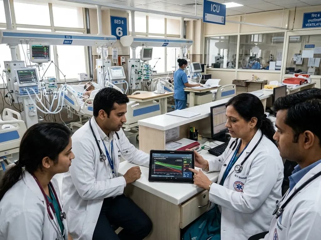
A multicenter study validates a machine learning-guided recalibrated SOFA score, demonstrating significantly improved ICU mortality discrimination while preserving the simple, additive, point-based structure critical for bedside decision-making.
Last week
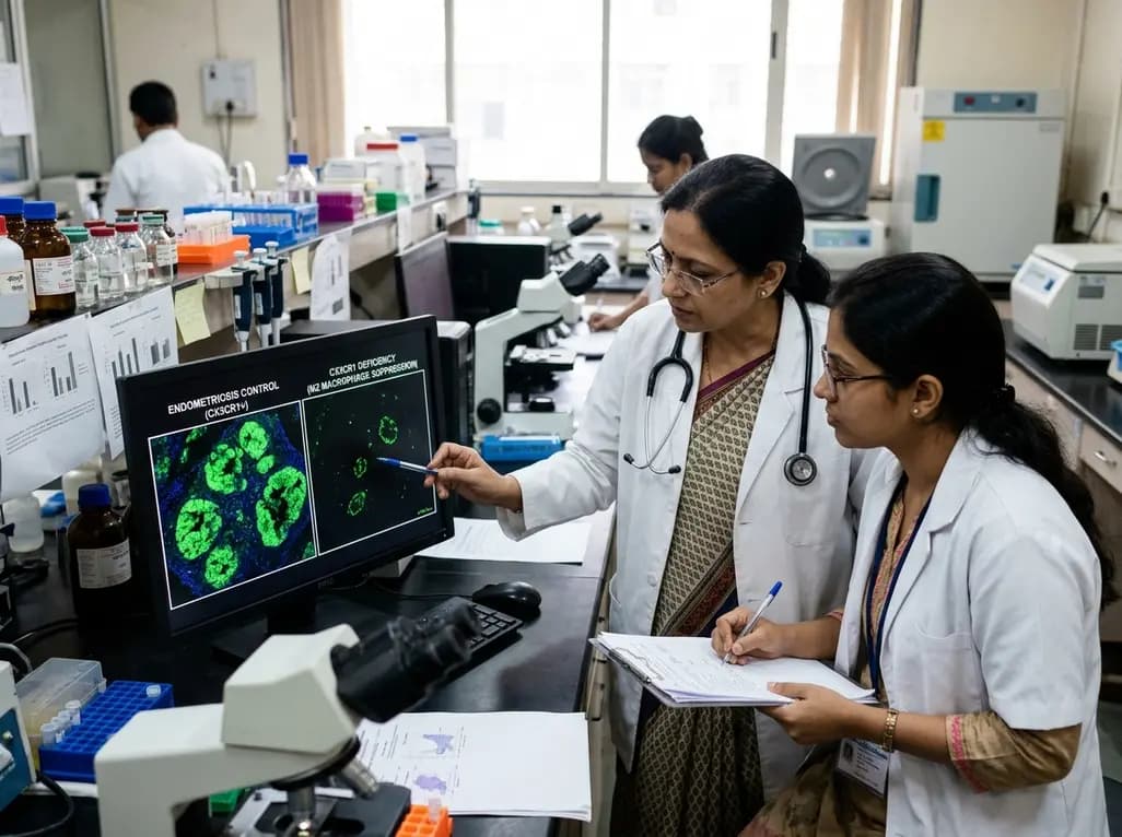
Groundbreaking experimental research demonstrates that CX3CR1 deficiency significantly attenuates endometriotic lesion growth by suppressing pro-repair M2 macrophage polarization. This discovery identifies the CX3CL1-CX3CR1 chemokine axis as a promising non-hormonal target for managing chronic endometriosis.
Yesterday

Researchers have engineered novel light-on photoswitchable adrenoceptor agonists targeting β1-receptors. By remaining inactive until illuminated, these precision molecules offer reversible optical control, tailored relaxation kinetics, and distinct efficacy profiles, paving the way for targeted cardiovascular therapies.
Last week

A single-cell transcriptomic atlas reveals how peripheral nerve injuries cause dorsal root ganglion remodeling. By evaluating proximal nerve crush and resection, researchers identify distinct glial and immune programs that resolve neuropathic pain and promote regeneration.
4 weeks back

A breakthrough 3D-printed piezoelectric collagen hydrogel activated by ultrasound accelerates diabetic wound repair, demonstrating 98.5% wound closure, robust antibacterial action, and enhanced vascularization without toxic chemical crosslinkers.
Last week
