

Loading, please wait...
Loading, please wait...
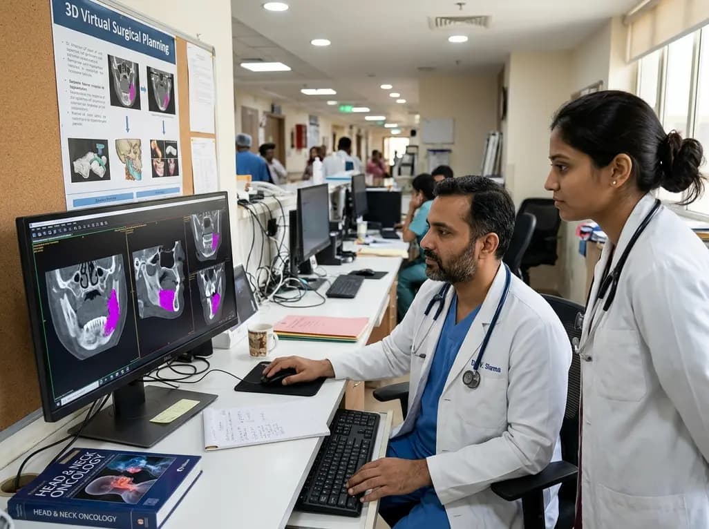
Surgical oncology requires precise anatomical boundaries when managing advanced oral head and neck malignancies. Specifically, accurate identification of mandibular bone invasion in oral squamous cell carcinoma determines whether a patient undergoes marginal cortical shaving or extensive segmental mandibulectomy. In routine practice, head and neck surgeons and radiologists manually delineate osteolytic cortical defects on computed tomography scans. However, manual contouring remains labor-intensive, operator-dependent, and prone to inter-observer variability. Consequently, automated solutions are urgently needed to streamline virtual surgical planning.
Assessing tumor spread through bone remains a complex task in advanced oral oncology. Head and neck malignancies often infiltrate adjacent cortical and cancellous architecture in subtle patterns, including erosive cortical saucerization and invasive trabecular permeation. Furthermore, dental restorations frequently introduce severe metal beam hardening artifacts on computed tomography scans. These streaks obscure cortical borders, hinder tumor evaluation, and distort cancellous bone density profiles. Therefore, clinicians spend considerable time scrolling through multiplanar cross-sectional images to trace subtle osteolytic margins. Moreover, inaccurate margins directly threaten patient safety. If a surgeon underestimates bony involvement, positive microscopic margins cause severe locoregional recurrence. Conversely, overestimating bone invasion leads to excessive bone resection, prolonged operative reconstruction time, and permanent speech or swallowing morbidity. Thus, surgeons require reproducible, objective tools that rapidly pinpoint malignant infiltration within bone before entering the operating room.
To resolve manual delineation bottlenecks, researchers developed an automated pipeline combining volumetric convolutional neural networks with specialized segmentation frameworks. Specifically, investigators trained a three-dimensional MedNeXt neural network implemented within the robust nnU-Net environment. The architecture processed preoperative computed tomography examinations from fifty patients with histopathologically confirmed bone involvement. Additionally, expert head and neck radiologists and surgeons established ground truth segmentations through rigorous multi-reader review. The neural network learned to delineate healthy cortical bone and pathologic osteolytic erosion simultaneously. By utilizing adaptive multi-scale convolutions, MedNeXt captured subtle grayscale differences between healthy cortical bone, reactive sclerosis, and active malignant bone lysis. Furthermore, the nnU-Net framework automatically configured preprocessing steps, patch sizes, and loss functions to handle spatial heterogeneity. Consequently, this computational approach establishes a standardized, automated methodology for parsing complex mandibular anatomy without human fatigue.
The algorithmic evaluation demonstrated remarkable differentiation between healthy skeletal structures and complex tumor lesions. First, the automated model segmented intact mandibular bone with exceptional accuracy, yielding a mean Dice similarity coefficient of 0.965. In contrast, segmenting invasive osteolytic tumor proved more challenging due to subtle contrast transitions and heterogeneous bone loss. For tumor invasion within bone, the deep learning network achieved a mean Dice similarity coefficient of 0.553 and a median of 0.623. Importantly, the model exhibited high precision at 0.742, although recall reached 0.483. In the independent holdout testing cohort, predicted invasion overlapped reference segmentations in ninety percent of test cases. Furthermore, eighty percent of test examinations achieved a Dice similarity coefficient above 0.20, while only two cases with extremely subtle cortical defects were missed. Thus, the model consistently pinpoints gross bone invasion while maintaining high positive predictive value across varied patient datasets.
Beyond volumetric overlap scores, surgical teams require precise spatial margins along anatomical curves to design custom cutting jigs. Therefore, researchers evaluated longitudinal invasion span differences and centerline-projected endpoint distances in the holdout testing cohort. In eighteen evaluable cases, the model demonstrated a median absolute invasion-span difference of 7.01 millimeters. Additionally, the network achieved a median symmetric endpoint distance of 5.88 millimeters along the projected mandibular centerline. These millimeter-level distances hold immense relevance for computer-aided design and computer-aided manufacturing workflows. Typically, oncologic surgeons add a safety margin of ten to fifteen millimeters beyond radiologically suspicious bone. Because the model predicts tumor margins with sub-centimeter accuracy, it directly aligns with standard macroscopic margins. Furthermore, centerline projections allow biomedical software to place virtual osteotomy planes automatically. As a result, surgical planners can rapidly position patient-specific reconstructive cutting guides without laborious manual slice-by-slice drawing.
Integrating artificial intelligence into surgical oncology workflows offers transformative benefits for multidisciplinary clinical teams. When oral squamous cell carcinoma invades the jaw, reconstructive surgeons often transfer a vascularized fibula free flap to restore speech, swallowing, and appearance. However, virtual surgical planning historically demanded several hours of technician time and manual clinician review. By introducing automated segmentation, clinical centers can dramatically shorten this planning interval. Consequently, oncology teams avoid surgical delays, ensuring patients receive timely ablative surgery within recommended oncologic windows. Moreover, objective automated segmentations reduce inter-operator variability across academic centers and community clinics. While prospective validation remains necessary, this technical framework represents a major step toward automated digital oncology workflows. Ultimately, coupling automated bone invasion detection with patient-specific cutting guides enhances surgical precision, protects clear margins, and improves post-treatment quality of life for oral cancer patients.
Artificial intelligence analyzes three-dimensional computed tomography scans using deep learning architectures like MedNeXt and nnU-Net. Specifically, the neural network rapidly scans cross-sectional imaging data to differentiate healthy mandibular cortical bone from osteolytic tumor destruction. By automating this laborious delineation, the algorithm provides consistent spatial boundaries of bone involvement. Consequently, surgical teams can visualize tumor extent faster, avoid observer bias, and design accurate patient-specific cutting guides for reconstructive operations.
Segmenting intact jawbone relies on high-density cortical bone boundaries that contrast sharply against surrounding soft tissues on computed tomography. In contrast, bone invasion presents ill-defined, heterogeneous osteolytic bone resorption and trabecular permeation. Furthermore, dental metal artifacts from crowns and fillings produce severe streak distortions across the lower jaw. Consequently, algorithms face significant noise when mapping cancer margins, resulting in lower Dice similarity scores for invasive lesions compared to healthy bone.
Automated segmentation rapidly converts radiological tumor extent into digital three-dimensional models for computer-assisted manufacturing. Reconstructive surgeons can immediately calculate longitudinal centerline margins and position patient-specific osteotomy guides without extensive manual drawing. Therefore, this technology substantially compresses preoperative preparation times and reduces surgical delays. In addition, standardized automated margins ensure adequate oncologic clearance while conserving healthy vascularized bone, thereby optimizing functional reconstructive outcomes and dental rehabilitation after cancer surgery.
Disclaimer: This content is for informational and educational purposes only. It does not constitute medical advice and should not be used as a substitute for professional clinical judgment, diagnosis, or treatment. Medical knowledge and healthcare guidelines continuously evolve; therefore, readers should independently corroborate all material. While the authors strive to present accurate and complete information, they assume no legal liability for any inaccuracies, omissions, or prospective outcomes resulting from the application of this information. In the event of a clinical emergency, contact emergency medical services or visit the nearest healthcare facility immediately. Professional medical consultations should never be ignored or delayed based on information found on this platform. If you have inquiries regarding medical claims, guidelines, or regulatory details, seek guidance from qualified healthcare experts or relevant governing authorities. Refer to the latest local and national guidelines for clinical practice.
References

Read summarized clinical updates, watch expert medical content, and earn CME certifications right from your smartphone.
Open your smartphone camera to scan and install.





A new proof-of-concept study demonstrates that deep learning can automate CT segmentation of mandibular bone invasion in oral squamous cell carcinoma. This AI approach streamlines virtual surgical planning, helping head and neck oncologists accurately delineate bone margins and plan CAD/CAM reconstructive workflows.
Yesterday
A multicenter study validates a machine learning-guided recalibrated SOFA score, demonstrating significantly improved ICU mortality discrimination while preserving the simple, additive, point-based structure critical for bedside decision-making.
Last week
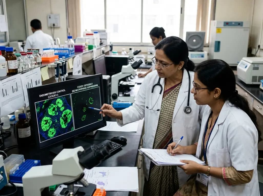
Groundbreaking experimental research demonstrates that CX3CR1 deficiency significantly attenuates endometriotic lesion growth by suppressing pro-repair M2 macrophage polarization. This discovery identifies the CX3CL1-CX3CR1 chemokine axis as a promising non-hormonal target for managing chronic endometriosis.
Yesterday

Researchers have engineered novel light-on photoswitchable adrenoceptor agonists targeting β1-receptors. By remaining inactive until illuminated, these precision molecules offer reversible optical control, tailored relaxation kinetics, and distinct efficacy profiles, paving the way for targeted cardiovascular therapies.
Last week

A single-cell transcriptomic atlas reveals how peripheral nerve injuries cause dorsal root ganglion remodeling. By evaluating proximal nerve crush and resection, researchers identify distinct glial and immune programs that resolve neuropathic pain and promote regeneration.
4 weeks back

A breakthrough 3D-printed piezoelectric collagen hydrogel activated by ultrasound accelerates diabetic wound repair, demonstrating 98.5% wound closure, robust antibacterial action, and enhanced vascularization without toxic chemical crosslinkers.
Last week
