.webp&w=384&q=75)

Loading, please wait...
Loading, please wait...

Mechanical loading profoundly regulates articular cartilage metabolism and joint structural integrity. Clinicians increasingly track walking speed joint biomarkers to evaluate how physical activity influences joint tissues in real time. Articular cartilage lacks intrinsic vascular networks, depending heavily on cyclic mechanical compression for nutrient delivery and matrix turnover. Consequently, ambulation serves as an essential physiologic stimulus for chondrocyte viability. However, the precise biological threshold where healthy mechanical loading becomes tissue stress remains an active research question. When individuals walk at varying velocities, tibiofemoral contact forces and shear stresses change substantially. Therefore, analyzing circulating biomarkers before and immediately following locomotion offers valuable non-invasive insights into cartilage dynamics. Recent investigations have isolated key molecules, including cartilage oligomeric matrix protein and matrix metalloproteinase-3, to quantify acute joint perturbation. Understanding these dynamic responses enables sports medicine physicians and orthopedic surgeons to distinguish adaptive turnover from subclinical tissue damage. Furthermore, this mechanistic perspective helps clinicians design tailored walking regimens that maintain joint health without causing structural harm.
To determine how gait speed impacts cartilage biology, researchers executed a repeated-measures crossover study in healthy human volunteers. Participants completed three distinct laboratory visits under standardized environmental conditions. Importantly, the investigators standardized pre-test physical activity to eliminate confounding mechanical strain on joint tissues. At each visit, participants rested completely supine for 45 minutes before phlebotomists drew baseline resting blood samples. Following this equilibration period, participants performed a structured 30-minute treadmill walking protocol. During each session, investigators altered walking velocity across three experimental conditions: slow walking, moderate walking, or brisk walking. Immediately after completing the 30-minute exercise trial, researchers collected a second venous blood sample to record acute mechanical perturbations. Subsequently, participants returned to a supine resting posture for an additional 45 minutes, after which phlebotomists drew a final blood sample. Through this systematic approach, the research team quantified serum concentrations of cartilage oligomeric matrix protein, matrix metalloproteinase-3, interleukin-6, and lubricin. Thus, this stringent experimental framework ensured that observed biomarker fluctuations reflected genuine load-dependent kinetics rather than postural fluid shifts.
Cartilage oligomeric matrix protein serves as a sensitive indicator of articular cartilage matrix turnover under acute mechanical loading. When cyclic compression deforms cartilage, interstitial fluid flows outward, carrying cleaved COMP fragments into the systemic circulation. In this investigation, treadmill ambulation induced significant elevations in serum COMP immediately post-walk across all tested conditions. Moreover, the magnitude of this biomarker elevation depended directly upon walking speed. Brisk ambulation produced greater serum COMP elevations than slow walking, demonstrating that higher speeds impart larger mechanical forces. Nevertheless, these elevated COMP concentrations decreased steadily during the subsequent 45-minute supine rest period, reflecting physiological clearance rather than structural injury. Concurrently, the study evaluated lubricin, also known as proteoglycan-4, which coats cartilage surfaces to reduce frictional wear. Although lubricin plays a critical lubricating role within synovial joints, acute walking speed variations produced modest, nuanced changes in circulating levels. Consequently, these findings indicate that while compressive strain markedly mobilizes structural matrix markers, surface boundary lubricants exhibit distinct regulatory kinetics during brief bouts of exercise.
Beyond matrix structural proteins, walking velocity substantially influences degradative enzymes and inflammatory signaling molecules. Matrix metalloproteinase-3 degrades proteoglycans and activates other collagenases within joint structures. In the current trial, walking speed altered serum MMP-3 concentrations in a velocity-dependent manner. Specifically, faster walking generated measurable shifts in circulating enzyme levels, highlighting acute mechanochemical coupling within articular tissues. In healthy joints, this transient enzymatic rise promotes healthy matrix remodeling by clearing micro-damaged proteins. Simultaneously, researchers quantified serum interleukin-6, a pleiotropic cytokine responsive to muscular work and mechanical load. Notably, acute treadmill walking elevated circulating IL-6 concentrations, with brisk walking producing more pronounced responses than slow strolls. While chronic IL-6 elevations signal pathological systemic inflammation, acute exercise-derived IL-6 functions primarily as an anti-inflammatory myokine. This cytokine enhances glucose disposal and downregulates pro-inflammatory cytokines such as tumor necrosis factor-alpha. Therefore, brisk ambulation stimulates a balanced biochemical environment where transient matrix metalloproteinase activity pairs with beneficial cytokine signaling, facilitating healthy tissue adaptation rather than unmitigated tissue degradation.
These molecular observations carry significant practical value for orthopedic and rehabilitation practices across India. Musculoskeletal disorders, particularly knee osteoarthritis, place an immense burden on aging and sedentary Indian adults. Clinicians routinely prescribe walking programs to address metabolic syndrome and joint stiffness. However, generic exercise recommendations frequently overlook how walking velocity alters joint mechanical loading. Brisk walking offers superior metabolic and cartilage benefits for healthy individuals, promoting synovial nutrient transport and cellular matrix maintenance. Conversely, patients with pre-existing knee malalignment or established osteoarthritis may struggle with elevated contact forces generated during rapid walking. For these individuals, brisk ambulation could accelerate cartilage breakdown instead of inducing adaptive remodeling. Therefore, Indian practitioners should adopt personalized walking prescriptions that specify pace, duration, and surface conditions. Clinicians should recommend moderate-paced ambulation paired with quadriceps strengthening for symptomatic patients, while reserving brisk intervals for healthy adults. Furthermore, incorporating planned rest periods allows mechanosensitive biomarkers to normalize, preserving joint health and optimizing long-term therapeutic outcomes.
Walking speed joint biomarkers reveal subtle, early metabolic changes in articular cartilage before structural joint narrowing appears on conventional radiographs. By measuring circulating proteins like cartilage oligomeric matrix protein and matrix metalloproteinases before and after standardized walking tests, clinicians can evaluate dynamic cartilage stress responsiveness. Consequently, this functional biochemical assessment identifies individuals with abnormal cartilage turnover or impaired mechanoadaptation, enabling early targeted physical therapy and lifestyle modifications to halt disease progression.
Serum cartilage oligomeric matrix protein rises transiently after brisk walking because increased gait velocity elevates intra-articular contact pressures and cyclic mechanical strain. Consequently, this physiological compression accelerates interstitial fluid exudation from cartilage into the synovial space, washing cleavage fragments into systemic circulation. In healthy joints, this transient elevation represents a normal metabolic adaptation rather than permanent tissue damage, and circulating biomarker concentrations typically return to baseline resting values within forty-five minutes of rest.
Yes, clinicians can successfully customize walking cadence to optimize joint loading and protect damaged cartilage during rehabilitation. Slower walking speeds reduce peak ground reaction forces and mechanical shear, making unhurried ambulation ideal for acute arthritis flare-ups or post-surgical recovery. Conversely, as joint stability improves, gradually increasing walking cadence stimulates proteoglycan synthesis and muscular endurance without overloading joint structures. Therefore, tailored cadence prescriptions provide an effective, non-invasive therapeutic strategy for long-term joint preservation.
Disclaimer: This content is for informational and educational purposes only... Refer to the latest local and national guidelines for clinical practice.
References

Read summarized clinical updates, watch expert medical content, and earn CME certifications right from your smartphone.
Open your smartphone camera to scan and install.





Investigating the influence of walking speed on mechanosensitive joint biomarkers reveals how gait velocity modulates COMP, MMP-3, IL-6, and lubricin kinetics. These findings provide vital physiological insights for orthopedics and sports medicine to personalize exercise prescriptions and preserve joint health.
Today
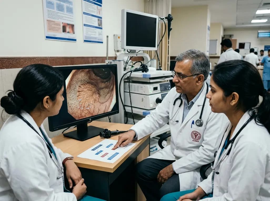
A post hoc decision tree analysis examined JNET Type 1 classification of serrated colorectal lesions. Experts prioritized ruling out anti-Type 1 features with low diagnostic entropy, whereas non-experts relied on mucosal spot patterns, highlighting key training targets for magnifying narrow-band imaging.
Today
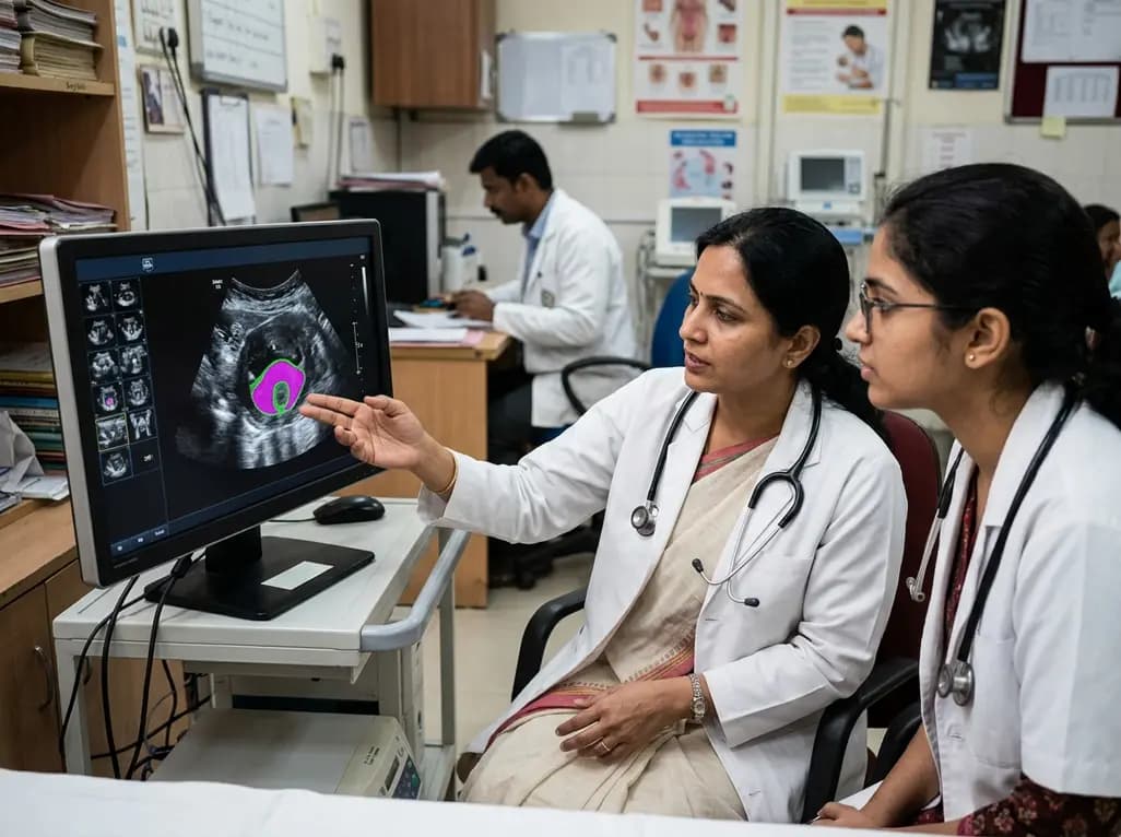
New evidence evaluates MoUNets, a static-weighted ensemble of UNet experts for automated levator hiatus segmentation in pelvic floor ultrasound. Learn how deep learning architectures perform in pelvic organ prolapse diagnostics and clinical practice.
Today

Mitochondrial deubiquitinases regulate organelle turnover by editing ubiquitin tags on damaged mitochondria. Discover how targeting these enzymes, notably USP30, offers therapeutic strategies for neurodegenerative, cardiovascular, and metabolic disorders.
Today

A cross-sectional study reveals that elevated METS-IR is independently associated with higher myopia odds in adolescents, showing biological synergy with obesity and highlighting the need for metabolic screening in pediatric eye care.
Today
