.webp&w=384&q=75)

Loading, please wait...
Loading, please wait...

Right-hemisphere stroke frequently precipitates debilitating cognitive sequelae, among which visuospatial neglect remains one of the most challenging disorders to treat. Patients experiencing this syndrome demonstrate a marked reduction in conscious engagement with contralesional space. Consequently, clinicians observe patients neglecting food on the left side of their plates or failing to notice obstacles situated in their left visual fields. While conventional clinical wisdom historically classified this impairment as a disorder of voluntary spatial orientation and high-level attention, the integrity of pre-attentive sensory mechanisms has remained unclear. Neurologists and neurorehabilitation specialists continue to seek objective biomarkers that explain why spatial recovery often plateaus prematurely.
Understanding visuospatial neglect requires delineating conscious spatial orienting from automatic sensory registration. Stroke lesions that produce neglect commonly disrupt vast white matter tracts and large-scale attention hubs, such as the right superior temporal gyrus, inferior parietal lobule, and ventral frontoparietal networks. Consequently, this structural disconnection disrupts the brain\'s internal salience maps. Clinicians regularly witness how stroke survivors exhibit severe functional impairment during daily mobility, reading, and self-care routines. However, whether structural damage also paralyzes pre-attentive sensory mechanisms remains an active question. Earlier literature posited that primary sensory input reached the visual cortex normally, leaving unconscious detection preserved. In contrast, emerging electrophysiological observations demonstrate that neglect impairs perceptual analysis far earlier along the visual pathway than previously suspected. Therefore, clinicians must re-examine the biological boundaries between sensory perception and conscious attention.
Event-related potentials provide an exquisite temporal window into sequential neural processing. Specifically, the early C1 component reflects initial feedforward afferent activity arriving in the primary visual cortex (striate area V1). Because the C1 waveform peaks between 50 and 90 milliseconds post-stimulus, it provides direct insight into baseline retinotopic processing. Following this initial peak, the C2 and N1 waveforms emerge around 100 to 200 milliseconds, representing subsequent ventral stream visual differentiation and structural encoding. Concurrently, the visual mismatch negativity, or vMMN, serves as a crucial index of automatic, pre-attentive change detection. The brain elicits a vMMN response when an incoming visual stimulus breaks an established temporal or spatial rule. Notably, the vMMN occurs automatically, even when an individual directs conscious attention elsewhere. Consequently, evaluating these distinct electrophysiological waveforms allows investigators to isolate exact processing stages damaged by cerebrovascular insults.
To pinpoint sensory deficits, investigators employed a sophisticated passive oddball paradigm across three well-defined cohorts. Specifically, the research group enrolled 17 individuals with right-hemisphere stroke exhibiting confirmed spatial neglect, 16 stroke control patients without neglect, and 17 age-matched healthy participants. The experimental setup presented concurrent, independent visual streams to the lower left and lower right quadrants of the visual field. Diamond patterns disappeared during regular OFF events and reappeared during ON events. Infrequent, unexpected shifts served as rule-violating deviant stimuli among repeated standard presentations. Importantly, participants completed an engaging central fixation task, thereby ensuring that peripheral visual changes remained unattended. Furthermore, researchers evaluated the neglect cohort across three separate clinical time points: prior to intensive inpatient rehabilitation, immediately post-rehabilitation, and at a four-month outpatient follow-up. This longitudinal framework permitted precise tracking of spontaneous and therapy-induced neuroplasticity.
The resulting neurophysiological data revealed striking differences between the patient cohorts. Healthy controls exhibited robust C1 amplitudes and demonstrated prominent vMMN responses to both left-sided and right-sided regularity violations. In stark contrast, patients with spatial neglect showed a total absence of the C1 component across all recording sessions. Stroke control patients without neglect retained an intermediate tendency toward preserved C1 waveforms. Meanwhile, subsequent C2 and N1 components emerged reliably across all three participant groups without significant statistical differences. This preserved C2/N1 activity confirms that intermediate structural visual transmission remained functionally viable. However, when evaluating the vMMN, patients with neglect failed to generate reliable mismatch responses to unexpected spatial changes. Stroke controls managed to produce reliable vMMN responses to left-sided OFF events, whereas neglect patients exhibited bilateral suppression of this pre-attentive mismatch signature.
Perhaps the most clinically sobering finding is the temporal rigidity of these neurophysiological deficits. Although neglect patients completed comprehensive physical and occupational rehabilitation programs, their electrophysiological profiles showed no measurable restoration. The C1 waveform remained entirely absent at discharge and at the four-month follow-up visit. Similarly, the vMMN failed to recover over time, indicating a lasting failure of automatic predictive visual processing. Standard rehabilitative interventions prioritize top-down visual search strategies, voluntary limb activation, and deliberate environmental scanning. While these interventions offer compensatory behavioral benefits, they apparently fail to repair low-level striate transmission or subcortical predictive loops. Consequently, persistent unconscious processing deficits may explain why patients frequently relapse into spatial errors once cognitive fatigue sets in. Clinicians must therefore recognize that behavioral compensation does not necessarily signify biological recovery within primary sensory circuits.
These electrophysiological findings mandate an evolution in stroke rehabilitation methodologies. Because standard top-down compensatory training leaves pre-attentive visual deficits unresolved, rehabilitation teams must explore neurotechnologies that directly modulate early sensory pathways. For instance, non-invasive neuromodulation, such as high-definition transcranial direct current stimulation, may help rebalance interhemispheric inhibition. Additionally, combining sensory stimulation with immersive virtual reality could stimulate early predictive processing networks. Neurologists should also integrate objective electrophysiological assessments into clinical practice rather than relying solely on paper-and-pencil cancellation tests. Recognizing pre-attentive impairment allows physicians to provide accurate prognostication to families. Ultimately, understanding that neglect involves deep-seated visual sensory failure will stimulate the development of targeted neuroplastic therapies.
Visual mismatch negativity reflects the brain\'s automatic ability to identify violations in established environmental regularities without conscious attention. Following a stroke, a diminished or absent mismatch negativity waveform signals severe breakdown in automatic sensory prediction, indicating that fundamental pre-attentive warning systems are functionally damaged.
Homonymous hemianopia is a sensory visual field defect caused by physical injury to the optic radiations or primary visual cortex, leaving spatial awareness intact. Conversely, spatial neglect represents an attentional failure to register, orient toward, or explore objects located across contralesional space despite intact peripheral optic pathways.
The C1 component originates primarily within retinotopic area V1 and depends on early feedforward cortical circuits. Because standard post-stroke rehabilitation emphasizes top-down voluntary visual scanning rather than primitive sensory reconstruction, damaged early sensory inputs and disrupted cortico-cortical connections frequently show limited spontaneous or therapy-induced neuroplasticity.
Disclaimer: This content is for informational and educational purposes only and should not be considered medical advice. Always consult a qualified healthcare provider for diagnosis and treatment. Refer to the latest local and national guidelines for clinical practice.
References

Read summarized clinical updates, watch expert medical content, and earn CME certifications right from your smartphone.
Open your smartphone camera to scan and install.





New neurophysiological research demonstrates that stroke-induced visuospatial neglect impairs early visual processing and automatic regularity violation detection, persisting despite standard rehabilitation.
Today
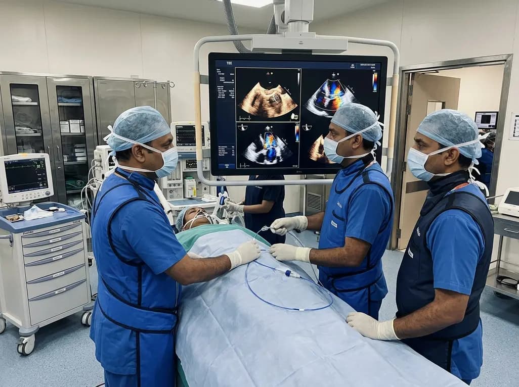
Transcatheter edge-to-edge repair (TEER) successfully treated severe mitral regurgitation from a posterior leaflet perforation. Guided by 3D multiplanar reconstruction, operators sandwiched the defect using two MitraClip devices, providing safe hemodynamic recovery without requiring open-heart surgery.
Today

A 32-year-old gravida at 38 weeks with anterior STEMI underwent emergent IVUS-guided PCI and successful cesarean delivery under uninterrupted DAPT, highlighting cardio-obstetric care.
Today

A systematic review and meta-analysis of 5,406 participants reveals that tirzepatide significantly reduces hsCRP by 34.3%, improves cardiorenal biomarkers, and enhances functional capacity and exercise endurance in patients with heart failure.
Today

A landmark biomechanical study demonstrates that sports bra support and running shoe cushioning interact significantly to influence tibial acceleration, breast motion, and comfort in female runners, providing crucial insights for clinical counseling in sports medicine and women's health.
Yesterday

New ADIPO-CKD study findings demonstrate that both GLP-1 receptor agonists and SGLT2 inhibitors significantly reduce perirenal adipose tissue thickness in chronic kidney disease patients, with GLP-1 therapy achieving greater reductions, highlighting a novel mechanism for cardiorenal metabolic protection.
Today
