

Loading, please wait...
Loading, please wait...
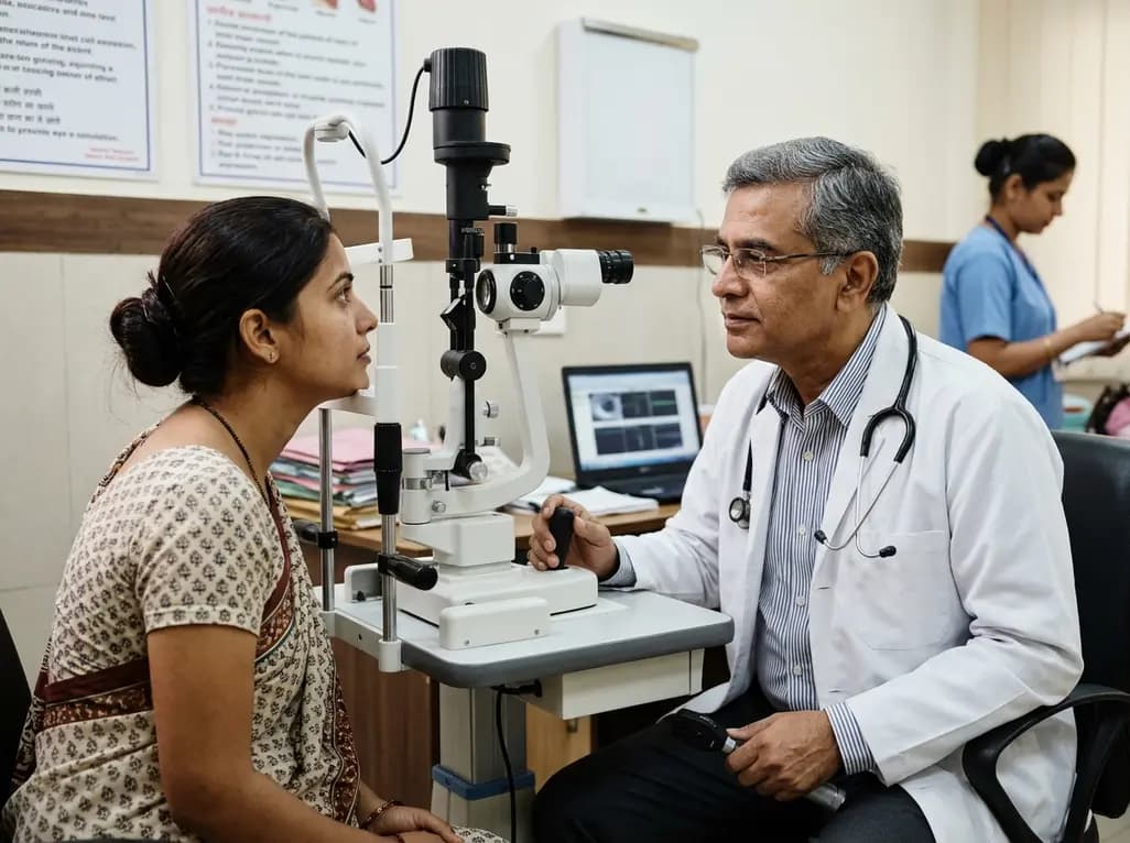
Clear visual acuity is often viewed as the primary benchmark of ocular health. However, achieving 20/20 clarity on a standard Snellen chart does not necessarily guarantee comfortable seeing throughout the day. In routine clinical settings, eye care practitioners frequently encounter patients who see sharply yet complain of profound ocular tiredness, brow ache, and intermittent blurring. This discrepancy highlights the critical concept of visual effort, which represents the physiological and cognitive energy required to acquire, sustain, and interpret visual stimuli. When individuals navigate complex visual environments, their eyes do not function like static cameras. Instead, the ocular motor system constantly adjusts focus across diverse viewing distances and variable lighting environments. Consequently, practitioners must recognize that optical clarity and visual comfort represent two distinct clinical dimensions.
Clinicians historically prioritized high-contrast central visual acuity when assessing ocular status. However, visual effort encompasses a far broader neuro-functional process than mere resolution alone. Specifically, visual effort reflects the continuous muscular and neural exertion needed to keep images crisp on the fovea during sustained tasks. When a patient reads low-contrast print, interprets spreadsheets, or drives in rain, the brain works harder to resolve subtle details. Consequently, visual processing speed decreases while muscular tension in the periorbital tissues escalates.
Moreover, refractive status plays a fundamental role in determining baseline visual strain. Uncorrected low-grade astigmatism or mild hyperopia often forces constant micro-accommodative hunting. Although the patient retains readable visual acuity, the ongoing effort drains neuro-visual reserves rapidly. Therefore, patients frequently report tension headaches and reading fatigue long before visual acuity measurably degrades. Furthermore, environmental degradation such as monitor glare or dim illumination amplifies the required neural compensation. Ultimately, assessing visual comfort requires eye care providers to evaluate how effortlessly a patient sees during real-world activities. Eye care professionals must therefore investigate how everyday viewing environments challenge binocular function. By recognizing visual effort as a functional vital sign, optometrists and ophthalmologists can deliver proactive care that protects visual stamina.
The ocular accommodation system relies on rapid coordination between the ciliary muscle, zonular fibers, and crystalline lens. When individuals shift focus to near screens or printed documents, the ciliary body contracts to increase lens curvature. Concurrently, the medial rectus muscles drive convergence to maintain single binocular vision. Under ancestral conditions, humans shifted gaze dynamically between distance and near targets. In contrast, modern office environments enforce continuous near-point fixation for hours at a time.
Therefore, sustained close-up demands provoke significant accommodative fatigue, also recognized clinically as accommodative asthenopia. When the ciliary muscle sustains static contraction, blood flow decreases and biochemical fatigue markers accumulate. Additionally, continuous near fixation disrupts the natural coupling between vergence and accommodation. As a result, the visual system experiences transient micro-fluctuations in focus, leading to intermittent blur and double vision. Furthermore, prolonged visual effort diminishes involuntary blink frequency by nearly sixty percent, causing tear film destabilization. Thus, muscular fatigue and ocular surface desiccation combine to compound patient discomfort. Clinicians must recognize these physiological mechanisms when addressing recurrent ocular complaints in intensive device users.
Real-world visual behavior rarely matches the static conditions of an examination chair. Modern occupations require individuals to navigate multiple digital displays, paperwork, handheld devices, and room-scale environments simultaneously. For example, a professional might glance from a laptop keyboard to an external monitor, before looking across a conference room. Each shift demands instantaneous saccadic eye movements, accurate vergence re-alignment, and rapid accommodative adjustments.
Consequently, viewing demands vary dramatically across different functional distances and occupational profiles. A software engineer experiences static, near-intermediate visual demands, whereas an active field worker alternates continuously between intermediate and far distances. When optical lenses fail to accommodate these dynamic gaze patterns, wearers compensate through repetitive neck movements and unnatural head tilting. Moreover, aberrant head posture produces secondary musculoskeletal tension across the cervical spine and shoulders. Therefore, comprehensive visual assessments must incorporate occupational ergonomics and natural gaze dynamics. Modern vision science emphasizes that spectacle lenses must provide expansive visual zones aligned with natural eye movement trajectories. When lenses match physiological gaze dynamics, patients experience fluid transitions and reduced visual strain throughout demanding workdays.
Spectacle lens technology has evolved far beyond traditional spherical corrections. Historically, standard corrective lenses focused exclusively on achieving foveal sharpness along the primary optical axis. However, peripheral optical aberrations frequently persist, inducing subtle optical distortions when the eye rotates off-center. Consequently, the brain must constantly filter peripheral blur, which silently heightens cognitive and visual effort.
To overcome these challenges, advanced ophthalmic lens manufacturers incorporate neuroscience principles into optical engineering. For instance, modern digital freeform manufacturing optimizes lens geometry according to natural visual habits and head-eye coordination metrics. Proprietary designs, including modern ClearMind lens technologies powered by neuro-visual insights, explicitly address real-world gaze patterns across multiple distances. By minimizing unwanted peripheral aberrations, these sophisticated designs deliver seamless visual transitions from near to far zones. Furthermore, recent wearer evaluations confirm that over ninety-six percent of patients report exceptionally crisp clarity with optimized lens profiles. In addition, anti-reflective coatings and tailored blue-attenuating substrates filter irritating glare from artificial sources. Ultimately, neuroscience-informed lens designs reduce neurological visual effort, allowing wearers to process visual data with effortless clarity.
Managing visual fatigue requires a multifaceted clinical strategy that extends beyond standard refraction. First, practitioners must conduct comprehensive binocular vision assessments, measuring accommodative amplitude, facility, and near point of convergence. Identifying subtle convergence insufficiency or accommodative lag enables practitioners to prescribe targeted refractive additions or functional vision therapy. Moreover, clinicians should recommend activity-specific lenses, such as dedicated computer lenses or ergonomic progressive designs, for patients experiencing high near-work loads.
Additionally, optometrists and ophthalmologists must actively counsel patients on visual hygiene and ergonomic optimization. Practitioners should advocate the 20-20-20 guideline, where individuals pause every twenty minutes to look twenty feet away for twenty seconds. This brief pause relaxes the ciliary muscle and restores normal blink dynamics. Furthermore, doctors should recommend ambient lighting adjustments to eliminate screen glare and reduce luminance contrast ratios. Preservative-free lubricating artificial tears provide immediate relief for concomitant dry eye symptoms, preserving optical surface regularity. Finally, scheduled annual vision evaluations guarantee timely optical updates, preventing the accumulation of chronic eye strain. By combining advanced optical correction with behavioral modifications, eye care practitioners effectively minimize visual effort and enhance patient quality of life.
Q1: How does visual effort differ from standard visual acuity?
Visual acuity measures the spatial resolving capacity of the eye under static, high-contrast conditions on an eye chart. In contrast, visual effort represents the continuous physiological and cognitive work required to maintain that clarity during dynamic daily activities. Even if a patient achieves perfect 20/20 acuity, uncorrected refractive micro-errors or binocular misalignments can force ocular muscles to overwork, causing persistent fatigue and headache.
Q2: Why do prolonged digital tasks cause excessive visual fatigue?
Prolonged digital tasks lock the ciliary muscles into sustained near accommodation while the medial rectus muscles continuously maintain convergence. Concurrently, focused screen attention reduces the spontaneous blink rate from roughly twenty times per minute to under eight. This sustained muscular contraction produces ocular ache, while tear film evaporation triggers surface inflammation. Consequently, users experience marked visual effort, ocular dryness, and intermittent blurry vision throughout the working day.
Q3: How do neuroscience-informed spectacle lenses minimize everyday eye strain?
Neuroscience-informed spectacle lenses analyze real-world gaze behavior, head-eye movement dynamics, and peripheral visual perception to optimize optical surface geometry. Traditional lenses often produce peripheral aberrations that force the brain to filter visual distortions. Modern designs eliminate these optical compromises across near, intermediate, and far fields. Consequently, wearers experience effortless gaze transitions, reduced micro-accommodative hunting, and significantly lower visual effort during intensive multi-screen occupational tasks.
Disclaimer: This content is for informational and educational purposes only. It does not constitute medical advice or replace professional judgment. Refer to the latest local and national guidelines for clinical practice.
References

Read summarized clinical updates, watch expert medical content, and earn CME certifications right from your smartphone.
Open your smartphone camera to scan and install.





Standard eye tests confirm 20/20 acuity, but true visual comfort depends on physiological effort. Discover how sustained accommodation, gaze dynamics, and ergonomic spectacle designs influence everyday visual performance and patient asthenopia in modern digital environments.
Today

A recent investigation reveals that GLP-1 induces IL-27 synthesis in macrophages via the PI3K/AKT signaling cascade. This axis regulates immune polarization and contextual inflammation, offering valuable perspectives for managing obesity-induced metabolic disease and chronic systemic inflammation.
Today
The GendAge weight loss study investigates how sex hormones, age, and menopausal status modulate caloric restriction response. By comparing metabolic adaptations across younger and older cohorts, this controlled feeding trial aims to refine precision dietary guidance for effective obesity management.
Today
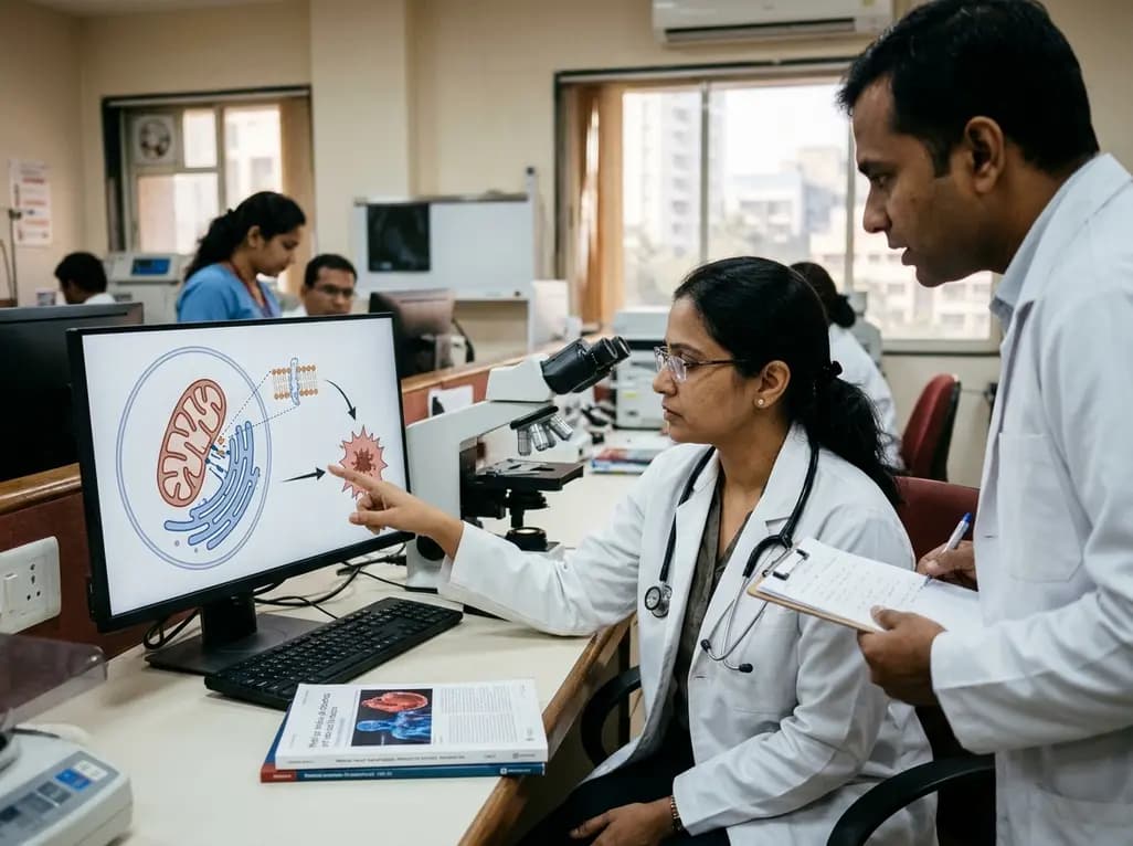
Recent research highlights how endoplasmic reticulum and mitochondrial interactions at contact sites regulate ferroptosis in cardiovascular diseases. This article explores lipid peroxidation, organelle crosstalk, and potential therapeutic strategies for ischemic heart disease and heart failure.
Today

A systematic review demonstrates that teriparatide significantly improves healing in medication-related osteonecrosis of the jaw (MRONJ), especially when combined with surgical and non-surgical therapies. This review explores clinical evidence, biological mechanisms, and treatment protocols for clinicians.
Today
An updated meta-analysis shows that 37% of patients remain euthyroid after stopping thyroid hormone therapy, with success rising to 41% in subclinical hypothyroidism. Clinicians can safely consider supervised deprescribing in selected individuals using structured monitoring.
Today
