

Loading, please wait...
Loading, please wait...

Surgical correction of adolescent idiopathic scoliosis (AIS) demands a balance between optimizing deformity correction and preserving spinal mobility. In thoracic curves, spine surgeons frequently debate how distal the fusion construct must extend. Choosing the touched vertebra as the lowest instrumented vertebra offers a motion-sparing alternative. However, this strategy can provoke postoperative complications, particularly distal add-on in scoliosis. This progressive loss of correction or curve extension distal to the instrumentation can compromise spinal balance and trigger secondary revision surgeries. Consequently, identifying precise biomechanical and axial predictors has become crucial for clinical decision-making.
The phenomenon of distal adding-on represents a well-recognized challenge following selective thoracic fusion in adolescent idiopathic scoliosis. Clinicians define this complication by progressive disc wedging, continuous increase in Cobb angle distal to the construct, or distal shift of the lower end vertebra. Therefore, selecting an appropriate lowest instrumented vertebra remains paramount to avoid postoperative failure. When surgeons instrument short of the stable landmark, residual forces transmit directly into adjacent motion segments. Consequently, the junctional discs experience abnormal mechanical shear stresses during spinal growth. Although preserving caudal lumbar motion segments remains a primary objective, underestimating curve extension often leads to progressive deformity. Recent investigations demonstrate an incidence of distal add-on reaching approximately 20.8% at three-year follow-up when surgeons select the touched vertebra as the lowest anchor. Furthermore, younger patients exhibiting high skeletal growth velocity experience significantly higher rates of this complication. Because spinal remodeling continues actively during adolescence, postoperative surveillance must monitor subtle alignment shifts early.
Selecting the lowest instrumented vertebra requires meticulous assessment of three-dimensional spinal parameters. Traditionally, surgeons selected either the stable vertebra or the neutral vertebra to anchor selective thoracic constructs securely. However, fusing down to the stable vertebra frequently consumes valuable mobile lumbar segments. As a result, patients face higher long-term risks of premature disc degeneration, chronic back stiffness, and functional limitations. To mitigate these adverse outcomes, spine specialists increasingly advocate selecting the touched vertebra, which the central sacral vertical line touches on coronal standing radiographs. Nevertheless, this motion-sparing approach introduces substantial mechanical variability into the transition zone. If surgeons underestimate subtle curve characteristics, mechanical imbalance accelerates distal junctional decompensation. Moreover, standard coronal measurements do not fully capture the torsional dynamics governing the thoracolumbar junction. Therefore, surgical teams must evaluate both coronal tilt and axial orientation before finalizing fusion boundaries in operating theaters.
While surgeons historically evaluated coronal cobb angles and sagittal alignment, recent biomechanical research highlights axial orientation as a key driver of junctional stability. Axial vertebral rotation discrepancy between the lowest instrumented vertebra and the adjacent uninstrumented vertebra creates severe torsional shear across the disc space. Consequently, when a construct rigidly fixes a rotated touched vertebra without harmonizing its axial plane to the uninstrumented caudal neighbor, persistent rotational torque develops. This torque gradually causes disc wedging and asymmetric remodeling of the vertebral ring apophysis. In addition, three-dimensional computed tomography studies confirm that axial rotational malalignment directly correlates with mechanical hardware strain. Therefore, evaluating axial vertebral rotation before surgery provides essential risk stratification. If marked rotational divergence exists across the planned junctional motion segment, selecting the touched vertebra alone substantially elevates the risk of distal add-on in scoliosis. Thus, axial alignment must guide instrumentation boundaries alongside standard coronal radiographs.
Spinal flexibility and the number of preserved motion segments significantly influence postoperative biomechanical equilibrium. Clinical cohort data indicate that patients developing distal adding-on preserve a higher average number of remnant motion segments compared to stable patients. Specifically, leaving greater caudal mobility increases the cumulative rotational and bending lever arms acting against the construct terminal. Furthermore, hypermobile lumbar discs absorb excessive compensatory motion when rigid thoracic instrumentation alters normal kinetic chain movement. Consequently, residual unfused segments experience concentrated torsional loads during daily physical activities. In contrast, fusing an additional segment distributes mechanical stresses more uniformly across stable lumbosacral anatomy. However, overextending spinal fusion sacrifices lumbar lordosis and flexibility unnecessarily. Spine surgeons must carefully weigh the physiological advantages of preserving mobile segments against the heightened risk of progressive distal decompensation. By analyzing skeletal maturity and flexibility bending films, clinicians can determine whether the remaining spinal column can compensate safely.
Translating these radiographic insights into clinical protocols enables surgeons to formulate individualized operative plans for thoracic scoliosis. First, spine teams should obtain upright full-spine radiographs alongside targeted axial rotation evaluations at the thoracolumbar junction. When surgeons identify minimal rotational discrepancy and substantial disc flexibility, choosing the touched vertebra remains a dependable, motion-sparing strategy. Conversely, if significant vertebral rotation discrepancy or significant coronal disc wedging exists at the transition level, surgeons should strongly consider extending instrumentation distally to the next vertebra. Additionally, surgeons must evaluate skeletal maturity using reliable grading systems such as Risser staging or Sanders maturity scores. Immature patients possessing high growth potential tolerate residual torsional stress poorly. Ultimately, precision deformity surgery requires a balanced synthesis of coronal, sagittal, and axial metrics. Adopting this holistic paradigm reduces revision rates while maximizing long-term spinal mobility.
Distal adding-on represents progressive deformity extension below the lowest instrumented vertebra following selective thoracic fusion. Surgeons identify it through progressive caudal disc wedging, increase in the unfused lumbar curve Cobb angle by more than five degrees, or distal migration of the lower end vertebra. This mechanical complication often results in spinal imbalance, asymmetric trunk posture, and eventual surgical revision.
Rotational discrepancy creates substantial torsional shear stress across the transition disc between the rigid construct and mobile spine. When surgeons fuse a vertebra that rotates substantially relative to the adjacent uninstrumented vertebra, residual torque forces asymmetric loading onto the junctional disc. Over time, these persistent forces induce progressive disc wedging and trigger distal curve decompensation.
Preserving lumbar motion segments protects long-term spine flexibility and reduces early degenerate disc disease risks. However, leaving excessive remnant motion segments adjacent to an axially malaligned lowest instrumented vertebra significantly increases adding-on risks. Surgeons must balance motion preservation against mechanical stability, extending fusion constructs only when high rotational discrepancy or growth potential threatens spinal alignment.
Disclaimer: This content is for informational and educational purposes only... Refer to the latest local and national guidelines for clinical practice.
References

Read summarized clinical updates, watch expert medical content, and earn CME certifications right from your smartphone.
Open your smartphone camera to scan and install.





A retrospective cohort study reveals that selecting the touched vertebra as the lowest instrumented vertebra in thoracic adolescent idiopathic scoliosis yields a 20.8% distal add-on rate, driven by remnant motion segments and axial rotational discrepancies.
Today
A case-control study reveals that severe poverty and silica dust exposure significantly increase tuberculosis risk among artisanal miners. This analysis reviews pathophysiology, clinical management, and targeted screening protocols relevant to clinicians treating occupational lung disease in high-burden settings.
Today

New research reveals that HPV16 E7 exosomes significantly upregulate PD-L1 on tumor-infiltrating lymphocytes in cervical cancer, driving local immune suppression and correlating with poorer patient survival. These findings expose key mechanisms of immune evasion with direct relevance to checkpoint blockade therapies.
Today
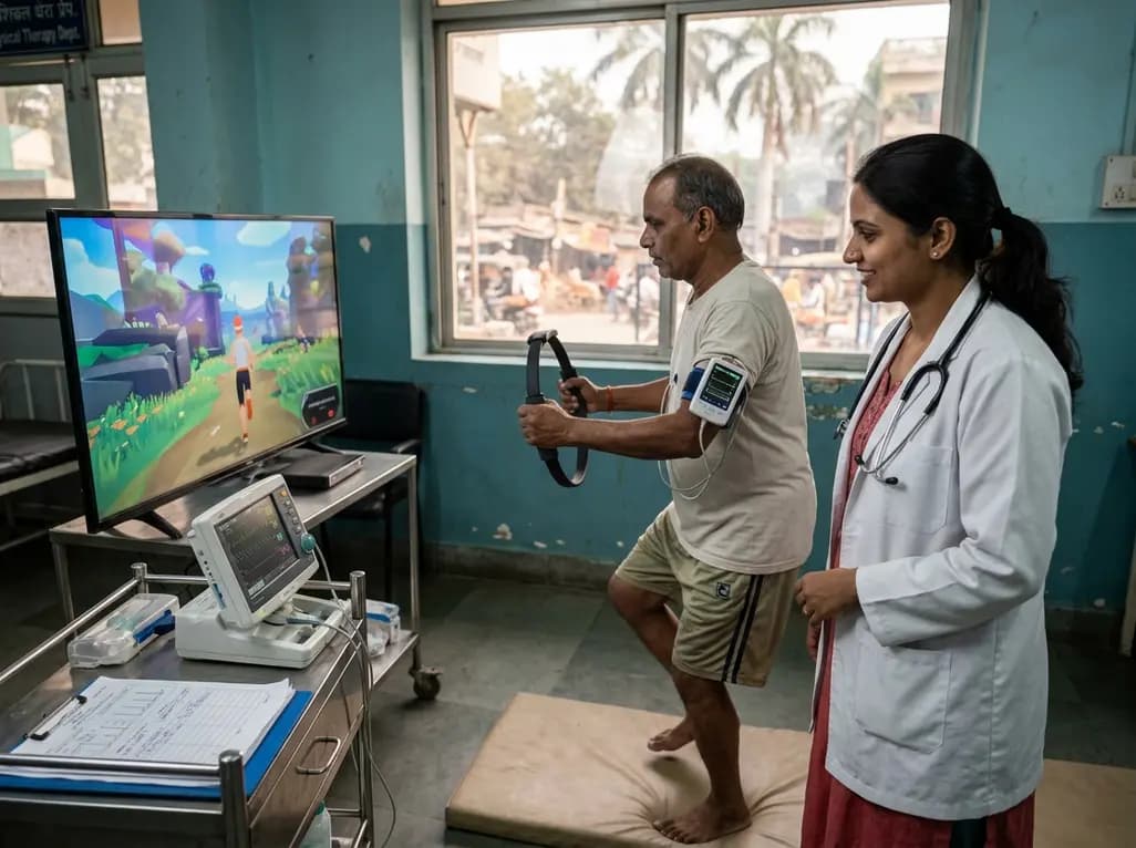
A clinical study evaluated Ring Fit Adventure in adults with chronic conditions, revealing that exergaming achieves moderate-to-vigorous exercise intensity while maintaining hemodynamic stability, high enjoyment, and zero adverse events.
Today
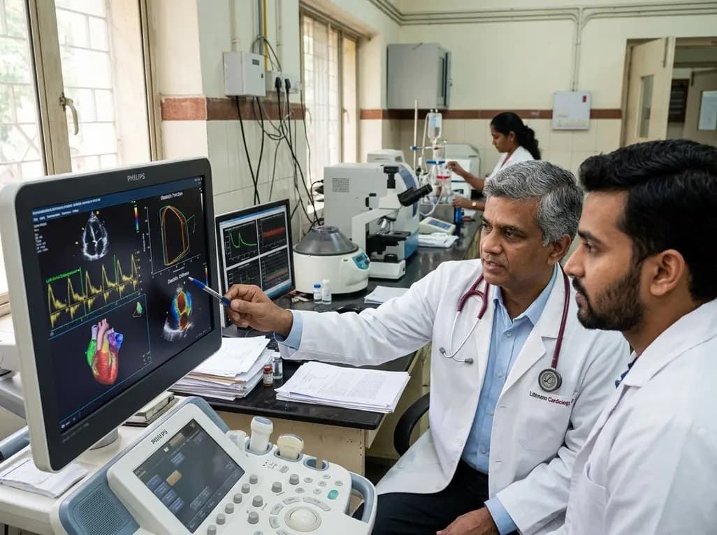
A study evaluates a cardiometabolic HFpEF model combining metabolic stress with chronic mineralocorticoid excess. The findings reveal profound diastolic stiffness, endothelial dysfunction, and pulmonary hypertension, illuminating key pathophysiological mechanisms for targeted cardiopulmonary therapies.
Today
