.webp&w=384&q=75)

Loading, please wait...
Loading, please wait...

Managing chronic, refractory symptoms in patients with painful diabetic neuropathy remains a significant clinical challenge for endocrinologists, neurologists, and pain medicine specialists worldwide. Conventional pharmacological interventions often fail to provide adequate analgesia, and escalating doses frequently precipitate intolerable side effects like sedation, dizziness, and peripheral edema. Consequently, interventional neuromodulation therapies, particularly pulsed radiofrequency directed toward the sympathetic nervous system, have gained substantial interest. A landmark study published by Wang and colleagues evaluates the comparative effectiveness and safety profile of ultrasound guidance versus conventional fluoroscopy for lumbar sympathetic ganglion pulsed radiofrequency, offering vital insights into modern interventional workflows.
Painful diabetic neuropathy affects up to one-third of individuals living with long-standing diabetes, fundamentally compromising sleep hygiene, emotional stability, and overall physical functioning. Persistent microvascular ischemia and metabolic alterations induce severe peripheral nerve sensitization, which ultimately amplifies sympathetic outflow to the lower extremities. Intervening at the lumbar sympathetic chain effectively reduces vasoconstriction, dampens central sensitization pathways, and alleviates intractable lower limb pain. Historically, clinicians have performed these interventional procedures under fluoroscopic guidance to confirm skeletal landmarks. However, fluoroscopy carries unavoidable occupational radiation exposure, requires dedicated lead aprons, and cannot directly visualize retroperitoneal soft-tissue architecture or adjacent major vessels in real time.
To determine whether high-resolution sonography provides equivalent therapeutic relief without radiographic trade-offs, investigators analyzed 228 patients experiencing refractory painful diabetic neuropathy. Researchers instituted a rigorous 1:1 propensity score-matching protocol, successfully balancing baseline clinical variables, pain severity, and underlying demographic traits between the ultrasound cohort and the fluoroscopy cohort. The primary non-inferiority margin was preset at 10% regarding the proportion of participants achieving at least 50% pain relief at one week following pulsed radiofrequency. Wang and colleagues observed that 67.8% of individuals in the ultrasound group achieved successful analgesia, demonstrating a mean difference of -0.9% (95% confidence interval: -9.5% to 7.8%) compared with fluoroscopy. Because the confidence boundary remained well within the predefined margin, the trial robustly confirmed clinical non-inferiority.
The therapeutic gains achieved immediately following sympathetic neuromodulation extended through intermediate follow-up intervals. At 4 weeks, 65.6% of patients in the ultrasound group maintained substantial relief compared to 66.7% in the fluoroscopy arm. Similarly, at 12 weeks post-procedure, successful symptom reduction persisted in 63.3% of ultrasound patients versus 64.0% of fluoroscopy patients, demonstrating no statistical divergence between guidance modalities. Both procedural approaches reliably increased distal lower-extremity skin temperature by approximately 5°C, confirming physiological sympatholysis. Furthermore, secondary evaluations revealed parallel reductions in Numerical Rating Scale pain scores, Neuropathy Impairment Score-Lower Limbs, Medical Outcomes Study sleep scores, Hospital Anxiety and Depression Scale measures, and Norfolk Quality of Life scores over the 12-week observation window.
While pain control proved equivalent, real-time dynamic ultrasound guidance provided critical practical and safety advantages over conventional fluoroscopy. Sonographic visualization enabled practitioners to observe the psoas muscle, vertebral body, inferior vena cava, and abdominal aorta continuously during needle advancement. This real-time tissue tracking led to significantly fewer needle passes and dramatically shortened total operative durations. Additionally, the ultrasound technique eradicated ionizing radiation exposure for operating room personnel and patients alike. Most importantly, continuous vascular visualization under ultrasound guidance produced a significantly lower rate of accidental intravascular puncture, shielding fragile diabetic patients from unnecessary systemic local anesthetic toxicity or retroperitoneal hematoma formation.
These findings hold profound practical relevance for healthcare delivery in India, where diabetes prevalence continues to surge and specialized radiation-shielded suites remain limited. Interventional pain practitioners in high-volume Indian centers often encounter logistical constraints, including overburdened cardiac catheterization units and fixed fluoroscopy suites. Point-of-care ultrasound machines offer portable, radiation-free utility across day-care procedure rooms, significantly mitigating procedural costs and eliminating recurring maintenance costs linked to fluoroscopic imaging tubes. Moreover, adopting ultrasound-guided pulsed radiofrequency protects healthcare providers from cumulative lifetime radiation exposure while widening timely access to durable, non-destructive neuromodulation for diabetic individuals unresponsive to first-line pharmacotherapies.
Pulsed radiofrequency delivers brief, high-voltage electrical bursts interrupted by silent resting phases, which prevents destructive thermal tissue coagulation. When targeted to the lumbar sympathetic ganglion, this non-neurolytic neuromodulation alters pain signaling, dampens ectopic discharges, and normalizes sympathetic tone. Consequently, blood flow to the lower limbs increases, relieving severe burning sensations without creating permanent nerve damage or post-procedural deafferentation dysesthesias.
Ultrasound provides real-time soft-tissue and vascular visualization that fluoroscopy cannot match. Using high-resolution sonography, clinicians track the exact needle trajectory adjacent to the psoas muscle, aorta, and inferior vena cava. This approach reduces accidental vascular puncture, shortens total procedure time, requires fewer needle repositioning maneuvers, and eliminates dangerous ionizing radiation exposure entirely for both the operating team and the patient.
Clinical data demonstrate that substantial pain relief, marked by at least a 50% reduction on validated pain scales, reliably persists for 12 weeks or longer. Furthermore, patients experience parallel improvements in nocturnal sleep quality, depressive symptoms, anxiety scores, and functional mobility. Long-term follow-up remains recommended to evaluate whether repeated sessions provide sustained analgesia across chronic disease trajectories.
Disclaimer: This content is for informational and educational purposes only. It does not constitute medical advice, diagnosis, or treatment recommendations. Always consult qualified healthcare professionals before initiating or modifying any clinical intervention. Refer to the latest local and national guidelines for clinical practice.
References
Wang L et al. Ultrasound-Guided Neuromodulation Is Non-inferior to Fluoroscopy Approach for Painful Diabetic Neuropathy: A Propensity Score Matching Analysis. J Ultrasound Med. 2026 Sep 11. doi: 10.1002/jum.70442. PMID: 42723570.
Pop-Busui R, Boulton AJ, Feldman EL, et al. Diabetic Neuropathy: A Position Statement by the American Diabetes Association. Diabetes Care. 2017;40(1):136-154. doi:10.2337/dc16-2042.
Manchikanti L, Kaye AD, Falco FJ, et al. Practice advisory for the safe management of interventional pain procedures in patients on anticoagulation and antiplatelet therapy. Pain Physician. 2018;21(3):E233-E261.

Read summarized clinical updates, watch expert medical content, and earn CME certifications right from your smartphone.
Open your smartphone camera to scan and install.





A propensity-matched study shows ultrasound-guided pulsed radiofrequency of the lumbar sympathetic ganglion is non-inferior to fluoroscopy for painful diabetic neuropathy, reducing needle passes, procedure time, and radiation exposure while achieving equal pain relief.
last month
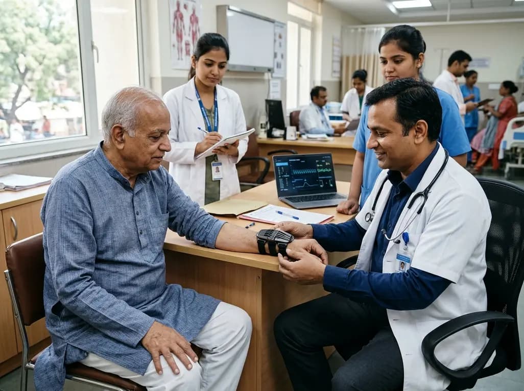
The TREM-AB open-label pilot study evaluated a wearable passive nonlinear vibration absorber for essential tremor. The Nonlinear Energy Sink bracelet reduced clinical Fahn-Tolosa-Marin tremor scores by 32.1% and electrophysiological tremor power by 49.2% with no adverse effects reported.
Today

A population-based study of 20,928 children reveals that small vulnerable newborns face sex-divergent mental capital outcomes at ages 6-7: term SGA girls show higher behavioral difficulties and lower resilience, while preterm non-SGA boys experience increased behavioral problems.
Today
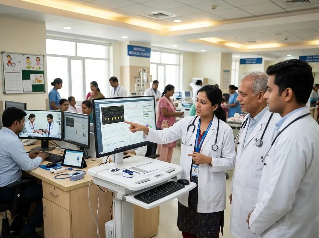
A landmark study details how an enterprise pediatric remote patient management model expanded from a single cardiology pilot to 16 diverse specialty programs by combining centralized digital operations with specialty clinical leadership.
Today

A comparative review of tension band wiring and intramedullary screw fixation in thumb metacarpophalangeal arthrodesis, evaluating union rates, hardware complications, and clinical outcomes.
Today

A breakthrough ultrathin multimodal sensor decouples physical hemodynamic monitoring from disposable electrochemical calibration, achieving long-term wearable glucose prediction with 81.36% accuracy and a 9.12% MARD. This offers a robust, drift-free solution for outpatient metabolic surveillance.
Today
