

Loading, please wait...
Loading, please wait...
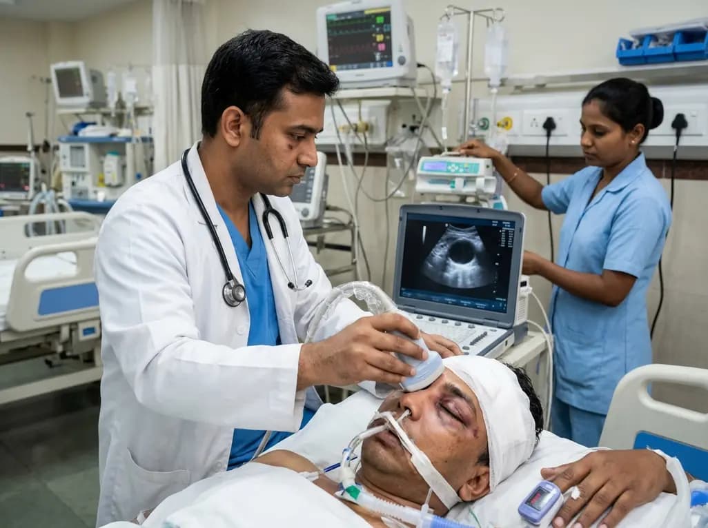
Severe traumatic brain injury demands continuous neuromonitoring to detect intracranial hypertension and avert secondary neuronal injury. In intensive care units, clinicians routinely evaluate pupillary reactivity to detect uncal herniation. However, severe maxillofacial trauma, extensive periorbital edema, and surgical dressings frequently prevent direct ocular inspection. Consequently, bedside ultrasonographic pupil assessment has emerged as a reliable non-invasive diagnostic solution. Furthermore, by evaluating ocular structures through closed eyelids, this technique enables objective pupillary evaluation when conventional examinations fail.
In neurocritical care, the pupillary light reflex serves as a vital surrogate marker for brainstem integrity. Because compression of the third cranial nerve rapidly alters pupil size and constriction velocity, serial neurological checks remain indispensable. Historically, physicians relied on manual penlight examinations, but subjective visual checks suffer from notable interobserver variability. Automated pupillometers addressed this issue by delivering objective metrics, including constriction velocity and constriction percentage. Nevertheless, massive facial trauma, soft tissue lacerations, and severe eyelid swelling frequently obstruct optical pupillometry devices. In severe blunt trauma, periorbital hematomas often seal the eyelids shut completely. Forcing swollen eyelids open risks causing corneal abrasions, aggravating tissue trauma, or provoking vagal responses. In addition, post-operative head bandaging often impedes physical access to the ocular surface. Therefore, clinicians require a non-invasive tool that bypasses swollen tissues entirely. Bedside ultrasound offers a seamless solution because acoustic waves penetrate soft tissues effortlessly. Hence, scanning through closed eyelids overcomes physical obstructions, allowing objective pupillometry at the bedside.
To validate this novel technique, researchers conducted a prospective study involving 45 adult patients admitted to the intensive care unit with severe traumatic brain injury. The study cohort presented with a mean age of 67.1 years and included 36 male subjects. Investigators sequentially performed direct optical pupillometry and transpalpebral ocular ultrasound on both eyes within a strict three-minute window. Furthermore, clinicians conducted all assessments under standardized low-light conditions to eliminate ambient optical bias. For the direct approach, investigators recorded video footage of the exposed pupil alongside a calibrated reference scale. Conversely, for the ultrasonographic examination, operators placed a high-frequency linear probe across the closed eyelid using acoustic gel. The primary investigative endpoint was the statistical agreement between modalities for pupillary constriction percentage. Additionally, secondary outcomes included agreement regarding resting baseline pupil diameter and minimum post-stimulation pupil diameter. By adhering to this standardized comparative protocol, the investigators minimized physiological fluctuations, thereby ensuring reliable comparative data across all enrolled subjects.
The trial demonstrated exceptional diagnostic consistency between ultrasound imaging and direct pupillometry. Specifically, ultrasonographic pupil assessment exhibited outstanding agreement with direct measurements when calculating the percentage of pupillary constriction. The median difference between the two techniques was only 0.44 percent, demonstrating no statistically significant variation across subjects. Moreover, Bland-Altman statistical analysis confirmed remarkably narrow limits of agreement, reinforcing the high clinical reliability of transpalpebral measurements. Ultrasonography also demonstrated strong correlations with direct pupillometry for resting and minimum pupil diameters, with coefficient values ranging from 0.600 to 0.873. Consequently, these findings verify that acoustic imaging precisely captures subtle dynamics of the pupillary light reflex. Even minor decreases in pupillary reactivity frequently indicate rising intracranial pressure or impending transtentorial herniation. Because sonographic imaging provides quantitative data comparable to calibrated video pupillometry, clinicians can confidently detect neurovascular deterioration. Therefore, ocular ultrasonography functions as a validated diagnostic surrogate when direct optical inspection remains impossible.
Executing ocular sonography requires meticulous technique to safeguard orbital structures while obtaining crisp anatomical views. First, the practitioner selects a high-frequency linear transducer, which provides superior spatial resolution for superficial structures. Next, the operator adjusts the ultrasound system preset to ophthalmic mode, thereby ensuring low mechanical and thermal indices. This step protects the retina and lens from acoustic energy exposure. Subsequently, the clinician applies a generous acoustic gel cushion over the closed eyelid, avoiding direct probe pressure on the globe. Applying excessive pressure must be strictly avoided because it risks elevating intraocular pressure or aggravating underlying globe injuries. The clinician positions the transducer in a transverse orientation to identify the anterior chamber, iris, and pupil. After measuring the baseline resting diameter, the operator stimulates the eye with a bright light source. Meanwhile, the clinician records real-time pupillary constriction on B-mode or M-mode sonography. Finally, software calipers measure the minimum constricted diameter to compute total constriction percentage rapidly.
Bedside point-of-care ultrasound already occupies an indispensable role in contemporary emergency departments and neurocritical care units. Clinicians routinely evaluate optic nerve sheath diameter to screen for intracranial hypertension. Therefore, incorporating pupillary light reflex assessment into the same ocular ultrasound examination represents a logical, highly efficient workflow enhancement. Instead of switching diagnostic devices, the intensivist evaluates optic nerve sheath diameter and pupillary dynamics within minutes during a single bedside session. Furthermore, this dual assessment offers valuable complementary data regarding elevated intracranial pressure and brainstem perfusion. In emergency trauma bays, rapid sonographic pupillometry guides immediate surgical triage before neuroimaging occurs. Similarly, in neurosurgical intensive care units, serial ultrasound tracking identifies delayed cerebral edema or postoperative hematoma expansion. Because point-of-care ultrasound machines are ubiquitous in modern hospitals, this methodology requires no expensive proprietary pupillometry hardware. Consequently, critical care units can implement this protocol immediately, establishing standardized training to optimize patient care and improve neurological outcomes.
Clinicians use a high-frequency linear ultrasound probe positioned gently over the closed eyelid with abundant acoustic gel. The operator visualizes the iris and fluid-filled anterior chamber in real time. After measuring the baseline resting pupil diameter, a bright light source stimulates the eye. The operator then records the resulting pupil constriction using M-mode or B-mode sonography. Finally, electronic calipers measure the minimum diameter to calculate constriction percentage accurately.
Transpalpebral ultrasound is particularly valuable when severe periorbital edema, facial hematomas, or soft-tissue lacerations prevent patients from opening their eyes. Automated optical pupillometers require an unobstructed optical path to image the pupil directly. Forcing swollen eyelids open risks causing corneal abrasions and secondary tissue trauma. In contrast, ultrasonography transmits acoustic waves through swollen eyelids, allowing non-invasive assessment of pupillary size and light responsiveness without manipulating fragile injured periorbital structures.
Clinicians must avoid applying physical pressure onto the globe to prevent acute intraocular pressure elevation and mechanical damage. Furthermore, practitioners should verify that the ultrasound machine runs in dedicated ophthalmic mode to minimize thermal and mechanical indices. Finally, suspected penetrating eye injuries or open globe fractures represent absolute contraindications to ocular sonography, because any external probe pressure could cause extrusion of intraocular contents, permanent visual impairment, and severe ocular infection.
Disclaimer: This content is for informational and educational purposes only and does not constitute medical advice, diagnosis, or treatment. Healthcare professionals should exercise their independent clinical judgment when managing patients. Refer to the latest local and national guidelines for clinical practice.
References

Read summarized clinical updates, watch expert medical content, and earn CME certifications right from your smartphone.
Open your smartphone camera to scan and install.





Ultrasonographic pupil assessment provides an accurate, objective alternative to direct pupillometry in severe traumatic brain injury when periorbital edema prevents direct eye visualization. A prospective ICU study confirms its strong concordance in measuring pupillary constriction and resting pupil diameter.
Today

A comprehensive meta-analysis examines the safety and efficacy of cannabinoid-based interventions in Parkinson's disease. While experimental interest remains high, randomized controlled trials show no clear therapeutic benefits alongside significant risks of acute confusion and dizziness in vulnerable patients.
Today

AIIMS researchers suggest yoga may alleviate gut dysbiosis in Polyendocrine Metabolic Ovarian Syndrome (PMOS) by modulating the gut-brain axis, lowering chronic inflammation, and enhancing parasympathetic tone to support metabolic and hormonal equilibrium.
Today

A prospective cohort study demonstrates that feeding tubes, formula exposure, aortic cross-clamp duration, and socioeconomic deprivation disrupt the neonatal gut microbiome in congenital heart disease, emphasizing the need for targeted nutritional and perioperative gut-protective strategies.
Today
Deep intramuscular vascular malformations in pediatric Klippel-Trenaunay syndrome can mimic Maffucci syndrome. Multimodality imaging with Doppler ultrasound and MRI, along with histopathology, ensures prompt differential diagnosis, targeted surgical debulking, and optimized conservative surveillance.
Today
Modern management of adrenal incidentalomas is shifting away from universal testing toward risk-stratified biochemical evaluation. Guidelines suggest omitting metanephrine testing in lipid-rich adenomas (≤10 HU), while maintaining autonomous cortisol testing to prevent cardiometabolic complications.
Today
