.webp&w=384&q=75)

Loading, please wait...
Loading, please wait...

Managing progressive renal decline in patients with chronic hyperuricemia remains a major therapeutic hurdle in routine clinical practice. In particular, hyperuricemic nephropathy frequently drives chronic kidney disease even when conventional urate-lowering therapies achieve serum target goals. Consequently, secondary tubular epithelial injury often continues unabated across vulnerable cohorts. Researchers have recently identified thioredoxin-interacting protein as a decisive molecular link that triggers regulated cell death. Therefore, understanding this upstream pathway provides vital insights into preserving nephron architecture beyond routine serum urate reduction.
Clinicians frequently encounter patients who experience relentless renal function decline despite optimal allopurinol or febuxostat dosing. Currently, standard management focuses primarily on clearing soluble urate and dissolving microcrystalline deposits. However, high circulating urate levels persistently trigger cellular stress before systemic crystallization occurs. Furthermore, persistent urate exposure damages renal tubular epithelial cells by inducing excessive oxidative stress. As a result, sterile inflammatory cascades and tubulointerstitial fibrosis gradually destroy functional nephrons. Standard xanthine oxidase inhibitors rarely arrest these intracellular cascades once sustained metabolic stress occurs. In addition, existing clinical guidelines offer minimal secondary protection against ongoing tubular epithelial necrosis. Thus, researchers have shifted attention toward non-apoptotic cell death mechanisms. In particular, ferroptosis has emerged as an indispensable pathway mediating acute and chronic tubular degeneration. Consequently, targeting upstream regulatory nodes that initiate ferroptosis offers substantial hope for salvage therapy. By interrupting this iron-dependent destructive cycle, physicians may soon halt insidious parenchymal damage. Moreover, preserving tubular cell survival prevents chronic maladaptive repair pathways. Ultimately, addressing these unresolved metabolic drivers fulfills a critical clinical priority in nephrology.
Recent investigative findings identify thioredoxin-interacting protein, known as TXNIP, as a crucial master regulator. Specifically, investigators analyzed renal biopsy specimens obtained from human patients presenting with hyperuricemic renal disease. Notably, TXNIP expression was markedly elevated in these diseased tissues compared to healthy controls. Furthermore, immunohistochemical evaluation demonstrated predominant TXNIP localization within renal tubular epithelial cells. To confirm these clinical observations, researchers examined two distinct murine models of hyperuricemia. Consistently, both drug-induced and genetically engineered mice exhibited robust tubular TXNIP upregulation. Moreover, cellular models revealed that hyperuricemic stress directly stimulates intracellular reactive oxygen species generation. Consequently, elevated oxidative stress dissociates TXNIP from thioredoxin, allowing uninhibited TXNIP to activate deleterious signaling. In addition, the genetic deletion of TXNIP in double-knockout mice completely abolished the destructive renal phenotype. Therefore, these rigorous genetic experiments establish TXNIP as an indispensable driver of hyperuricemic pathology. In contrast to passive bystander markers, TXNIP actively coordinates tubular dysfunction during sustained urate overload. Furthermore, transcriptomic profiling revealed broad suppression of downstream inflammatory mediators following TXNIP disruption. Thus, blocking TXNIP signaling represents an attractive molecular strategy to shield vulnerable tubular segments.
Ferroptosis is an iron-dependent form of non-apoptotic cell death driven by toxic lipid peroxide accumulation. Mechanistically, renal tubular epithelial cells remain exceptionally susceptible to ferroptotic injury due to their high metabolic activity. In hyperuricemic models, elevated TXNIP directly accelerates lipid peroxidation while disrupting essential endogenous antioxidant defense systems. Specifically, TXNIP upregulation significantly depletes intracellular glutathione, leaving cells vulnerable to free radical assault. Furthermore, TXNIP suppresses the system xc- cystine/glutamate antiporter subunit xCT and downregulates glutathione peroxidase 4. Simultaneously, TXNIP increases the expression of acyl-CoA synthetase long-chain family member 4, also known as ACSL4. Consequently, elevated ACSL4 enriches cellular membranes with polyunsaturated fatty acids, promoting massive lethal peroxidation. Moreover, transmission electron microscopy demonstrated severe ultrastructural defects, including shrunken mitochondria with condensed membrane densities. In contrast, inhibiting TXNIP restored glutathione levels and rescued GPX4 activity across experimental models. In addition, TXNIP suppression markedly reduced iron deposition within proximal tubular segments. Accordingly, these biochemical alterations prevented mitochondrial outer membrane rupture and halted cellular disintegration. Therefore, TXNIP serves as the crucial upstream checkpoint regulating the entire ferroptotic death cascade in hyperuricemic kidneys.
To evaluate translational pharmacological applications, researchers investigated verapamil, an established L-type calcium channel blocker. Interestingly, previous cardiovascular studies established that verapamil potently suppresses cellular TXNIP expression in various tissues. When administered to hyperuricemic animals, verapamil effectively suppressed renal TXNIP expression and halted tubular ferroptosis. Consequently, treated animals demonstrated marked preservation of glomerular filtration and significant reductions in serum creatinine. Furthermore, histological analysis revealed dramatic attenuation of tubular dilation, protein cast formation, and interstitial fibrosis. Importantly, verapamil conferred these substantial renoprotective benefits without significantly lowering serum urate levels. Thus, the drug bypassed systemic hyperuricemia to protect tubular cells directly against intracellular oxidative injury. Additionally, verapamil therapy normalized mitochondrial morphology and dramatically reduced urinary biomarkers of tubular stress. Because verapamil already holds regulatory approval for hypertension, its clinical repurposing presents fewer developmental obstacles. However, clinicians must carefully consider individual hemodynamic profiles, heart rate, and drug interactions before off-label application. In particular, monitoring for bradycardia and peripheral edema remains prudent during any clinical translation. Nevertheless, these convincing preclinical findings demonstrate the feasibility of pharmacologically targeting TXNIP-driven tubular destruction.
These landmark experimental findings offer transformative perspectives for managing nephropathy in gouty patients. Currently, nephrologists and rheumatologists rely almost exclusively on lowering systemic urate to preserve renal function. However, clinical trials consistently demonstrate that serum urate reduction alone cannot reverse established tubulointerstitial scarring. Therefore, clinicians must adopt a dual therapeutic approach that simultaneously lowers urate and protects tubular viability. In this regard, combining conventional xanthine oxidase inhibitors with TXNIP-targeted ferroptosis inhibitors represents an innovative paradigm. Moreover, evaluating urinary ferroptosis markers and cellular TXNIP levels could help identify patients at elevated risk. In addition, ongoing prospective studies should determine whether calcium channel blockers yield superior renal outcomes in hyperuricemic hypertension. Clinicians should also recognize that tubular epithelial integrity dictates long-term prognosis in chronic kidney disease. Consequently, mitigating ferroptosis could prevent progressive nephron dropout and delay the eventual need for dialysis. Furthermore, these mechanistic discoveries encourage investigators to explore dedicated small-molecule TXNIP inhibitors in future human trials. As research progresses, targeted anti-ferroptotic therapies may redefine standard guidelines across global nephrology practices. Ultimately, targeting the TXNIP-ferroptosis axis bridges an urgent unmet need in contemporary renal medicine.
Thioredoxin-interacting protein acts as an indispensable upstream regulator linking hyperuricemia to renal tubular ferroptosis. High cellular urate stimulates oxidative stress, causing TXNIP upregulation in tubular epithelial cells. Consequently, increased TXNIP suppresses the cystine antiporter xCT and glutathione peroxidase 4 while upregulating ACSL4. This molecular imbalance leads to severe glutathione depletion, intracellular iron overload, and massive lipid peroxidation. Ultimately, TXNIP activation accelerates tubular degeneration, inflammation, and progressive interstitial fibrosis.
Verapamil confers renoprotection by pharmacologically suppressing tubular TXNIP expression rather than clearing circulating serum urate. By inhibiting TXNIP, verapamil effectively restores cellular glutathione content and preserves protective GPX4 enzyme levels. In addition, the drug prevents mitochondrial membrane condensation and attenuates iron-mediated lipid peroxidation in tubular segments. Therefore, verapamil prevents structural tubular necrosis, minimizes local inflammation, and halts interstitial scarring. These actions preserve overall kidney function without altering systemic urate concentration.
Conventional urate-lowering therapies primarily inhibit xanthine oxidase to suppress systemic uric acid production. However, these agents do not directly block autonomous intracellular injury pathways once persistent hyperuricemia activates them. Specifically, elevated intracellular urate induces oxidative stress, TXNIP upregulation, and ferroptotic cell death cascades that perpetuate independent of serum urate levels. Consequently, tubular epithelial cell loss and interstitial fibrosis continue progressing. Therefore, direct cytoprotective interventions are necessary to supplement standard systemic urate-lowering regimens.
Disclaimer: This content is for informational and educational purposes only... Refer to the latest local and national guidelines for clinical practice.
References
Xi Y et al. Identification of Thioredoxin-Interacting Protein as an Upstream Regulator of Ferroptosis in Hyperuricemic Nephropathy. Antioxid Redox Signal. 2026 Oct 07. doi: 10.1177/15230864261494911. PMID: 42844223.
Zhou Y, Wang L, Chen X, et al. Molecular interplay between TXNIP and GLUT9 underlies uric acid transport dysregulation in vitro under hyperuricemic stress. Cell Mol Biol Lett. 2025;30(1):88.
Zhang X, Liu J, Bai C, et al. Palmitic acid enhances the sensitivity of ferroptosis via endoplasmic reticulum stress mediated the ATF4/TXNIP axis in polycystic ovary syndrome. Phytomedicine. 2025;142:156777.
Martin-Sanchez D, Ruiz-Andres O, Poveda J, et al. Ferroptosis, but not necroptosis, is important in nephrotoxic folic acid-induced AKI. J Am Soc Nephrol. 2017;28(4):1202-1214.

Read summarized clinical updates, watch expert medical content, and earn CME certifications right from your smartphone.
Open your smartphone camera to scan and install.





A breakthrough study identifies thioredoxin-interacting protein (TXNIP) as a key upstream driver of tubular ferroptosis in hyperuricemic nephropathy. Pharmacological suppression by verapamil halts renal injury independently of serum urate levels, presenting a novel therapeutic paradigm for chronic kidney disease.
Today

A large UK cohort study of 1.4 million women shows that pregnancy complications, including gestational diabetes, hypertension, and preterm birth, substantially raise long-term risks for hypertension, chronic kidney disease, type 2 diabetes, and cardiovascular events, highlighting early postpartum intervention needs.
Today
A cross-sectional study reveals that hypertension knowledge, illness perceptions, depression, and substance use significantly predict self-care behaviors. Addressing mental health and cognitive beliefs in primary care is crucial for optimal blood pressure control and cardiovascular disease prevention.
Today
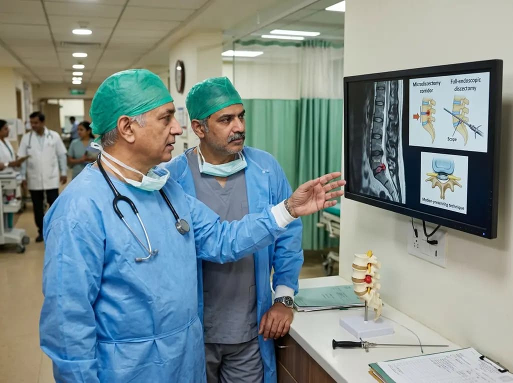
This clinical overview examines recent advancements in lumbar disc herniation surgery, comparing microscopic and full-endoscopic discectomy while highlighting biportal techniques and emerging motion-preserving innovations for spine specialists.
Today

Small interfering RNA (siRNA) therapeutics harness RNA interference to degrade disease-causing messenger RNA. By delivering hepatocyte-targeted, durable gene silencing, agents targeting PCSK9, LPA, and angiotensinogen offer potent cardiovascular risk reduction with infrequent, biannual dosing schedules.
Today
