.webp&w=384&q=75)

Loading, please wait...
Loading, please wait...
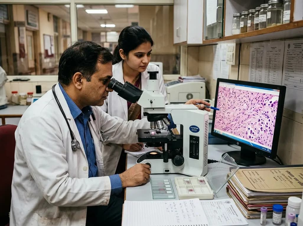
Meningiomas represent the most prevalent primary intracranial neoplasms encountered in adult neurology and neurosurgery. Although pathologists classify the majority of these tumors as histologically benign, postoperative recurrence remains a formidable challenge. Clinicians increasingly recognize that neoplastic progression depends heavily on the surrounding immune microenvironment. In particular, tumor-associated macrophages have emerged as critical modulators of local invasiveness, tissue remodeling, and treatment resistance. Understanding how these myeloid cells drive early tumor relapse provides vital opportunities to refine post-surgical risk stratification and patient surveillance protocols.
The tumor microenvironment in intracranial neoplasms comprises diverse stromal, vascular, and immune components that dynamically interact with neoplastic cells. Among these infiltrating immune populations, tumor-associated macrophages constitute a major cellular fraction. In healthy cerebral tissue, resident macrophages maintain homeostatic surveillance and clear cellular debris. However, tumors actively recruit circulating monocytes and polarize them toward distinct functional phenotypes.
Historically, researchers categorized macrophages into pro-inflammatory M1 and immunosuppressive M2 subsets. In the meningioma microenvironment, local cytokine signals induce an M2-like state. Consequently, these polarized macrophages support tissue remodeling rather than initiating antineoplastic immune attacks. Moreover, they secrete potent angiogenic factors, including vascular endothelial growth factor, which stimulates neo-vascularization. Additionally, macrophages release matrix metalloproteinases that degrade extracellular matrix barriers. This enzymatic breakdown facilitates dural infiltration and bone invasion. As a result, microscopic tumor remnants often survive within an immunosuppressive, macrophage-rich niche.
In a comprehensive retrospective investigation of 146 primary meningioma cases, researchers evaluated the presence of morphologically identifiable tumor-associated macrophages. Overall, investigators detected macrophage infiltration in 30 out of 146 cases, representing a prevalence of 20.5 percent. Notably, patient age demonstrated a statistically significant relationship with macrophage presence. Patients with macrophage-positive tumors were significantly younger than those with macrophage-negative tumors.
Conversely, the analysis showed no statistically significant correlations between macrophage positivity and patient sex, tumor size, or anatomical location. Furthermore, standard histopathological parameters, including the World Health Organization grade and the Simpson resection grade, did not significantly differ according to macrophage presence. This lack of correlation with traditional grading systems represents a crucial finding. Specifically, it indicates that microenvironmental immune infiltration operates as an independent biological axis. Therefore, conventional histology alone may overlook high-risk features harbored within the inflammatory stroma of young patients.
The study demonstrated a striking difference in tumor recurrence trajectories between macrophage-positive and macrophage-negative cohorts. Patients harboring macrophage-positive meningiomas experienced significantly more frequent and substantially earlier recurrences than their macrophage-negative counterparts. Multivariate Cox proportional hazards regression analysis confirmed that the presence of tumor-associated macrophages serves as an independent predictor of disease recurrence.
Importantly, this predictive power remained statistically robust after adjusting for standard clinical confounders. Nevertheless, the study found no statistically significant association between macrophage infiltration and overall survival or disease-free survival. This divergence likely reflects the indolent overall course of meningiomas, where repeated resections and salvage therapies extend survival despite local relapse. Consequently, recurrence-free survival emerges as the most clinically relevant endpoint when evaluating inflammatory biomarkers. Identifying macrophage-rich tumors at initial resection provides neurosurgeons with actionable prognostic information, allowing tailored surveillance regimens before gross clinical progression occurs.
Pathologists in this investigation identified macrophage infiltration primarily through morphological examination on standard hematoxylin-eosin stained tissue sections. Morphological evaluation relies on identifying characteristic cytological features, including eccentric nuclei, abundant foamy cytoplasm, and distinct spatial clustering. However, the authors appropriately highlighted morphological assessment as a major methodological limitation of their work.
Standard light microscopy cannot reliably differentiate between functional macrophage polarizations. For instance, morphologically identical cells may express diametrically opposed functional phenotypes. In modern neuropathology, immunohistochemical markers such as CD68, CD163, and CD206 offer superior sensitivity and specificity for identifying pro-tumoral M2 phenotypes. Furthermore, automated digital pathology and multiplex tissue cytometry enable precise quantification of immune cell densities across entire tumor sections. By implementing standardized digital thresholds, pathologists can minimize inter-observer variability. Consequently, while standard morphology provides a valuable initial screening tool, future clinical protocols should integrate validated immunohistochemical panels.
The identification of macrophage-driven recurrence risk carries immediate practical implications for neuro-oncology multidisciplinary teams. Currently, clinical guidelines dictate postoperative follow-up intervals based predominantly on histological grade and surgical resection completeness. However, low-grade tumors harboring dense macrophage infiltrates frequently defy their benign classification by recurring prematurely.
Therefore, clinicians should consider incorporating immune microenvironment assessment into holistic postoperative risk algorithms. For patients diagnosed with macrophage-positive meningiomas, neurosurgeons and radiation oncologists might justify shortening the initial neuroimaging surveillance interval. Rather than waiting twelve months for follow-up magnetic resonance imaging, an earlier scan at six months may capture subclinical regrowth. Moreover, early identification of high-risk biological features aids in determining whether adjuvant radiation therapy warrants consideration after subtotal resection. Thus, multidisciplinary tumor boards can utilize these insights to engage patients in shared decision-making regarding proactive surveillance.
As researchers uncover the complex interplay between neoplastic cells and the immune stroma, novel therapeutic opportunities continue to emerge. Historically, systemic therapies for refractory meningiomas have yielded limited clinical efficacy. However, targeting tumor-associated macrophages presents an attractive therapeutic strategy to disrupt the supportive pro-tumoral niche.
Pharmacological agents targeting the colony-stimulating factor-1 receptor have demonstrated remarkable ability to deplete or reprogram tumor-associated macrophages in solid malignancies. Additionally, novel antibody-drug conjugates directed against macrophage scavenger receptors are entering clinical evaluation. By eliminating immunosuppressive M2 macrophages, these agents can restore antitumor immune surveillance and enhance concurrent radiotherapy efficacy. Furthermore, large-scale multicenter clinical trials are urgently needed to validate standardized prognostic scoring systems. Such studies should combine spatial transcriptomics and genomic sequencing to profile the meningioma immune ecosystem. Ultimately, these translational endeavors will bridge the gap between microscopic discoveries and personalized precision therapies.
Tumor-associated macrophages stimulate tumor recurrence by releasing pro-angiogenic cytokines, growth factors, and matrix metalloproteinases that remodel the extracellular matrix. Furthermore, these immune cells foster an immunosuppressive microenvironment. Consequently, neoplastic cells evade immune surveillance and exhibit aggressive proliferation, which ultimately accelerates tumor progression and relapse.
Younger patients frequently demonstrated higher macrophage infiltration within their meningioma tissues. Although the precise biological driver requires further exploration, younger individuals often sustain robust baseline inflammatory responses. Therefore, active immune cell recruitment into the tumor stroma occurs more readily, potentially driving earlier meningioma recurrence in this specific patient subset.
Standard hematoxylin and eosin staining identifies macrophage infiltration based on classic cytological features such as foamy cytoplasm. However, routine morphology cannot distinguish between anti-tumoral M1 and pro-tumoral M2 phenotypes. Therefore, specialized immunohistochemical markers like CD68 and CD163 provide far greater precision for comprehensive clinical risk stratification.
Disclaimer: This content is for informational and educational purposes only and should not be considered medical advice. Always consult a qualified healthcare professional regarding any medical condition or treatment. Refer to the latest local and national guidelines for clinical practice.
References

Read summarized clinical updates, watch expert medical content, and earn CME certifications right from your smartphone.
Open your smartphone camera to scan and install.





A retrospective cohort study reveals that morphologically identified tumor-associated macrophages act as an independent risk factor for meningioma recurrence, highlighting the role of the inflammatory tumor microenvironment in early relapse and informing postoperative neurosurgical surveillance strategies.
last month

Explore advances in precision toxicity control for immune checkpoint immunotherapy. Discover how mechanistic cytokine blockade, cellular targets, and emerging biomarkers mitigate adverse events without compromising antitumor efficacy.
Today
Integrating adult genetic services into reproductive and pregnancy care substantially alters clinical management. A study reveals that diagnostic genetic evaluations directly inform maternal, fetal, and neonatal care in nearly 57% of tested patients, highlighting the value of preconception and antenatal genomics.
Today
A cross-sectional study in Nepal evaluates cardiovascular risk factors among adults working above 4,000 meters. The findings reveal unexpected rates of diabetes, hypertension, and mountain sickness, underscoring the urgent need for targeted cardiometabolic screening in high-altitude occupational cohorts.
Today
An economic evaluation shows that combining physiotherapist-delivered stress inoculation training with exercise is cost-effective compared with exercise alone for acute whiplash injury, improving functional recovery and lowering downstream healthcare expenditure.
Today
Preclinical findings demonstrate that dietary flavonoid hyperoside ameliorates high-fat diet-induced type 2 diabetes by stimulating brown adipose tissue thermogenesis, promoting white fat browning, dampening adipose macrophage inflammation, and restoring gut microbiota homeostasis.
Today
