.webp&w=384&q=75)

Loading, please wait...
Loading, please wait...

Drug-resistant epilepsy represents a substantial therapeutic dilemma worldwide, with focal cortical dysplasia serving as one of its most prevalent etiologies. Complete surgical resection offers these individuals their best opportunity for sustained seizure freedom. However, standard diagnostic protocols frequently fail because conventional magnetic resonance imaging cannot visualize subtle architectural anomalies. Innovative molecular diagnostics such as TSPO-PET imaging have emerged to illuminate these concealed lesions by targeting cellular inflammation. Consequently, this advanced approach equips multidisciplinary teams with unprecedented diagnostic precision during comprehensive presurgical evaluations.
Focal cortical dysplasia represents localized disruptions of normal cortical development, featuring disoriented laminar architecture, dysmorphic neurons, and aberrant balloon cells. In clinical practice, these malformations generate intrinsic epileptogenicity that routinely defies multiple anti-seizure medications. Consequently, curative management necessitates precise surgical excision of the entire epileptogenic zone. However, structural neuroimaging remains normal in nearly one-third of affected individuals. These MRI-negative cases present profound localization dilemmas for neurologists and neurosurgeons alike.
Furthermore, standard high-resolution MRI protocols often fail when dysplastic lesions reside deep within cortical sulci or exhibit minimal architectural thickening. When structural imaging yields non-lesional findings, clinical teams must deploy secondary functional tools. Interictal electroencephalography and scalp video telemetry frequently produce non-localizing or broad vector fields. In addition, invasive intracranial electroencephalography requires a reasoned preliminary anatomical hypothesis before electrode placement. Without clear structural targets, blind intracranial explorations carry increased procedural risk and lower postoperative seizure freedom rates. Therefore, closing this diagnostic gap demands molecular modalities that expose functional pathophysiology rather than macroscopic morphology alone.
To overcome structural diagnostic limitations, researchers developed TSPO-PET imaging to evaluate neuroinflammatory alterations within epileptogenic tissue. The 18-kDa translocator protein resides predominantly on the outer mitochondrial membranes of activated microglia, monocyte-derived macrophages, and reactive astrocytes. Under baseline physiological conditions, healthy cerebral parenchyma displays modest, homogenous TSPO expression. However, sustained epileptic networks and focal architectural dysplasias evoke localized immune cascades that substantially upregulate translocator protein density.
Using specialized second-generation radioligands such as fluorine-18-labeled DPA714, molecular positron emission tomography can visualize this localized microglial proliferation in vivo. Consequently, focal tracer accumulation demarcates the epicenters of chronic neuroinflammation and hyperexcitability. Furthermore, the molecular tracer rapidly crosses the blood-brain barrier and selectively binds to the mitochondrial receptor complex with notable affinity. This direct pathophysiological targeting allows clinicians to evaluate cellular disease activity in real time. Because intense neuroinflammation tightly mirrors intrinsic epileptogenic networks, this molecular strategy pinpoints dysplastic margins that remain completely imperceptible on traditional structural scans.
Historically, presurgical evaluation protocols have utilized fluorodeoxyglucose positron emission tomography to identify cerebral metabolic abnormalities. In typical cases, FDG-PET demonstrates regional glucose hypometabolism across the epileptogenic cortex. However, interictal hypometabolic zones frequently extend far beyond the actual structural lesion, encompassing widespread networks of functional diaschisis. This broad hypometabolism often obscures the true surgical boundary, leading to either incomplete resection or unnecessarily extensive tissue removal.
In contrast, novel clinical investigations demonstrate that translocator protein molecular scanning provides superior lesion-to-background contrast. Specifically, quantitative comparative analyses show that TSPO tracers generate a statistically significant increase in conspicuous signal contrast compared to FDG-PET. Furthermore, TSPO-PET imaging successfully identified distinct hypermetabolic activation in thirteen out of twenty-four patients whose anatomical MRI scans appeared completely unremarkable. By highlighting localized microglial activation rather than broad metabolic depression, the molecular technique delineates compact pathological foci. Thus, clinicians gain an extraordinarily specific visual marker that eliminates the diagnostic ambiguity common to conventional metabolic evaluations.
Electrophysiological concordance remains the gold standard when validating any emerging neuroimaging biomarker for epilepsy surgery. Notably, patients undergoing invasive stereotactic electroencephalography monitoring exhibited robust spatial agreement between high-level tracer activation and electrographic seizure onset zones. Ictal discharges originated directly within or adjacent to the cortical regions defined by increased tracer binding. Consequently, these invasive recordings confirm that localized translocator protein upregulation directly reflects true primary epileptogenic networks.
In addition, postoperative histopathological examinations validated the structural substrate underlying these molecular imaging findings. Researchers identified distinct translocator protein activation across different dysplastic subtypes, observing universal high-level tracer uptake in focal cortical dysplasia type IIb cases. Pathologists also confirmed tracer activation in representative type IIa specimens. Notably, type IIb lesions harbor prominent balloon cells and dysplastic neurons that actively release proinflammatory cytokines. As a result, these cellular components drive extensive microglial recruitment, providing a concrete biological basis for robust radiotracer binding. These histopathological correlations confirm that molecular neuroimaging accurately maps severe cellular disruptions.
To assess tracer distribution quantitatively, investigators established a standardized metric designated as the Highlight Index. Semiquantitative analyses demonstrate that this index correlates directly with key clinical markers of disease severity. Specifically, patients experiencing higher daily seizure frequencies demonstrated significantly greater tracer uptake. In addition, individuals suffering from focal to bilateral tonic-clonic seizures exhibited higher Highlight Index values compared to patients with less severe seizure phenotypes.
Furthermore, temporal factors profoundly influence the intensity of localized molecular signals. Statistical evaluations revealed a significant negative correlation between the scan-to-seizure interval and the magnitude of tracer uptake. Patients who experienced seizures shortly before undergoing imaging scans displayed markedly higher Highlight Index values. This temporal association occurs because recurrent ictal discharges provoke immediate waves of microglial activation and secondary neuroinflammation. However, even during prolonged interictal periods, baseline cellular dysplasia preserves elevated tracer binding above normal background levels. Consequently, clinicians must consider recent seizure timing when interpreting molecular intensity and planning imaging schedules.
Integrating translocator protein molecular imaging into contemporary clinical pathways promises to revolutionize surgical planning for difficult cases. By unmasking previously invisible malformations, this modality directly converts non-lesional cases into surgically targetable candidates. Therefore, patients historically deemed poor surgical candidates due to unrevealing MRI scans can now access potentially curative resections. Moreover, accurate non-invasive target identification minimizes unnecessary invasive intracranial monitoring arrays, thereby reducing patient morbidity and overall healthcare expenditure.
Additionally, refined boundary delineation enables neurosurgeons to perform tailored cortical resections that maximize pathological tissue removal while sparing eloquent brain areas. Because postoperative seizure freedom correlates strictly with complete surgical resection of the dysplastic cortex, this molecular technique directly enhances long-term prognostic outcomes. Neurologists and neurosurgeons must now collaborate to incorporate these molecular findings into multimodal consensus conferences. Ultimately, the broader clinical adoption of neuroinflammatory molecular scanning represents a paradigm shift toward precision medicine in refractory epilepsy management.
TSPO-PET imaging utilizes radiotracers such as 18F-DPA714 to target the 18-kDa translocator protein, an established marker of activated microglia and reactive neuroinflammation. Because focal cortical dysplasia often harbors persistent localized inflammatory cascades, this molecular modality highlights dysplastic tissue with exceptional contrast, even when conventional high-resolution structural MRI scans fail to identify anatomical lesions.
While FDG-PET detects broad regional glucose hypometabolism that often spans beyond the true epileptogenic focus, TSPO-PET imaging directly targets neuroinflammatory glial responses. Consequently, TSPO-PET provides a significantly sharper contrast signal against normal brain background tissue. This distinctive signal contrast enables clinicians to pinpoint small, discrete epileptogenic cortical boundaries with markedly superior diagnostic clarity.
Clinical data reveal an inverse correlation between tracer uptake and the duration elapsed since the most recent seizure. Specifically, shorter intervals between an ictus and scanning produce a significantly elevated Highlight Index. Recurrent seizures amplify localized neuroinflammation and microglial activation, which temporarily magnifies TSPO expression within the underlying dysplastic epileptogenic zone.
Disclaimer: This content is for informational and educational purposes only... Refer to the latest local and national guidelines for clinical practice.
References

Read summarized clinical updates, watch expert medical content, and earn CME certifications right from your smartphone.
Open your smartphone camera to scan and install.





A clinical study demonstrates that 18-kDa translocator protein (TSPO) PET imaging using 18F-DPA714 successfully detects MRI-negative focal cortical dysplasia. By capturing localized neuroinflammation, TSPO-PET outperforms FDG-PET, correlates with stereo-EEG seizure zones, and refines presurgical epilepsy mapping.
Today
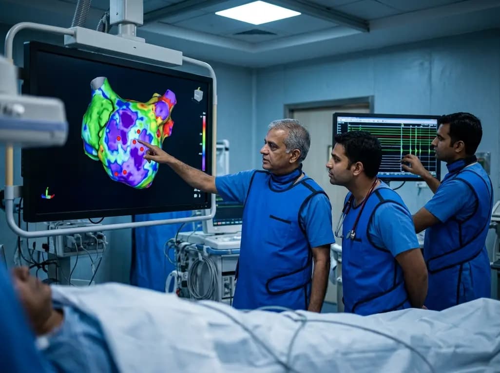
A study of 1,428 AF ablation patients reveals that over 60% of left atrial non-PV triggers arise within low-voltage areas, with anterior and roof triggers colocalizing entirely within fibrotic substrate. Identifying and ablating these sites is critical for long-term arrhythmia freedom.
Today

A comprehensive meta-analysis of over 278,000 women demonstrates that maternal atopic dermatitis significantly increases risks of premature rupture of membranes and low birth weight. Discover essential clinical insights on disease mechanisms, serial antenatal growth monitoring, and safe therapeutic interventions.
Today
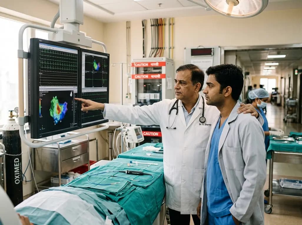
A pioneering study highlights the first United States experience using the FARAPOINT focal pulsed-field ablation catheter for refractory ventricular tachycardia. The catheter achieved complete acute non-inducibility across tested patients without procedural complications, marking a milestone in ventricular ablation.
Today

A biomechanical study shows that augmenting thin fascia lata grafts with a reinforced polyester patch doubles ultimate tensile strength and stiffness, matching the performance of thick grafts for superior capsule reconstruction while reducing donor-site morbidity.
Today

A cross-sectional study reveals that elevated METS-IR is independently associated with higher myopia odds in adolescents, showing biological synergy with obesity and highlighting the need for metabolic screening in pediatric eye care.
Today
