.webp&w=384&q=75)

Loading, please wait...
Loading, please wait...
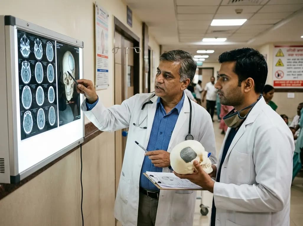
Decompressive craniectomy remains a life-saving neurosurgical intervention for refractory intracranial hypertension resulting from severe traumatic brain injury, ischemic stroke, and intracranial hemorrhage. However, removing a large cranial bone flap disrupts normal intracranial physiology and dynamics. Consequently, patients frequently develop secondary disturbances in cerebrospinal fluid hydrodynamic circulation and cerebral blood flow. Clinicians have long debated the optimal timing of cranioplasty to reverse these pathological changes safely. While surgeons historically deferred cranial reconstruction for several months to minimize infectious complications, modern clinical evidence suggests that early restoration offers superior neurological, motor, and cognitive advantages.
Removal of a rigid cranial vault exposes the cerebral cortex directly to atmospheric pressure. Consequently, this exposure alters normal intracranial compliance and cerebral perfusion pressure. Over time, patients may manifest the syndrome of the trephined, which presents as postural headaches, motor deficits, fatigue, and marked cognitive decline. Furthermore, impaired venous outflow and altered cerebrospinal fluid resorption exacerbate these clinical problems. Therefore, cranial reconstruction does not merely serve a cosmetic or protective purpose. Cranioplasty actively re-establishes a closed, rigid intracranial system, thereby restoring physiological atmospheric barriers and improving cerebral vascular autoregulation. Modern perfusion studies confirm that reconstructing the skull defect promptly enhances microvascular blood flow across both ipsilateral and contralateral hemispheres.
Recent systematic reviews and meta-analyses show that the timing of cranioplasty directly influences long-term neurological rehabilitation. Specifically, early cranioplasty, typically defined as reconstruction within three months of craniectomy, correlates with significantly greater functional recovery compared to delayed procedures. Standardized assessment tools, including the Barthel Index, Functional Independence Measure, and Karnofsky Performance Scale, consistently demonstrate superior improvements in patients who undergo earlier cranial repair. Additionally, earlier intervention appears to halt prolonged cortical compression, which facilitates faster motor rehabilitation and daily living independence. Therefore, waiting extended periods may inadvertently delay critical neurorehabilitation milestones during the optimal period of neuroplasticity.
Cognitive rehabilitation represents a vital clinical endpoint for neurosurgical patients surviving traumatic brain injury. Notably, pooled meta-analyses highlight significant improvements in Mini-Mental State Examination scores among patients receiving early reconstruction. In contrast, delayed skull repair frequently prolongs cognitive deficits and executive dysfunction due to sustained perturbations in cerebral perfusion. Subgroup analyses focusing exclusively on traumatic brain injury cohorts reveal even more pronounced functional gains following early intervention. Furthermore, investigators have evaluated ultra-early cranioplasty performed within thirty-five to forty-five days post-injury. While ultra-early cohorts display encouraging trends toward rapid cognitive recovery, larger prospective cohorts are necessary to confirm whether ultra-early surgery provides incremental benefits over standard early reconstruction within twelve weeks.
Surgeons historically feared that early cranioplasty would substantially increase perioperative infection risks, surgical site complications, and epidural collections. However, contemporary meta-analyses reveal no statistically significant difference in overall complication rates between early and late cranial reconstruction cohorts. In fact, delayed surgical intervention frequently correlates with higher rates of autologous bone flap resorption and severe tissue scarring. Consequently, late operations can complicate surgical dissection and extend operative durations. Nevertheless, clinicians must monitor patients closely for hydrocephalus and extra-axial fluid collections, which may present slightly more often in earlier surgical windows. Therefore, strict aseptic surgical protocols, thorough intracranial monitoring, and careful patient selection remain imperative regardless of timing.
In Indian healthcare settings, traumatic brain injuries from road traffic accidents represent a significant clinical burden in neurotrauma units. Consequently, optimizing neurorehabilitation pathways through timely cranial reconstruction is crucial for maximizing patient independence and reducing long-term caregiver dependency. Multidisciplinary teams, including neurosurgeons, intensivists, and neuro-rehabilitation specialists, should evaluate patients for cranioplasty as soon as cerebral edema resolves and systemic infections clear. Furthermore, selecting between autologous bone preservation and synthetic implants, such as titanium mesh or polyetheretherketone (PEEK), should depend on anatomical feasibility and resource availability. Ultimately, adopting an early cranioplasty strategy within three months post-craniectomy offers substantial physiological, cognitive, and functional benefits for eligible neurotrauma patients across tertiary healthcare centers.
In modern neurosurgical literature, early cranioplasty refers to skull reconstruction performed within three months (typically under 86 to 90 days) following decompressive craniectomy. In contrast, late cranioplasty occurs after three months, whereas ultra-early cranioplasty is generally undertaken within 35 to 45 days after the initial decompression procedure.
Comprehensive meta-analyses demonstrate that early cranioplasty does not significantly increase overall postoperative infection rates compared to delayed reconstruction. While clinicians must ensure complete resolution of initial cerebral edema and intracranial infections, performing cranial repair within three months maintains a safe complication profile while facilitating earlier neurological recovery.
Cranioplasty restores a closed, rigid intracranial compartment, which eliminates direct atmospheric pressure on the cerebral hemisphere. Consequently, this structural restoration normalizes cerebrospinal fluid circulation, improves cerebral vascular autoregulation, and enhances hemispheric blood flow, directly alleviating syndrome of the trephined symptoms and boosting cognitive function scores.
Disclaimer: This content is for informational and educational purposes only. It is not intended to provide medical advice or to replace independent clinical judgment. Healthcare professionals must evaluate every patient individually. Refer to the latest local and national guidelines for clinical practice.
References
Roa Castro JA et al. Timing of cranioplasty after decompressive craniectomy and neurological recovery: A systematic review and meta-analysis. Neurosurg Rev. 2026 Jun 09. doi: 10.1007/s10143-026-04361-3. PMID: 42260006.
Ochoa Hernandez D et al. Cranioplasty Timing After Decompressive Craniectomy: A Meta-Analysis of 4703 Patients. Neurosurgery. 2026 Mar 19.
Thamilmaran A et al. Optimal timing of cranioplasty post-decompressive craniectomy in traumatic brain injury: a systematic review, meta-analysis, and overview of ongoing trials. Acta Neurochir (Wien). 2026 Jan 08;168(1):32. doi: 10.1007/s00701-025-06759-2.

Read summarized clinical updates, watch expert medical content, and earn CME certifications right from your smartphone.
Open your smartphone camera to scan and install.





A meta-analysis evaluates the timing of cranioplasty after decompressive craniectomy, demonstrating that early cranial reconstruction (≤3 months) significantly enhances functional independence and cognitive recovery across clinical cohorts.
last month

A scientific statement highlights the crucial role of nutrition in managing chronic heart failure. It emphasizes GLIM-based screening, individualized dietary patterns like the Mediterranean diet, appropriate protein and energy intake, avoiding unnecessary fluid restriction, and targeted iron supplementation.
Today

A nationwide South Korean study shows underreporting of smoking among women dropped from 57.4% to 36.4% between 2008 and 2021. However, over one-third of female smokers still conceal their habit, highlighting the critical role of cotinine verification in clinical risk assessment.
Today

PepsiCo and Monster Beverage have challenged the FSSAI ban on the energy drink label in Indian courts. While companies highlight supply disruptions, regulatory actions spotlight severe cardiovascular and metabolic risks associated with excessive caffeine, sugar, and taurine consumption, especially in adolescents.
Today

A new study evaluates the rheological stability of decellularized peripheral nerve matrix hydrogels under varied storage conditions. Findings show -80°C storage and freeze-drying preserve gelation kinetics and mechanical stiffness, offering key insights for shipping and translational clinical applications.
Today

A qualitative meta-synthesis highlights the complex interplay between gender dysphoria, minority stress, and eating disorders in transgender and gender-diverse individuals. Clinicians must recognize these distinct drivers to offer supportive, gender-affirming, and interdisciplinary eating disorder care.
Today
