.webp&w=384&q=75)

Loading, please wait...
Loading, please wait...

Atypical intracranial meningiomas present a unique management challenge for neurosurgeons and neuro-oncologists worldwide. These intermediate-grade lesions exhibit aggressive biological behavior, characterized by rapid local proliferation, tissue invasiveness, and high recurrence rates compared to benign counterparts. Consequently, prompt clinical decision-making remains vital. Emerging large-scale evidence emphasizes that the timing of atypical meningioma surgery plays a pivotal role in dictating long-term overall survival. Clinicians frequently encounter diagnostic and scheduling hurdles that inadvertently prolong the preoperative interval. Therefore, understanding the measurable prognostic impact of these delays is essential to optimize clinical care pathways. Recent data show that timely operative intervention offers meaningful therapeutic benefits, particularly for individuals who cannot achieve complete gross resection.
World Health Organization Grade II atypical meningiomas represent approximately fifteen to twenty percent of all diagnosed meningiomas. Unlike benign grade I tumors, these neoplasms demonstrate increased mitotic activity, prominent brain invasion, pattern loss, and spontaneous necrosis. Because these tumors proliferate rapidly, progressive neurological deficits often develop within shorter intervals. Neurosurgeons must balance comprehensive preoperative assessment with swift intervention. However, patients frequently experience diagnostic delays due to non-specific initial presentations, such as intermittent headaches, insidious cognitive changes, or late-onset seizures. Additionally, logistical delays associated with referral coordination, comprehensive high-resolution neuroimaging, and surgical scheduling can compound the diagnosis-to-treatment timeline. Thus, delays permit persistent tumor expansion, which inevitably complicates subsequent microdissection and vascular preservation during neurosurgical procedures.
Recent multicenter retrospective evidence from the National Cancer Database confirms that extending the interval between initial diagnosis and definitive surgical resection directly compromises survival. In an extensive cohort of over five thousand patients, multivariable proportional hazards modeling demonstrated a three percent rise in mortality for every additional month of delay. Consequently, prolonged preoperative wait times act as an independent predictor of premature death. While an average diagnosis-to-surgery interval was less than one month across high-volume centers, prolonged wait times generated cumulative adverse prognostic effects. Furthermore, researchers controlled for critical confounding variables, including baseline patient age, systemic frailty, tumor dimension, facility classification, and adjuvant therapies. Therefore, treating physicians must prioritize prompt intervention to avoid preventable decrements in patient longevity.
The survival impact of surgical timing becomes especially pronounced when stratified by the extent of resection achieved in the operating room. For patients undergoing complete gross-total resection, minor surgical postponements showed less statistical association with altered survival outcomes. Conversely, patients who underwent subtotal resection experienced a four percent increase in mortality risk per month of preoperative delay. Hence, residual tumor tissue combined with prolonged initial tumor proliferation creates a dual oncological disadvantage. Subtotal resections occur frequently in complex anatomical locations, including the petroclival region, cavernous sinus, and posterior fossa. Because residual neoplastic cells in Grade II tumors harbor elevated proliferative capacity, preoperative delay permits extensive microinvasion into surrounding neurovascular structures. Consequently, early operation prevents excessive local infiltration, thereby maximizing the safety and effectiveness of subsequent cytoreduction.
Optimizing clinical pathways requires a coordinated multidisciplinary approach across neurosurgical, neuro-oncological, and neuroradiological services. Clinicians must establish expedited referral pipelines when preliminary imaging exhibits features suggestive of atypia. These features include heterogeneous contrast enhancement, prominent peritumoral vasogenic edema, indistinct brain-tumor interfaces, and elevated diffusion restriction. Furthermore, preoperative optimization protocols should proceed concurrently rather than sequentially. For example, cardiopulmonary assessments, baseline laboratory investigations, and pre-anesthetic clearance must occur rapidly. In healthcare ecosystems facing resource constraints or high patient loads, prioritizing intracranial lesions with aggressive radiological markers reduces avoidable attrition. In addition, prompt multidisciplinary tumor board reviews ensure timely postoperative planning, expediting required adjuvant stereotactic or fractionated radiotherapy without unnecessary lag periods.
The clinical findings also illuminate significant sociodemographic disparities that influence surgical timing and survival trajectories. Analyses reveal that male sex, African American race, significant systemic comorbidities, and tumor dimensions exceeding sixty millimeters correlate with heightened mortality rates. Conversely, treatment delivered at dedicated academic cancer centers independently associates with superior overall survival. Academic facilities often maintain specialized neuro-oncology multidisciplinary teams and integrated operative scheduling systems that minimize administrative friction. Therefore, expanding access to specialized tertiary centers and standardizing fast-track surgical booking protocols remain imperative. By systematically eliminating institutional and socioeconomic barriers, healthcare systems can ensure that all patients receive equitable, rapid access to life-prolonging cranial surgery.
World Health Organization Grade II atypical meningiomas exhibit aggressive features, including increased mitotic activity, brain invasion, high cellularity, and spontaneous necrosis. Unlike benign Grade I tumors, atypical meningiomas grow substantially faster, recur more frequently following resection, and carry a significantly higher risk of patient morbidity and overall mortality.
Preoperative delays directly correlate with increased mortality, presenting a three percent higher risk of death for each month between diagnosis and resection. Delays allow tumor growth and tissue invasion, which dramatically worsens outcomes, particularly in patients who ultimately undergo subtotal surgical resection rather than complete tumor removal.
Clinicians should urgently evaluate meningiomas showing pronounced peritumoral edema, irregular non-spherical borders, intratumoral necrosis, heterogeneous contrast enhancement, or evidence of direct parenchymal invasion. These imaging characteristics strongly suggest atypical or malignant histology, requiring expedited neurosurgical referral and prompt operative intervention to maximize long-term survival.
Disclaimer: This content is for informational and educational purposes only. It is not intended to be a substitute for professional medical advice, diagnosis, or treatment. Always seek the advice of your physician or other qualified health provider with any questions you may have regarding a medical condition. Refer to the latest local and national guidelines for clinical practice.
References

Read summarized clinical updates, watch expert medical content, and earn CME certifications right from your smartphone.
Open your smartphone camera to scan and install.





A comprehensive National Cancer Database analysis reveals that delaying atypical meningioma surgery increases mortality risks. Prompt surgical resection, particularly when complete excision is challenging, proves essential for optimizing long-term survival outcomes.
Today
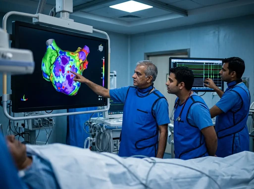
A study of 1,428 AF ablation patients reveals that over 60% of left atrial non-PV triggers arise within low-voltage areas, with anterior and roof triggers colocalizing entirely within fibrotic substrate. Identifying and ablating these sites is critical for long-term arrhythmia freedom.
Today

A comprehensive meta-analysis of over 278,000 women demonstrates that maternal atopic dermatitis significantly increases risks of premature rupture of membranes and low birth weight. Discover essential clinical insights on disease mechanisms, serial antenatal growth monitoring, and safe therapeutic interventions.
Today

A pioneering study highlights the first United States experience using the FARAPOINT focal pulsed-field ablation catheter for refractory ventricular tachycardia. The catheter achieved complete acute non-inducibility across tested patients without procedural complications, marking a milestone in ventricular ablation.
Today

A biomechanical study shows that augmenting thin fascia lata grafts with a reinforced polyester patch doubles ultimate tensile strength and stiffness, matching the performance of thick grafts for superior capsule reconstruction while reducing donor-site morbidity.
Today

A cross-sectional study reveals that elevated METS-IR is independently associated with higher myopia odds in adolescents, showing biological synergy with obesity and highlighting the need for metabolic screening in pediatric eye care.
Today
