.webp&w=384&q=75)

Loading, please wait...
Loading, please wait...

Epileptic spasms represent severe seizures that typically emerge during early childhood and often resist conventional pharmacotherapy. For decades, investigators debated whether these debilitating events originate purely within focal cortical regions or involve widespread subcortical pathways. Recent neurophysiological research now confirms robust thalamocortical network activation during clinical spasms. Historically, clinicians linked infantile epileptic spasms syndrome with characteristic high-voltage slow-wave complexes on surface electroencephalography. However, scalp recordings cannot accurately define deep subcortical dynamics. Consequently, medical teams faced substantial barriers when determining why localized cortical lesions provoke generalized motor jerks. Animal models previously suggested subcortical pacemaker involvement, but human in vivo electrophysiological data remained scarce. Modern intracranial stereotactic electroencephalography now bridges this diagnostic divide. By sampling cortical regions alongside specific deep thalamic nuclei, clinicians observe real-time neural recruitment across distributed circuits. Furthermore, identifying these precise connections reshapes our fundamental understanding of ictogenesis. As a result, pediatric neurologists can better appreciate how focal disruptions rapidly recruit subcortical circuits. This conceptual shift provides immediate therapeutic hope for children suffering from intractable epileptic spasms.
To uncover these subcortical mechanisms, researchers conducted an intracranial stereotactic electroencephalography study in pediatric candidates undergoing presurgical evaluation. The research team evaluated four pediatric and adolescent patients, aged 2.7 to 16.9 years, who suffered from refractory epileptic spasms. Notably, neurosurgeons implanted depth electrodes directly into both suspected cortical seizure-onset zones and key thalamic nuclei. These subcortical targets included the anterior nucleus, centromedian nucleus, and pulvinar. Across the monitoring period, the investigators recorded and systematically analyzed 84 individual epileptic spasms. Initially, clinicians performed rigorous visual inspection and time-frequency signal decomposition. They specifically assessed slow-wave complexes, which define the classic electrographic footprint of epileptic spasms. In addition, the researchers divided neural signals into two distinct bands: slow rhythms (0.5-10 Hz) and fast oscillations (10-80 Hz). Therefore, the team could isolate directional interactions between cortical regions and subcortical nuclei. This comprehensive approach allowed unprecedented visualization of deep nuclear firing patterns during active clinical seizures. Moreover, advanced mathematical modeling tracked signal propagation across each seizure event. Consequently, the protocol captured both local nuclear changes and broader inter-structural synchrony.
Quantitative signal processing demonstrated striking electrophysiological synchrony between the thalamus and cortex during every analyzed spasm. First, visual analysis confirmed characteristic high-voltage slow-wave complexes within the thalamic depth channels. Furthermore, power spectral density analyses revealed significant signal power surges in both slow and fast frequency bands at the thalamus. To determine functional connectivity, the investigators calculated undirected coherence between the seizure-onset zone and thalamic contacts. This coherence increased substantially during spasms compared to pre-ictal baseline recordings. Next, the investigators utilized spectral Granger causality to evaluate directed causal influences between these structures. Interestingly, slow-band connectivity exhibited symmetric, bidirectional information flow. Inflow from the cortex to the thalamus mirrored outflow from the thalamus back to the cortex. However, fast-band connectivity displayed marked asymmetry. Inflow of fast frequencies from the cortical seizure-onset zone into the thalamus significantly exceeded thalamic outflow. Thus, cortical nodes predominantly drive high-frequency rhythms into subcortical structures. Simultaneously, the thalamus and cortex collaboratively sustain the high-voltage slow-wave envelope through balanced bidirectional communication. Consequently, these complementary rhythms illustrate an active, coordinated circuit rather than passive volume conduction.
Beyond directional connectivity, the investigators explored how different frequency oscillations coordinate locally through cross-frequency phase-amplitude coupling. Specifically, they utilized the modulation index to measure how slow-wave phase modulates high-frequency oscillation amplitude. During epileptic spasms, the modulation index rose significantly within thalamic recording sites. Moreover, cortical channels exhibited powerful phase-amplitude interactions during every motor seizure. Importantly, the researchers discovered a strong clinical correlation. Patients exhibiting higher cortical modulation indices displayed significantly more pronounced motor manifestations of epileptic spasms. In contrast, weaker cross-frequency coupling produced subtle clinical movements. This finding provides crucial mechanistic insight into the variable semiology of infantile spasms. In typical clusters, individual spasms often fluctuate in visual intensity from subtle eye deviations to massive tonic contractions. The authors hypothesize that cortical fast activity probabilistic recruitment drives symptomatogenic motor areas during the spasm slow-wave envelope. Because thalamocortical networks modulate this interaction, subcortical recruitment directly shapes phenotypic expression. Therefore, quantitative coupling metrics may serve as reliable neurophysiological markers for assessing seizure burden and clinical severity. Consequently, clinicians gain an objective physiological measure reflecting clinical spasm intensity.
These neurophysiological discoveries carry profound clinical implications for pediatric epilepsy surgery and neuromodulation protocols. Historically, pediatric patients with non-localized, multifocal, or non-resectable epileptic spasms faced limited surgical options. Because resective surgery often proves unfeasible in widespread developmental encephalopathies, neurostimulation offers a viable therapeutic alternative. Specifically, deep brain stimulation and responsive neurostimulation can target critical subcortical relay nodes. The demonstration that the centromedian nucleus and anterior thalamic nucleus participate actively during spasms validates them as therapeutic targets. By delivering electrical stimulation directly to the thalamus, clinicians can disrupt pathologic synchrony across corticothalamic loops. Furthermore, electrical pulses may desynchronize the bidirectional slow waves that propagate clinical spasms across both hemispheres. Closed-loop neuromodulation systems could also detect early cortical fast activity and trigger thalamic stimulation before widespread recruitment occurs. In addition, thalamic modulation may reduce interictal epileptiform discharges and restore physiological sleep architecture. Consequently, neuromodulation emerges as a promising, circuit-based therapy for children with drug-resistant epileptic spasms. Ultimately, mapping these pathways allows teams to transition from empirical pharmacotherapy toward precision network medicine.
The thalamus functions as a critical subcortical node that coordinates widespread ictal activity during epileptic spasms. Rather than acting as a passive bystander, it exhibits prominent slow-wave complexes, spectral power surges, and synchronized engagement with cortical seizure-onset zones, helping sustain the electrographic and motor manifestations of each spasm.
Directed connectivity analyses reveal symmetric bidirectional communication between the thalamus and cortex in slow frequency bands (0.5-10 Hz). In contrast, fast band (10-80 Hz) connectivity demonstrates significant asymmetry, characterized by predominant high-frequency inflow from the cortical seizure-onset zone into the thalamus, confirming that cortical drivers project fast rhythms subcortically.
Phase-amplitude coupling quantifies how slow-wave phase modulates high-frequency oscillation amplitude across the brain. Elevated coupling within cortical channels directly correlates with more pronounced clinical spasm movements. Therefore, measuring this modulation index provides clinicians with an objective neurophysiological biomarker that reflects seizure severity and helps assess therapeutic responses to treatment.
Disclaimer: This content is for informational and educational purposes only. It is not intended as medical advice, diagnosis, or treatment. Always consult qualified healthcare providers before modifying clinical regimens. Refer to the latest local and national guidelines for clinical practice.
References

Read summarized clinical updates, watch expert medical content, and earn CME certifications right from your smartphone.
Open your smartphone camera to scan and install.





A stereotactic EEG study confirms thalamocortical network activation during epileptic spasms, revealing bidirectional slow waves and fast-band cortical inflow.
Today
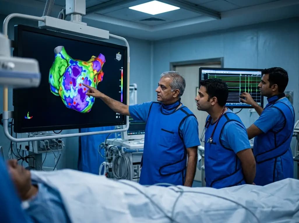
A study of 1,428 AF ablation patients reveals that over 60% of left atrial non-PV triggers arise within low-voltage areas, with anterior and roof triggers colocalizing entirely within fibrotic substrate. Identifying and ablating these sites is critical for long-term arrhythmia freedom.
Today

A comprehensive meta-analysis of over 278,000 women demonstrates that maternal atopic dermatitis significantly increases risks of premature rupture of membranes and low birth weight. Discover essential clinical insights on disease mechanisms, serial antenatal growth monitoring, and safe therapeutic interventions.
Today

A pioneering study highlights the first United States experience using the FARAPOINT focal pulsed-field ablation catheter for refractory ventricular tachycardia. The catheter achieved complete acute non-inducibility across tested patients without procedural complications, marking a milestone in ventricular ablation.
Today

A biomechanical study shows that augmenting thin fascia lata grafts with a reinforced polyester patch doubles ultimate tensile strength and stiffness, matching the performance of thick grafts for superior capsule reconstruction while reducing donor-site morbidity.
Today

A cross-sectional study reveals that elevated METS-IR is independently associated with higher myopia odds in adolescents, showing biological synergy with obesity and highlighting the need for metabolic screening in pediatric eye care.
Today
