.webp&w=384&q=75)

Loading, please wait...
Loading, please wait...
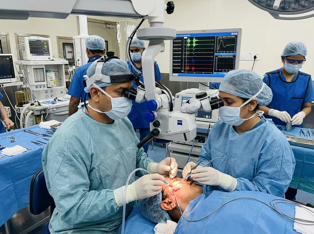
Facial nerve schwannomas represent rare benign primary tumors originating from Schwann cells that envelop the seventh cranial nerve. Because these neurogenic lesions can develop anywhere along the intra-axial or extra-axial course of the nerve, clinical presentation varies significantly among patients. Common presenting symptoms include progressive facial palsy, facial spasms, conductive or sensorineural hearing impairment, and otalgia. Managing these complex lesions presents a unique clinical dilemma for neurosurgeons and otolaryngologists alike. Historically, radical surgical excision was the primary therapeutic objective; however, total resection often resulted in permanent complete facial paralysis and severe functional deficits. Recent clinical advances have prompted a major paradigm shift toward function-centered management strategies. These contemporary facial nerve schwannoma surgery approaches aim to strike an optimal balance between long-term oncological control and functional nerve preservation. Medical evidence from recent contemporary series highlights that customized, patient-tailored interventions significantly improve functional outcomes without compromising safety. Clinicians must thoroughly evaluate anatomical tumor extent, baseline nerve function, and neurophysiological status when formulating treatment plans. Adapting surgery to patient-specific parameters ensures better quality of life and minimizes long-term morbidity for individuals diagnosed with facial nerve schwannomas.
The implementation of a function-centered surgical framework fundamentally changes how surgical teams address facial nerve schwannomas. Instead of applying a uniform radical resection protocol to every patient, clinicians carefully stratify surgical techniques based on preoperative clinical presentation and intraoperative diagnostic assessments. A function-centered approach categorizes patients based on the degree of pre-existing facial weakness evaluated through standardized grading systems like the House-Brackmann scale. For patients presenting with mild to moderate facial palsy, surgeons prioritize nerve-preserving surgery, which typically involves bony decompression and nonradical tumor debulking. This conservative intervention relieves compression within rigid bony canals while leaving the continuity of the nerve trunk intact. Conversely, in patients presenting with severe or complete facial paralysis, complete tumor removal combined with immediate primary nerve reconstruction becomes the preferred surgical intervention. Nerve reconstruction techniques include direct end-to-end anastomosis or interposition nerve grafting using peripheral donor nerves such as the greater auricular or sural nerves. By tailoring the intervention directly to the patient's existing neurological status, surgical teams maximize the potential for functional recovery. This nuanced decision-making process ensures that patients with acceptable baseline motor function do not suffer unnecessary postoperative neurological deficits.
Selecting the appropriate surgical procedure requires meticulous evaluation of pre-existing neurological deficits and tumor localization. Clinical guidelines recommend nerve-preserving techniques primarily for patients who retain moderate facial motor function, defined typically as House-Brackmann grades lower than four. In these instances, complete surgical resection carries an unacceptably high risk of converting partial motor function into total facial paralysis. Bony decompression of the fallopian canal relieves mechanical entrapment and reduces microvascular compromise without damaging viable nerve fibers. In contrast, patients who present with severe baseline dysfunction, corresponding to House-Brackmann grades five or six, have minimal preserved native nerve fiber continuity. For these individuals, radical tumor extirpation combined with primary nerve reconstruction offers the highest probability of restoring meaningful facial tone and voluntary motor reinnervation. Reconstruction allows nerve axons to regenerate across the graft, typically achieving a outcome around House-Brackmann grade three or four over time. Furthermore, tumor localization plays a crucial role in determining technical feasibility. Tumors confined within the bony temporal bone present distinct anatomical boundaries that facilitate decompressive techniques. Consequently, understanding the complex interplay between clinical grading and tumor location allows surgeons to execute surgical procedures that preserve baseline function while effectively managing disease progression.
Intraoperative electromyographic monitoring serves as an indispensable diagnostic tool during facial nerve schwannoma surgery. Continuous electrophysiological evaluation provides real-time functional feedback regarding the physiological integrity of the facial nerve during operative manipulation. By placing recording electrodes in target facial muscles, such as the orbicularis oculi and orbicularis oris, surgical teams monitor both spontaneous mechanical irritation responses and electrically evoked compound muscle action potentials. Favorable intraoperative electromyography responses indicate the presence of functional, conducting nerve fibers running across or within the tumor mass. When robust electrical responses are present, surgeons receive immediate feedback confirming that nerve-preserving maneuvers remain safe and effective. Conversely, absent or severely blunted electrophysiological responses signal profound loss of axonal continuity, supporting the decision to perform total tumor resection and nerve reconstruction. Beyond guiding structural preservation, continuous intraoperative monitoring minimizes inadvertent mechanical or thermal injury during bony decompression and tumor debulking. Incorporating real-time monitoring significantly enhances surgical precision, reduces procedure-related morbidity, and optimizes long-term functional recovery for patients undergoing complex skull base and temporal bone interventions.
Recent long-term clinical data from contemporary surgical series demonstrate the clinical efficacy of adopting a function-centered management model. Patients undergoing nerve-preserving surgery demonstrate high rates of functional preservation, with over eighty-seven percent maintaining stable or improved postoperative facial nerve function. On average, patients in the nerve preservation group show noticeable improvements in House-Brackmann clinical grades following surgical decompression. Similarly, patients undergoing complete tumor resection with interposition nerve grafting exhibit meaningful recovery, moving from severe baseline paralysis toward moderate functional recovery. Nevertheless, clinicians must recognize that subtotal tumor removal or nonradical debulking requires vigilant long-term surveillance. Long-term follow-up studies reveal that approximately twenty-seven percent of patients undergoing nerve-preserving management may eventually require supplementary therapeutic interventions due to tumor progression or recurrent symptoms. Secondary management options include salvage microsurgery, targeted stereotactic radiosurgery, or specialized facial reanimation procedures. Consequently, established clinical follow-up protocols combining serial magnetic resonance imaging and neurophysiological testing remain essential components of care. Comprehensive long-term monitoring ensures timely detection of disease progression, enabling clinicians to intervene appropriately while maintaining optimal patient outcomes and quality of life over extended periods.
Identifying clear prognostic factors helps surgical teams predict clinical outcomes and counsel patients effectively prior to surgery. In contemporary clinical analyses, several key clinical variables correlate strongly with superior postoperative outcomes following nerve-preserving surgery. First, anatomical tumor location serves as a critical prognostic determinant; tumors strictly confined within the temporal bone, such as intrameatal, tympanic, or mastoid segments, exhibit significantly better functional outcomes compared to extensive multi-segmental or intracranial lesions. Second, patient age significantly influences nerve recovery potential, as younger patients generally demonstrate enhanced neuroplasticity and axon regeneration capacity following decompression. Third, favorable intraoperative electromyography parameters consistently predict long-term retention of facial motor control. Recognizing these favorable indicators allows multidisciplinary medical teams to refine patient selection and establish realistic functional expectations. Patients who present with localized intra-temporal tumors, younger age, and robust electrophysiological responses represent ideal candidates for nerve-preserving procedures. Conversely, patients lacking these favorable features require comprehensive preoperative discussion regarding the potential necessity of reconstruction or secondary therapies. Incorporating evidence-based prognostic stratification ultimately enhances clinical decision-making and standardizes management pathways for this challenging pathology.
Nerve-preserving surgery, consisting of bony decompression and subtotal tumor removal, aims to maintain native nerve continuity in patients with mild to moderate pre-existing facial weakness. By relieving pressure within the rigid temporal bone without severing functional nerve fibers, this approach preserves baseline motor function and prevents immediate severe paralysis. It provides an effective balance between tumor control and quality of life.
Conventional nerve reconstruction is indicated for patients who present with severe or complete facial paralysis prior to surgery. In these cases, native axonal continuity is already severely compromised. Surgeons perform complete tumor resection followed by nerve grafting or anastomosis. This intervention offers the best opportunity to re-establish nerve continuity and regain functional facial muscle tone over long-term follow-up.
Intraoperative electromyography provides real-time electrophysiological monitoring of facial nerve function during surgical procedures. By recording muscle action potentials, monitoring alerts surgeons to mechanical stress on nerve fibers and confirms the functional viability of the nerve. Favorable electromyographic responses support the decision to perform nerve-preserving decompression, whereas absent signals guide decisions toward primary resection and nerve reconstruction.
Disclaimer: This content is for informational and educational purposes only... Refer to the latest local and national guidelines for clinical practice.
References

Read summarized clinical updates, watch expert medical content, and earn CME certifications right from your smartphone.
Open your smartphone camera to scan and install.





Facial nerve schwannomas are rare benign tumors presenting unique surgical challenges. A contemporary series evaluates function-centered surgical strategies, combining intraoperative EMG monitoring with tailored nerve preservation or reconstruction to optimize long-term clinical outcomes and facial function.
last month

A study verifies the quantitative risk assessment model using EC1.6 values from LLNA: BrdU-ELISA to predict skin sensitization from medical devices. Comparing FreeStyle Libre and Libre 2, researchers confirmed how reducing isobornyl acrylate below acceptable levels prevents allergic contact dermatitis.
Today

A recent longitudinal study evaluated the link between prenatal loss of control eating and cardiovascular health using Life's Essential 8 frameworks. While direct associations across pregnancy were non-significant, the high prevalence of dysregulated eating highlights critical implications for obstetric practice.
Today
Explore modern perioperative management protocols for clinical xenotransplantation. Learn how costimulation blockade, anti-inflammatory perfusion, and coagulopathy rescue overcome interspecies hurdles.
Today
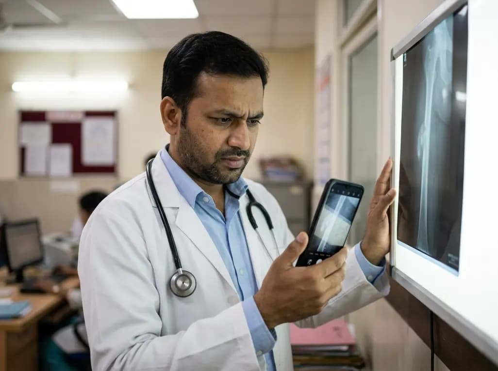
A retrospective study evaluating a locally deployed vision language model (Qwen3-VL) reveals that smartphone-captured radiographs and retrieval-augmented generation can paradoxically reduce diagnostic accuracy in bone tumor referrals due to optical noise and textual hallucinations.
Today
A real-world quasi-experimental study reveals that web-based apps fostering healthy habit formation significantly boost physical activity and dietary quality in adults. Discover key clinical insights on digital adherence, dose-response effects, and patient-specific modifiers in preventive medicine.
Today
