

Loading, please wait...
Loading, please wait...

Preserving autonomic nerve integrity remains a cornerstone of successful lumbosacral reconstruction. Surgeons frequently encounter challenging retroperitoneal anatomy during a superior hypogastric plexus ALIF exposure at the L5-S1 junction. Anterior lumbar interbody fusion provides exceptional disc height restoration, high fusion rates, and reliable indirect neural decompression. However, iatrogenic injury to autonomic fibers often leads to retrograde ejaculation, an adverse event that profoundly undermines postoperative satisfaction. Therefore, understanding micro-anatomical relationships and plexus mobilization limits is critical for modern spine specialists.
Autonomic nerve dysfunction following anterior lumbar exposure can drastically degrade a patient's quality of life. The superior hypogastric plexus supplies sympathetic innervation to the bladder neck and internal urethral sphincter. Consequently, disruptive mechanical stretching or electrocautery injury impairs coordinated sphincter closure during emission. Semen subsequently flows retrogradely into the urinary bladder instead of antegrade through the urethra. Male patients experience distressing sexual dysfunction and unexpected secondary infertility. Published clinical literature demonstrates variable rates of postoperative retrograde ejaculation, ranging from under two percent in gentle retroperitoneal approaches to over fifteen percent in extensive transperitoneal dissections. Moreover, the psychological burden on working-age men can generate substantial clinical dissatisfaction even when radiographic arthrodesis succeeds. Surgical teams must therefore prioritize proactive neural preservation over rapid exposure. Careful blunt handling, minimal diathermy, and evidence-based mobilization protocols protect fragile autonomic networks effectively. By recognizing the true clinical weight of these injuries, reconstructive surgeons can systematically adapt operative strategies before initiating intervertebral disc preparation.
Recent anatomical evaluations reveal consistent structural arrangements of the autonomic plexus anterior to the lumbosacral junction. The superior hypogastric plexus reliably forms just below the aortic bifurcation, descending across the anterior surface of the fifth lumbar vertebral body. Dissection demonstrates that this neural meshwork consistently crosses the anterior disc space at L5-S1. Importantly, the plexus does not rest symmetrically across the geometric midline. Instead, the nerve fibers exhibit a significant left-sided shift in their longitudinal axis. Morphometric measurements reveal that this asymmetric orientation occurs in direct continuity with left lumbar splanchnic contributions. Specifically, the third left lumbar splanchnic nerve regularly joins the superior hypogastric complex, pulling its dominant bulk toward the left anterolateral sacral promontory. Meanwhile, the right margin of the plexus frequently lies closer to the median line. Recognizing this innate leftward distribution allows access surgeons to anticipate neural location accurately before incising the prevertebral soft tissue mantle.
Surgeons must also appreciate that the superior hypogastric plexus presents distinct structural configurations. Cadaveric analyses categorize the neural architecture into three distinct morphology patterns: single cord, plexiform meshwork, and loose fiber arrangements. Both single cord and plexiform patterns appear in over forty percent of cases each, whereas loose, dispersed fibers occur in approximately sixteen percent. Furthermore, the feasibility of lateral mobilization depends directly on these branching patterns. In roughly two-thirds of individuals, surgeons can mobilize the intact plexus away from the disc space safely. However, in approximately one-third of surgical exposures, lateral retraction is physically unachievable. This restriction arises when the plexus divides prematurely into hypogastric nerves directly over the L5-S1 interspace or displays an exceptionally broad anatomical footprint. Attempting forced unilateral retraction across such wide or branching structures risks severe tension tears. Consequently, surgeons must inspect the morphological configuration before applying lateral instruments.
When mobilization is anatomically feasible, directionality dictates operative safety. Because the plexus naturally shifts toward the left, surgical teams should preferentially retract the mobilized nerve bundle toward the left side. Dissection data demonstrates that left-sided mobilization safely achieves up to 15.3 mm of lateral displacement from the midline. In sharp contrast, right-sided retraction remains severely restricted, achieving an average displacement of only 5.3 mm before encountering excessive tension. This asymmetry confirms that pulling the plexus rightward risks substantial traction neuropathy. Nevertheless, if anatomical width or premature distal bifurcation prevents safe unilateral movement, surgeons require an alternative strategy. Under these challenging circumstances, experts recommend gently splitting the superior hypogastric plexus directly down the midline. Once longitudinally separated along natural fibrous planes, clinicians mobilize the respective left and right halves laterally. This bilateral division preserves individual nerve conduit continuity, exposes the central annular window, and avoids dangerous stretch neuropraxia.
Mobilizing autonomic fibers demands meticulous coordination with retroperitoneal vascular structures. The middle sacral artery and accompanying veins travel in intimate contact with the posterior leaf of the prevertebral fascia, directly beneath the superior hypogastric network. Inadvertent vascular laceration prompts brisk hemorrhage, which quickly obscures delicate nerve filaments. Inexperienced operators might hastily apply monopolar cautery to control bleeding, inadvertently causing irreversible thermal coagulation of adjacent sympathetic fibers. Therefore, surgical teams should systematically identify, clip, and divide the middle sacral vessels using cold instruments or delicate bipolar coagulation prior to full disc exposure. Additionally, the common iliac veins lie closely adjacent to the lateral border of the exposure corridor. Maintaining clear surgical planes between the vascular adventitia and neural fascia ensures uninhibited operative visibility. By systematically securing local hemostasis without extensive electrocautery, spine specialists simultaneously protect autonomic integrity and prevent life-threatening vascular complications.
Achieving superior clinical outcomes in lumbar fusion requires disciplined execution during every anterior exposure. Before entering the operating room, clinicians must counsel male patients regarding the risks of retrograde ejaculation and discuss elective sperm banking when family planning remains relevant. Intraoperatively, the surgical team should adhere to strict nerve-sparing protocols. Surgeons should avoid monopolar electrocautery near the aortic bifurcation and sacral promontory, relying instead on blunt dissection and warm saline sponges. When exposing the L5-S1 disc, the access surgeon identifies the prevertebral plexus, observes its structural layout, and attempts gentle leftward mobilization. If dense adherence or anatomical bifurcation prevents lateral translation, the team performs a controlled midline release to preserve bilateral fibers. By embedding these evidence-based techniques into routine spine care, surgeons minimize autonomic morbidity, enhance functional recovery, and deliver predictable clinical results.
The superior hypogastric plexus delivers essential sympathetic innervation to the internal urethral sphincter. When surgery damages these fibers, the bladder neck fails to close during emission. As a result, seminal fluid flows backwards into the urinary bladder instead of exiting antegrade through the penile urethra.
Retroperitoneal exposures generally allow gentle blunt mobilization of the peritoneum and prevertebral nerves, keeping rates of retrograde ejaculation below two percent. Conversely, transperitoneal approaches involve direct peritoneal incision, extensive electrocautery, and aggressive retraction, which significantly increase the incidence of autonomic nerve trauma.
A surgeon should split the plexus along the midline when premature bifurcation into bilateral hypogastric nerves occurs directly over the L5-S1 disc. Splitting is also necessary when a wide, rigid plexus prevents safe leftward retraction, thereby avoiding severe mechanical traction injury.
Disclaimer: This content is for informational and educational purposes only... Refer to the latest local and national guidelines for clinical practice.
References
Khadanovich A et al. Anatomy of the superior hypogastric plexus and its relevance to anterior lumbar interbody fusion. J Neurosurg Spine. 2025 Jul 01. doi: 10.3171/2025.1.SPINE241365. PMID: 40279716.
Burkus JK, Gornet MF, Dickman CA, Zdeblick TA. Retrograde ejaculation after anterior lumbar interbody fusion: transperitoneal versus retroperitoneal exposure. Spine (Phila Pa 1976). 2003;28(10):989-992.
Carragee EJ, Mitsunaga KA, Hurwitz EL, Scuderi GJ. Retrograde ejaculation after anterior lumbar interbody fusion with and without bone morphogenetic protein-2 augmentation: a 10-year cohort controlled study. Spine (Phila Pa 1976). 2013;38(4):300-305.

Read summarized clinical updates, watch expert medical content, and earn CME certifications right from your smartphone.
Open your smartphone camera to scan and install.





Understanding superior hypogastric plexus anatomy during ALIF at L5-S1 is crucial for preventing retrograde ejaculation. Discover dissection techniques, lateral mobilization thresholds, and nerve preservation strategies based on recent cadaveric findings.
Today

The Food Safety and Standards Authority of India has issued an urgent advisory against food business operators violating mandatory labelling standards for nutraceuticals and botanicals. Regulators prohibited therapeutic claims while mandating clear advisory warnings to prevent patient toxicity and medical misuse.
Today

Biotechnology researchers in India are developing an automated liquid biopsy platform to detect cervical malignancy from blood samples. Supported by the MAHA MedTech Mission, this initiative applies nanopore sequencing and piRNA biomarker detection to eliminate invasive speculum exams and expand rural screening.
Today
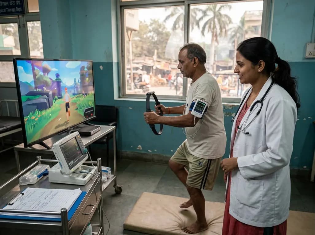
A clinical study evaluated Ring Fit Adventure in adults with chronic conditions, revealing that exergaming achieves moderate-to-vigorous exercise intensity while maintaining hemodynamic stability, high enjoyment, and zero adverse events.
Today

A retrospective cohort study reveals that selecting the touched vertebra as the lowest instrumented vertebra in thoracic adolescent idiopathic scoliosis yields a 20.8% distal add-on rate, driven by remnant motion segments and axial rotational discrepancies.
Today
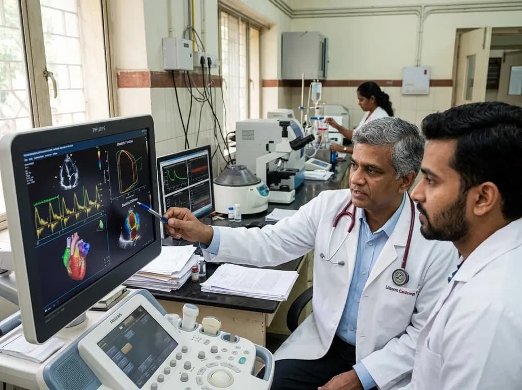
A study evaluates a cardiometabolic HFpEF model combining metabolic stress with chronic mineralocorticoid excess. The findings reveal profound diastolic stiffness, endothelial dysfunction, and pulmonary hypertension, illuminating key pathophysiological mechanisms for targeted cardiopulmonary therapies.
Today
