.webp&w=384&q=75)

Loading, please wait...
Loading, please wait...

Acute large vessel occlusive stroke remains a leading cause of mortality and long-term adult disability worldwide. Furthermore, approximately one-quarter of acute ischemic stroke presentations lack a definitive source upon initial diagnostic evaluation. Endovascular revascularization now yields retrieved biological specimens that offer direct insight into clot biology. Clinicians increasingly evaluate stroke thrombus histopathology to decipher the exact pathophysiological origins of acute vascular occlusions. By quantifying cellular architecture and biochemical matrices, pathologists illuminate fundamental differences between diverse vascular mechanisms. Consequently, post-thrombectomy structural clot analysis bridges acute procedural recanalization and personalized secondary prevention strategies. As a result, stroke teams gain actionable diagnostic information to optimize patient outcomes.
Mechanical thrombectomy yields intact biological specimens that reflect acute intravascular pathophysiology. In a comprehensive investigation of 132 acute stroke patients, researchers conducted quantitative histomorphometry on extracted occlusive material. Standard hematoxylin and eosin staining revealed substantial compositional heterogeneity across all recovered specimens. On average, fibrin and platelet complexes constituted the largest single structural fraction, averaging 51.46% of total clot volume. Meanwhile, erythrocytes represented approximately 40.97% of the thrombus mass, forming tightly packed cellular islands. Leukocytes comprised a much smaller fraction, showing a median value of 6.88%.
Beyond standard hematological stains, investigators applied advanced immunohistochemical assays to detect specific inflammatory elements. They quantified CD3-positive T lymphocytes and citrullinated histone H3 markers of neutrophil extracellular traps within the fibrin mesh. Consequently, the analysis demonstrated a median CD3-positive T-cell density of 20.64 cells per square millimeter. Neutrophil extracellular traps occupied a median area of 1.03% across evaluated sections. Interestingly, pathologists observed no intact cholesterol crystals or macroscopic calcifications within these endovascular samples. Therefore, acute neurovascular occlusions consist predominantly of dynamic cellular and proteinaceous networks rather than inert debris. These quantitative baselines provide essential reference points for investigating causal vascular mechanisms in acute ischemic stroke.
Stroke etiology fundamentally influences clot composition and mechanical behavior. When investigators classified cases according to established TOAST criteria, distinct histological signatures emerged between mechanistic subgroups. Specifically, cardioembolic thrombi exhibited a significantly higher proportion of fibrin and platelets compared to large artery atherosclerosis emboli. In contrast, atherosclerotic thrombi contained a substantially higher proportion of red blood cells. Statistical comparisons confirmed that these compositional differences reached high statistical significance. Thus, fibrin-rich matrices define embolic fragments that originate within turbulent, low-flow intracardiac chambers.
Conversely, shear-dependent atherothrombosis favors local erythrocyte entrapment within expanding platelet aggregates. However, inflammatory markers behaved differently across these stroke categories. Proportions of neutrophil extracellular traps and CD3-positive T cells did not differ significantly between cardioembolic and atherosclerotic occlusions. This stability suggests that sterile inflammation accompanies thrombus maturation regardless of primary anatomical origin. Nevertheless, the stark divergence in erythrocyte and fibrin-platelet ratios provides a reliable distinguishing benchmark. Clinicians can therefore leverage cellular fractions to reconstruct the primary pathophysiological event that triggered acute vessel occlusion.
Determining stroke etiology remains a persistent challenge in acute vascular neurology. Approximately one-quarter of all ischemic presentations fall into the cryptogenic or undetermined category after standard hospital workups. Fortunately, stroke thrombus histopathology offers direct biological evidence to help resolve these diagnostic dilemmas. In the evaluated cohort, thrombi retrieved from patients with undetermined etiology displayed an architectural profile nearly identical to cardioembolic clots. Specifically, they exhibited high fibrin-platelet densities and correspondingly reduced red blood cell volumes.
This striking morphological resemblance suggests that a substantial proportion of cryptogenic strokes originate from occult cardiac sources. Paroxysmal atrial fibrillation often evades detection during brief in-hospital telemetry monitoring. Consequently, patients without an identified etiology may leave the acute care setting without appropriate anticoagulation therapy. Histopathological confirmation of a cardioembolic-like clot structure alerts treating physicians to search diligently for paroxysmal arrhythmias. Furthermore, this histological insight justifies extended ambulatory cardiac monitoring or implantable loop recorders. By decoding the physical clot, pathologists provide decisive diagnostic clues that redirect secondary prevention strategies toward targeted anticoagulation.
Thrombus composition correlates closely with neurovascular anatomy and procedural complexity. In this clinical cohort, researchers noted marked differences in presentation patterns between anterior and posterior cerebral circulations. Cardioembolic strokes occurred far more frequently in the anterior circulation than large artery atherosclerosis occlusions. Moreover, the absolute density of CD3-positive T cells per unit area was significantly higher in anterior circulation clots. This anatomical variation highlights how local hemodynamics and vascular geometry shape leukocyte recruitment during acute thromboembolism.
Additionally, underlying etiology significantly impacted endovascular recanalization timelines. Operators achieved successful vessel recanalization significantly faster in cardioembolic occlusions than in large artery atherosclerosis cases. Atherosclerotic occlusions required prolonged procedural times, primarily because local vessel stenosis and unstable plaque morphology impede device deployment. In addition, red blood cell-rich atherosclerotic thrombi often fragment or interact tenaciously with damaged endothelial surfaces. Conversely, cohesive fibrin-rich cardioembolic clots frequently engage well with modern stent retriever devices. Therefore, understanding the interplay between clot histology and arterial location helps interventionalists anticipate technical hurdles during urgent revascularization procedures.
The primary clinical goal of etiological classification is the prevention of recurrent neurological events. Traditional diagnostic protocols rely on remote vascular imaging, echocardiography, and rhythm monitoring. However, these conventional modalities frequently miss intermittent embolic triggers. Incorporating routine quantitative histopathology into standard neurointerventional workflows transforms discarded thrombectomy material into valuable diagnostic tissue. As interventional neuroradiologists retrieve clots, pathology laboratories can systematically quantify fibrin, platelets, and red cell fractions.
Moreover, identifying a cardioembolic histotype in a cryptogenic patient directly influences therapeutic decision-making. If histological analysis reveals overwhelming fibrin-platelet dominance, clinicians can justify prolonged cardiac rhythm monitoring. Consequently, stroke teams may identify previously occult atrial fibrillation before a second debilitating stroke occurs. Conversely, erythrocyte-predominant thrombi reinforce the need for aggressive antiplatelet therapy and intensive plaque-stabilizing lipid management. Furthermore, future stroke management pathways may integrate digital slide scanning and artificial intelligence to deliver rapid automated clot phenotyping. Ultimately, routine histopathological evaluation bridges interventional mechanical recanalization with individualized, long-term pharmacological neuroprotection.
Cardioembolic thrombi exhibit significantly higher fibrin and platelet proportions alongside markedly lower erythrocyte densities compared to large artery atherosclerosis clots. Furthermore, atherosclerotic occlusions demonstrate substantial red blood cell enrichment. Consequently, quantitative histological evaluation provides objective structural markers that reliably differentiate between these two primary stroke etiologies in clinical practice.
Quantitative analysis shows that thrombi from strokes of undetermined etiology share architectural profiles with confirmed cardioembolic occlusions, featuring elevated fibrin-platelet content and low erythrocyte counts. Therefore, researchers hypothesize that many cryptogenic presentations stem from paroxysmal, undetected cardiac arrhythmias. Consequently, these findings justify intensified cardiac monitoring, such as prolonged ambulatory electrocardiography, following mechanical thrombectomy.
Fibrin-rich cardioembolic thrombi generally yield faster recanalization times during mechanical thrombectomy. Conversely, large artery atherosclerosis occlusions require significantly longer procedural duration to achieve complete revascularization. This disparity occurs because underlying endothelial ulceration, in situ plaque disruption, and variable clot friction impede device engagement, whereas organized cardioembolic fragments often respond more predictably to stent retrieval.
Disclaimer: This content is for informational and educational purposes only... Refer to the latest local and national guidelines for clinical practice.
References

Read summarized clinical updates, watch expert medical content, and earn CME certifications right from your smartphone.
Open your smartphone camera to scan and install.





A study of 132 acute ischemic stroke patients undergoing mechanical thrombectomy demonstrates that quantitative thrombus histopathology distinguishes cardioembolic from atherosclerotic occlusions. Clots of undetermined origin mirrored cardioembolic thrombi, highlighting opportunities for targeted secondary prevention.
Today
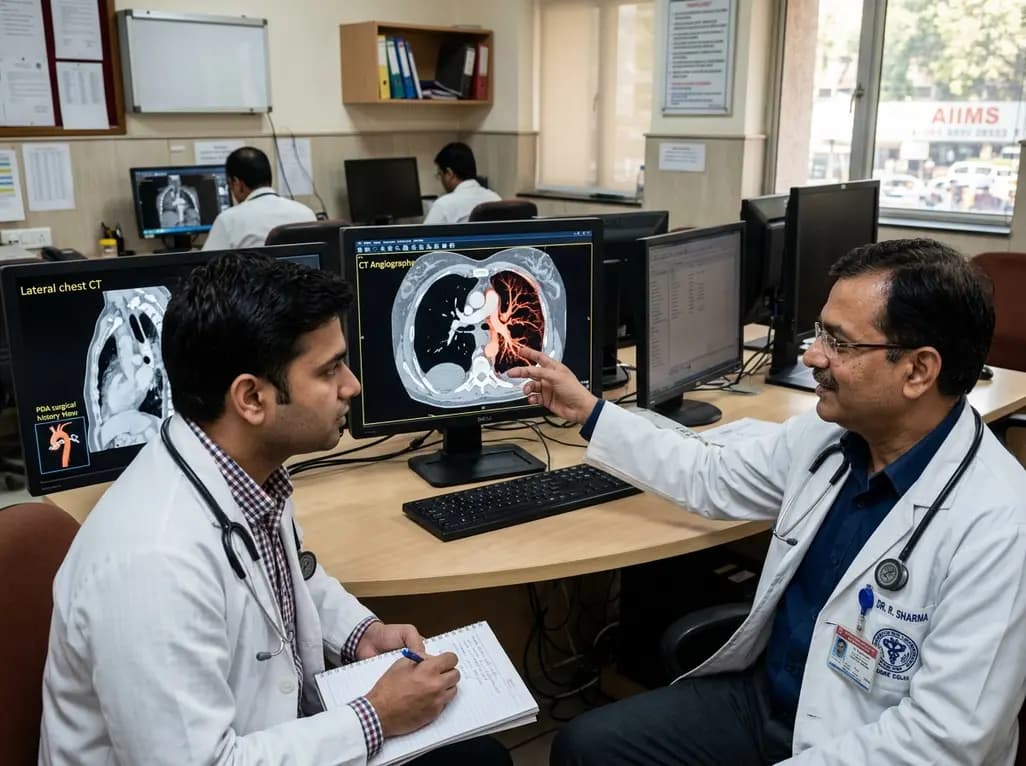
Congenital unilateral absence of the pulmonary artery can remain undetected until adulthood. This case report examines a young male presenting with hemoptysis after prior PDA surgery, highlighting the critical role of CT angiography in mapping systemic collaterals and guiding appropriate clinical intervention.
Today

Perimenopausal obstructive sleep apnea often goes undetected due to atypical polysomnographic presentations and symptom overlap with menopause. This review details its neurocognitive mechanisms, polysomnography markers, and precision management strategies.
Today

Mitochondrial deubiquitinases regulate organelle turnover by editing ubiquitin tags on damaged mitochondria. Discover how targeting these enzymes, notably USP30, offers therapeutic strategies for neurodegenerative, cardiovascular, and metabolic disorders.
Today

A rare case of a pediatric pathological femoral fracture caused by occult telangiectatic osteosarcoma demonstrates the risks of misdiagnosis after trauma, where intramedullary nailing prompted iatrogenic tumor dissemination and required modular total endoprosthetic reconstruction.
Today
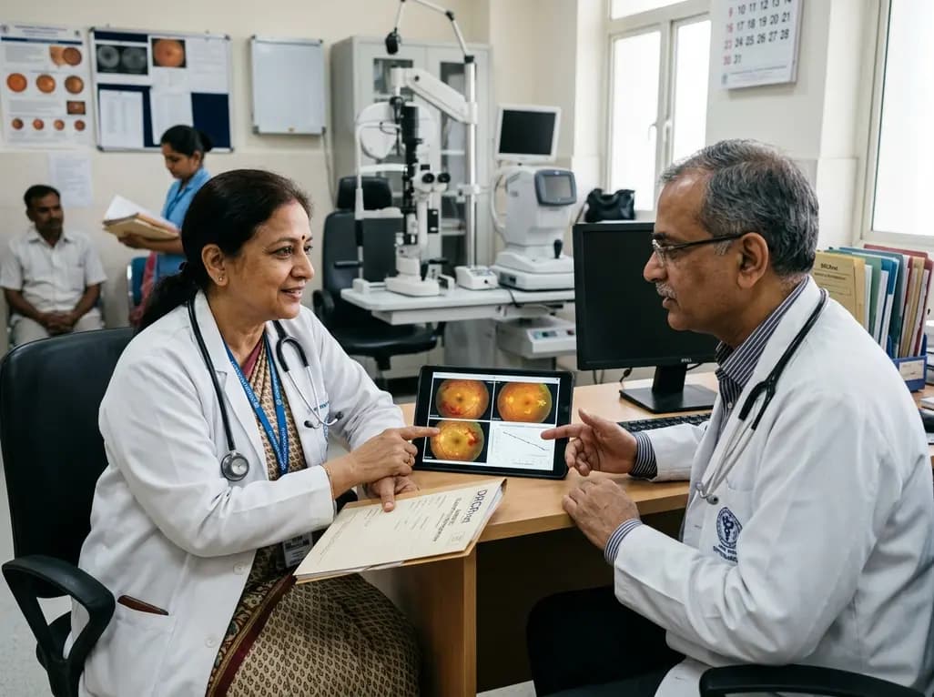
A comprehensive analysis of DRCRnet studies reveals that 15.1% of participants discontinue diabetic retinopathy clinical trials. Identifying baseline demographic and clinical predictors helps ophthalmologists and endocrinologists mitigate real-world treatment dropouts and protect long-term visual outcomes.
Today
