.webp&w=384&q=75)

Loading, please wait...
Loading, please wait...

Status epilepticus represents a critical medical emergency that demands rapid seizure termination and immediate diagnostic precision. Clinicians frequently order extensive diagnostic panels, yet the yield of individual tests has remained poorly defined. Recent prospective evidence from the multicenter ICTAL registry now clarifies the precise role of status epilepticus etiologic investigations. By establishing pretest probability strata, intensive care specialists can identify underlying pathologies faster, minimize unnecessary testing, and implement targeted therapies without delay.
The prospective ICTAL registry evaluated 1,123 critically ill adult patients across 26 intensive care units in France between 2018 and 2025. Overall, motor-predominant seizures occurred in 90% of cases, and the median patient age was 60 years. Critically, investigators assessed a structured diagnostic protocol comprising neuroimaging, routine blood chemistry, lumbar puncture, toxicology screening, antiseizure medication assays, and autoimmune or paraneoplastic panels. Rather than viewing diagnostic testing through an unselected lens, the researchers analyzed each modality against pretest clinical probabilities. Consequently, this stratified method revealed that diagnostic yield depends heavily on clinical suspicion at admission. First-line investigations confirmed suspected etiologies in 8.5% to 44.5% of high pretest probability presentations. Furthermore, these tests reclassified etiologies in 22.1% to 35.1% of patients. Therefore, clinical history and bedside evaluations provide vital context before initiating broad laboratory and imaging tests. When clinicians align diagnostic ordering with clinical likelihood, they optimize resource utilization in intensive care settings. Conversely, indiscriminate testing without clinical stratification often creates diagnostic ambiguity and delays targeted therapy.
The ICTAL study highlights substantial variation in confirmatory yield among standard diagnostic modalities. Specifically, toxicology screening achieved the highest confirmation rate, verifying alcohol, toxic, or iatrogenic triggers in 44.5% of suspected cases. Similarly, antiseizure medication assays confirmed precipitating subtherapeutic levels in 44.0% of patients with known epilepsy. Meanwhile, specialized paraneoplastic and autoimmune encephalitis panels confirmed diagnoses in 40.0% of patients with suspected noninfectious encephalitis. Emergency neuroimaging also demonstrated strong diagnostic utility. Non-contrast brain computed tomography confirmed acute pathology in 28.0% of vascular presentations and 27.7% of intracranial tumor presentations. In contrast, standard laboratory investigations confirmed metabolic derangements in 21.4% of patients with suspected metabolic encephalopathies. Lumbar puncture confirmed central nervous system infections in 17.2% of patients with suspected meningitis or meningoencephalitis. Therefore, these findings emphasize that specific tests excel in distinct clinical phenotypes. For instance, therapeutic drug monitoring and toxicological analyses deliver rapid answers for acute toxic or adherence-related crises. Consequently, critical care teams should prioritize these high-yield assays during initial patient stabilization.
Beyond confirming initial impressions, etiologic workups frequently uncover unexpected diagnoses that redefine clinical care. In this registry, brain magnetic resonance imaging served as the most powerful tool for diagnostic reclassification. Notably, brain MRI reclassified between 27.8% and 35.1% of patients across infective, metabolic, encephalitic, and remote sequelae strata. The highest reclassification rate occurred in suspected noninfectious encephalitis, where MRI revised the diagnosis in 35.1% of cases. Brain computed tomography also demonstrated meaningful reclassification, altering diagnostic categories in 22.1% to 27.9% of cases. Additionally, toxicology screening reclassified 32.4% of patients who initially presented with presumed metabolic decompensation. Because clinical presentations of encephalopathy and toxic exposures overlap significantly, toxicological evaluations often uncover occult ingestions. Furthermore, advanced neuroimaging regularly identifies subtle cortical dysplasia, focal ischemia, or inflammatory parenchymal changes missed on preliminary non-contrast computed tomography. As a result, early magnetic resonance imaging provides critical diagnostic clarity when the primary cause remains uncertain after preliminary bedside evaluations.
Diagnostic investigations are most valuable when their findings directly modify acute treatment plans. In the ICTAL registry, test results led directly to crucial management changes across multiple etiologic strata. Specifically, brain MRI triggered direct therapeutic adjustments in up to 58.3% of evaluated patients. Brain computed tomography altered therapeutic management in up to 56.9% of acute cases, primarily by directing neurosurgical consultation or secondary stroke prevention. Meanwhile, cerebrospinal fluid analysis via lumbar puncture prompted therapeutic modifications in 39.4% of patients, primarily guiding antimicrobial regimens. In addition, antiseizure medication assays modified therapeutic regimens in up to 35.1% of patients through dose adjustments or reloading re-administration. Paraneoplastic and autoimmune antibody testing prompted significant treatment changes in 33.3% of patients, frequently initiating immunotherapy or prompt malignancy screening. Consequently, these investigations do not merely assign retrospective diagnostic labels. Instead, they actively steer real-time clinical decisions, optimize pharmacotherapy, and improve resuscitation pathways in the intensive care unit.
These registry findings offer practical guidance for clinicians managing status epilepticus in Indian hospitals. In tertiary emergency rooms and medical intensive care units across India, resource availability and financial constraints often govern diagnostic choices. Consequently, adopting a probability-stratified diagnostic algorithm ensures cost-effective and clinically robust outcomes. Clinicians should immediately perform point-of-care metabolic testing, review antiseizure medication adherence, and evaluate toxic exposures before pursuing exhaustive laboratory panels. When patients exhibit refractory status or atypical focal features, clinicians must secure early neuroimaging. While computed tomography remains widely accessible for ruling out acute intracranial hemorrhage, brain MRI should be scheduled promptly when initial imaging is non-diagnostic. Furthermore, clinicians must maintain a low threshold for lumbar puncture whenever infectious or autoimmune encephalitis remains possible. Given the high prevalence of neuroinfections in India, stratified workups protect patients from empirical treatment delays. Moreover, this rational approach preserves critical intensive care resources.
Pretest clinical probability prevents indiscriminate testing and sharpens diagnostic accuracy during acute neurologic emergencies. In the ICTAL registry, first-line investigations confirmed suspected causes in up to 44.5% of targeted cases while reclassifying over 30%. Therefore, estimating pretest probability allows intensive care teams to prioritize high-yield tests, such as toxicology screening or drug assays, avoiding costly diagnostic delays and ensuring rapid therapeutic intervention.
Neuroimaging and targeted laboratory panels produce the highest frequency of acute management changes. Specifically, brain magnetic resonance imaging alters therapeutic management in up to 58.3% of cases, closely followed by brain computed tomography at 56.9%. Lumbar puncture directly influences management in 39.4% of patients by guiding antimicrobial selection. Additionally, antiseizure medication assays prompt dosage revisions or reloading in approximately 35.1% of tested patients.
While non-contrast brain CT quickly rules out gross hemorrhage or large structural lesions, brain MRI provides superior resolution for subtle pathologies. In the ICTAL cohort, brain MRI reclassified diagnoses in up to 35.1% of patients, particularly those with suspected noninfectious encephalitis or CNS infections. Consequently, MRI unmasks cortical dysplasia, early cytotoxic edema, subtle limbic inflammation, and ischemic territory involvement that computed tomography frequently misses.
Disclaimer: This content is for informational and educational purposes only... Refer to the latest local and national guidelines for clinical practice.
References

Read summarized clinical updates, watch expert medical content, and earn CME certifications right from your smartphone.
Open your smartphone camera to scan and install.





A prospective study from the ICTAL registry reveals how pretest probability dictates the diagnostic yield and therapeutic impact of investigations in status epilepticus. Neuroimaging, toxicology, and drug levels drove critical management changes in intensive care units.
Today
A real-world quasi-experimental study reveals that web-based apps fostering healthy habit formation significantly boost physical activity and dietary quality in adults. Discover key clinical insights on digital adherence, dose-response effects, and patient-specific modifiers in preventive medicine.
Today

A recent longitudinal study evaluated the link between prenatal loss of control eating and cardiovascular health using Life's Essential 8 frameworks. While direct associations across pregnancy were non-significant, the high prevalence of dysregulated eating highlights critical implications for obstetric practice.
Today

A cross-sectional analysis of NHANES 2013-2020 reveals that depressive symptoms and sleep disturbance significantly elevate the odds of cardiometabolic multimorbidity in adults with arthritis. Comprehensive clinical screening and integrated multi-organ interventions are vital to reduce overall cardiovascular burden.
Today
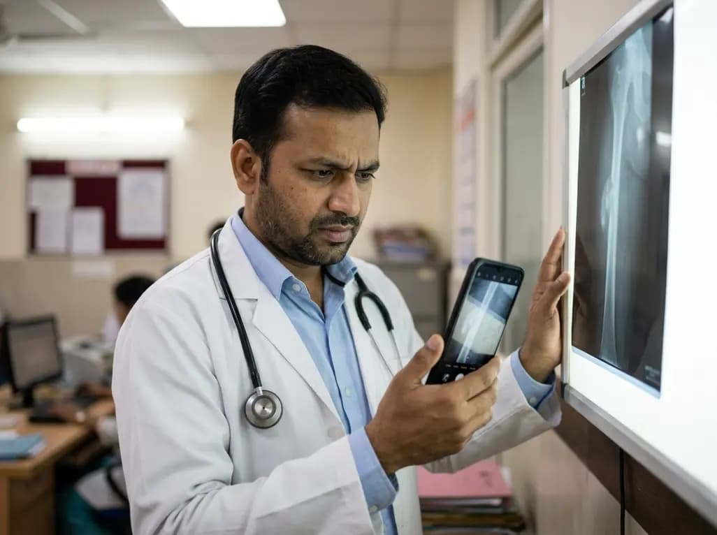
A retrospective study evaluating a locally deployed vision language model (Qwen3-VL) reveals that smartphone-captured radiographs and retrieval-augmented generation can paradoxically reduce diagnostic accuracy in bone tumor referrals due to optical noise and textual hallucinations.
Today
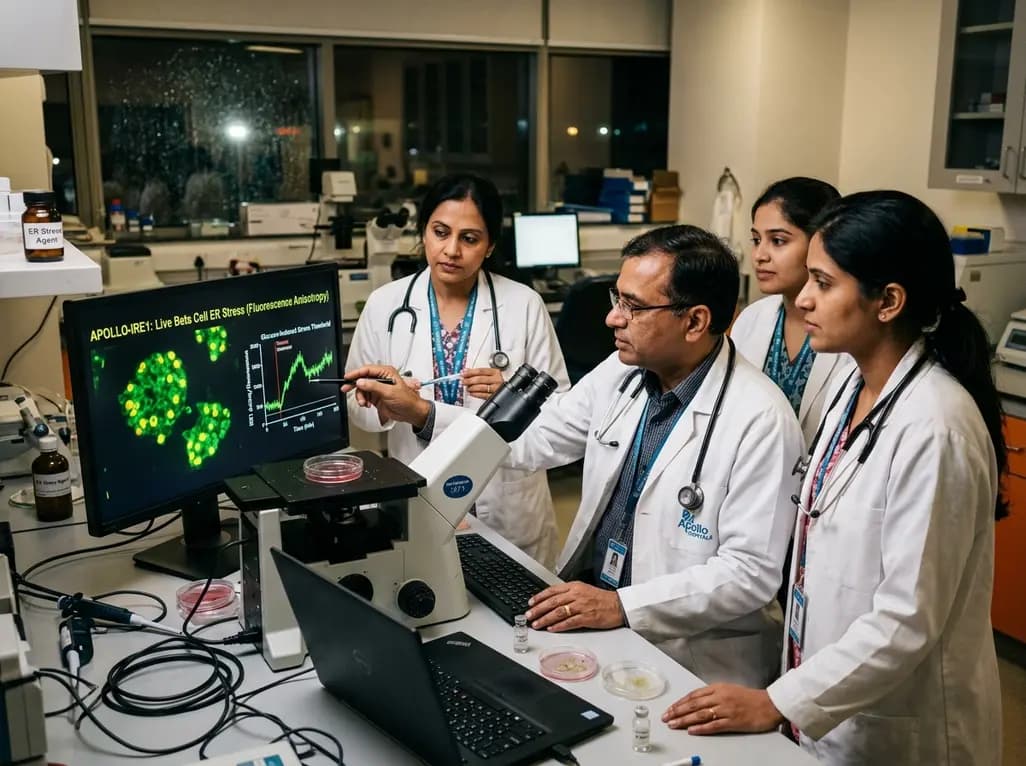
Apollo-IRE1 is a genetically encoded biosensor that monitors real-time endoplasmic reticulum stress dynamics in living pancreatic beta cells. By measuring IRE1 oligomerization through fluorescence anisotropy, it illuminates stress thresholds critical for understanding and treating diabetes.
Today
