.webp&w=384&q=75)

Loading, please wait...
Loading, please wait...

Modern interventional medicine increasingly relies on minimally invasive diagnostic techniques. Consequently, clinicians frequently collect concurrent fine-needle aspiration and core needle biopsy specimens during a single clinical encounter. However, traditional diagnostic workflows process these samples through separate laboratory pipelines. This procedural fragmentation frequently yields conflicting descriptive narratives and delayed therapeutic decisions. Therefore, standardizing small biopsy reporting using structured cytology frameworks has emerged as a critical imperative. This unified approach bridges communicative gaps, streamlines pathological evaluation, and directly aligns diagnostic precision with oncological patient care.
Interventional radiologists and pulmonologists routinely sample lesions using simultaneous fine-needle aspiration and core biopsies. For example, endobronchial ultrasound procedures and image-guided abdominal interventions generate dual tissue streams. Despite originating from the identical lesion, these specimens travel along distinct departmental tracks. Surgical pathologists evaluate core needle biopsies, whereas cytopathologists examine aspiration smears and cell blocks.
Consequently, clinicians receive two separate diagnostic documents with differing terminology. While the core biopsy report might describe nonspecific reactive atypia, the companion cytology report might designate the lesion suspicious for malignancy. This divergence generates significant clinical ambiguity. Furthermore, treating oncologists must reconcile asynchronous turnaround times and contradictory diagnostic thresholds. As a result, patient management faces preventable treatment delays and unnecessary repeat procedures. In resource-strained healthcare systems, such discrepancies strain clinical multidisciplinary tumor boards. Pathologists must therefore establish collaborative reporting protocols that unify concurrent specimens under one interpretive umbrella.
Over the past two decades, cytopathology has revolutionized diagnostic consistency through risk-stratified classification frameworks. Specifically, systems such as Bethesda for cervical and thyroid lesions, Paris for urinary specimens, and Milan for salivary glands established clear diagnostic categories. Each category links directly to an evidence-based risk of malignancy. Furthermore, these standardized systems provide clinicians with explicit clinical management recommendations.
In contrast to descriptive narratives, categorized cytology reports provide reproducible prognostic benchmarks. Clinicians understand immediately whether an atypical categorization mandates molecular profiling, active surveillance, or immediate surgical excision. Additionally, the World Health Organization and International Academy of Cytology expanded universal reporting schemes to pulmonary, pancreaticobiliary, and soft tissue specimens. These developments dramatically reduced inter-observer variability among pathologists globally. Consequently, structured cytology frameworks transformed ambiguous cellular descriptions into actionable clinical guidelines. Modern histologic diagnostics must now adopt these proven structural principles to achieve similar communicative clarity.
Although core needle biopsies preserve tissue architecture, small biopsy reporting remains plagued by non-standardized terminology. Pathologists frequently generate lengthy, free-text histological descriptions that lack definitive risk stratification. Consequently, oncologists encounter varied phrases like atypical proliferations, borderline features, or indeterminate lesions without uniform risk estimates.
To resolve this communication gap, pathologists propose adapting cytology-derived classification tiers to small tissue specimens. By establishing parallel diagnostic tiers, pathologists can assign calibrated malignancy risks to architectural and cytologic patterns. Moreover, adopting structured reporting templates eliminates subjective narrative discrepancies between institutional laboratories. This structural alignment ensures that histological findings correlate directly with companion aspiration smears. Therefore, clinicians receive uniform risk assessments regardless of whether diagnostic material comes from micro-cores or cell pellets. Ultimately, standardized reporting terminology fosters reproducible clinical research, multicenter clinical trials, and seamless data exchange across international healthcare networks.
The central philosophy of modern tissue triage revolves around the "one procedure-one reporting system" framework. Under this integrated model, pathologists review all concurrent specimens from a single clinical intervention together. Instead of distributing multiple standalone reports, the laboratory issues a single, comprehensive diagnostic document.
First, this unified document presents a consolidated diagnostic summary that synthesizes cytology and core histology. Pathologists cross-correlate architectural patterns in the core biopsy with cellular nuances in cytologic smears. Consequently, any diagnostic discordance undergoes immediate internal resolution before clinicians receive the final verdict. Additionally, unified reporting streamlines ancillary diagnostic testing, including immunohistochemistry and next-generation sequencing. By triaging cytologic smears, cell blocks, and core tissue concurrently, laboratories conserve limited tumor material efficiently. Furthermore, oncologists obtain a definitive, multi-parameter assessment without waiting for staggered individual reports. This consolidated workflow accelerates therapeutic initiation and enhances trust between diagnostic specialists and treating physicians.
Implementing a unified reporting system across high-volume pathology laboratories presents distinct operational and technical challenges. First, traditional departmental divisions separate surgical pathology from cytopathology divisions in large academic centers. Pathologists operate under different scheduling rosters, workstation locations, and administrative oversight. Consequently, cross-subspecialty collaboration requires restructuring daily laboratory workflows.
Second, specimen processing timelines differ fundamentally between tissue modalities. Rapid cytology smears often yield same-day diagnoses, whereas formalin-fixed paraffin-embedded core biopsies require prolonged fixation and sectioning. Therefore, holding cytology results until histology finishes could potentially delay preliminary clinical communications. To overcome this limitation, institutions can issue standardized interim communications while maintaining an integrated final document. Additionally, laboratory information systems must adapt to allow multi-user accessioning, dual-sign-out protocols, and synchronized digital image integration. Although these technical adjustments demand institutional investment, the clinical benefits far outweigh the initial operational disruption.
In Indian clinical environments, standardizing biopsy reporting holds profound value for patient care and resource optimization. Tertiary cancer centers and corporate healthcare networks across India increasingly handle massive volumes of complex oncological cases. Furthermore, many patients travel extensive distances across state borders to consult regional cancer specialists.
When such patients carry fragmented pathology reports with ambiguous descriptions, oncologists must frequently repeat biopsies. Consequently, repeat invasive sampling increases healthcare costs, complications, and emotional distress for Indian families. Implementing unified, risk-stratified reporting protocols prevents redundant diagnostic testing. Moreover, structured reporting formats facilitate accurate communication during multidisciplinary tumor board discussions in teaching hospitals. For tier-two and tier-three diagnostic centers, adhering to universal cytology-aligned criteria elevates diagnostic reliability. Ultimately, adopting integrated small tissue reporting strengthens telepathology consultations and nationwide cancer registries across India.
Separate reporting generates fragmented diagnostic information that confuses clinical oncologists. When cytology and core needle biopsy specimens from the same lesion travel through separate workflows, reports often contain discordant terminology or asynchronous release dates. Consequently, treating physicians face diagnostic ambiguity regarding malignancy risk. An integrated report reconciles these discrepancies internally, ensuring a cohesive clinical recommendation and eliminating treatment delays for patients undergoing cancer therapy.
Cytopathology established successful tiered reporting systems that link defined diagnostic categories directly to evidence-based malignancy risks. Systems like Bethesda and Milan provide clear clinical management algorithms. By applying these standardized tiers to small biopsies, pathologists eliminate ambiguous descriptive narratives. Furthermore, this approach harmonizes architectural tissue interpretation with cytologic findings, providing oncologists with predictable diagnostic certainty and standardized criteria for molecular biomarker testing across diverse institutional settings.
Laboratories overcome disparate processing timelines by establishing structured two-stage reporting protocols within their laboratory information systems. Pathologists can issue a rapid preliminary cytology evaluation for immediate clinical triage, especially following on-site evaluations. Meanwhile, the final definitive report merges complete histologic, immunohistochemical, and molecular data into one unified diagnostic record. Consequently, clinicians receive essential immediate updates without losing the diagnostic synthesis necessary for final therapeutic decision-making.
Disclaimer: This content is for informational and educational purposes only and should not be considered medical advice. Always consult a qualified healthcare professional regarding any medical condition or treatment. Refer to the latest local and national guidelines for clinical practice.
References

Read summarized clinical updates, watch expert medical content, and earn CME certifications right from your smartphone.
Open your smartphone camera to scan and install.





Standardized small biopsy reporting bridges the diagnostic gap between concurrent cytology and histology specimens. Adopting a unified, risk-stratified reporting framework enhances diagnostic clarity, prevents miscommunication, and streamlines clinical decision-making in oncology.
Today
A recent clinical case series highlights the synthetic polylactic acid matrix as an innovative local therapy for refractory pyoderma gangrenosum. The bioresorbable scaffold demonstrated complete ulcer closure and favorable scar quality across chronic wounds, presenting a viable regenerative modality.
Today

A nationwide South Korean study shows underreporting of smoking among women dropped from 57.4% to 36.4% between 2008 and 2021. However, over one-third of female smokers still conceal their habit, highlighting the critical role of cotinine verification in clinical risk assessment.
Today
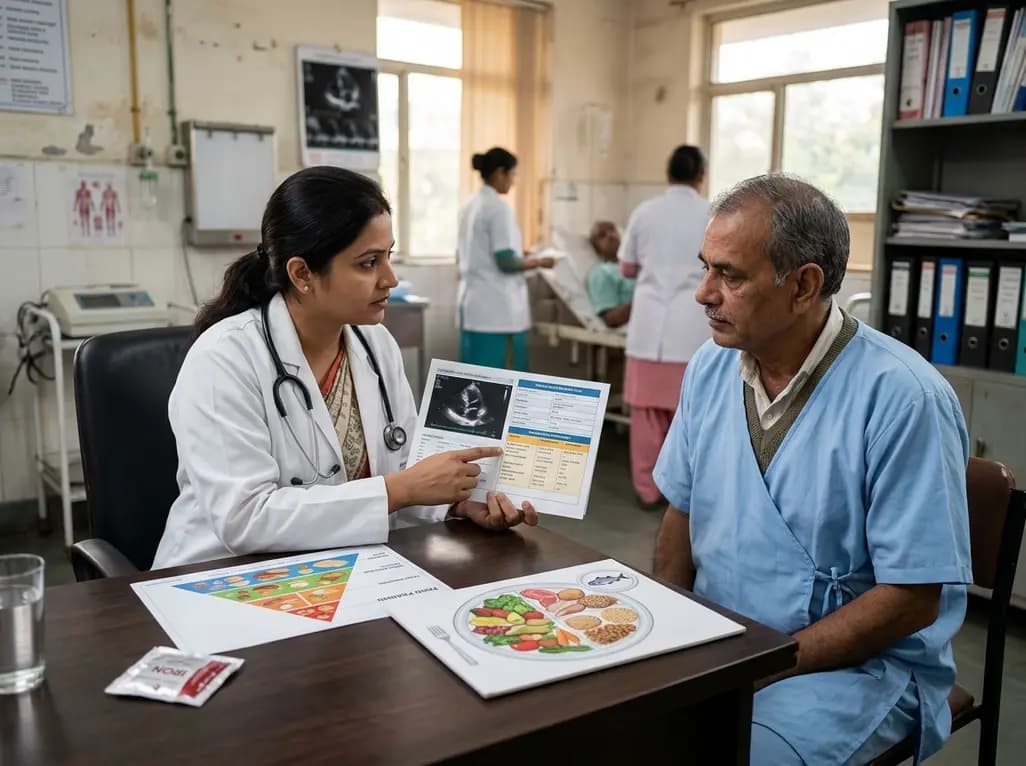
A scientific statement highlights the crucial role of nutrition in managing chronic heart failure. It emphasizes GLIM-based screening, individualized dietary patterns like the Mediterranean diet, appropriate protein and energy intake, avoiding unnecessary fluid restriction, and targeted iron supplementation.
Today

A new study evaluates the rheological stability of decellularized peripheral nerve matrix hydrogels under varied storage conditions. Findings show -80°C storage and freeze-drying preserve gelation kinetics and mechanical stiffness, offering key insights for shipping and translational clinical applications.
Today

A qualitative meta-synthesis highlights the complex interplay between gender dysphoria, minority stress, and eating disorders in transgender and gender-diverse individuals. Clinicians must recognize these distinct drivers to offer supportive, gender-affirming, and interdisciplinary eating disorder care.
Today
