.webp&w=384&q=75)

Loading, please wait...
Loading, please wait...

The human brain relies on intricate fluid mechanisms to eliminate neurotoxic metabolic waste accumulated during wakefulness. Recent neuroimaging discoveries highlight the critical role of glymphatic clearance dynamics in preserving cognitive longevity. The glymphatic system functions as a macroscopic waste clearance pathway, facilitating cerebrospinal fluid influx along periarterial spaces and interstitial fluid efflux. Although animal models show that this restorative exchange surges during sleep, human diurnal variability remains less understood. Consequently, understanding how daily sleep quantity modulates macroscopic fluid shifts offers urgent diagnostic value.
Synchronized slow-wave oscillations during deep non-rapid eye movement sleep expand the cerebral interstitial space. Consequently, this morphological transition significantly reduces hydraulic tissue resistance. Therefore, physiological sleep enables brisk convective cerebrospinal fluid flux throughout the brain parenchyma. In addition, uninterrupted rest facilitates the clearance of neurotoxic proteins, such as soluble amyloid-beta and tau. In contrast, sustained wakefulness suppresses this convective perivascular exchange. Specialized astrocytic water channels, designated as aquaporin-4, govern these microscopic fluid shifts. However, chronic sleep restriction compromises aquaporin-4 polarization, thereby impairing parenchymal waste elimination. Moreover, modern lifestyle patterns frequently disrupt natural diurnal cycles, misaligning peripheral tissue rhythms with the master circadian pacemaker. As a result, chronic sleep deprivation triggers prolonged neurofluid stagnation. Furthermore, persistent fluid stagnation promotes microglial activation, persistent neuroinflammation, and progressive synaptic degradation over time. Physicians must recognize that adequate sleep duration establishes the biological foundation for continuous metabolic detoxification.
To elucidate how habitual rest influences human glymphatic performance, investigators conducted an observational neuroimaging study. Specifically, researchers enrolled twenty-five healthy young adults with a mean age of 26.5 years. The protocol required each participant to complete three comprehensive multimodal magnetic resonance imaging sessions within a single day. Investigators acquired high-resolution scans at 8 a.m., 2 p.m., and 8 p.m. to track dynamic diurnal fluctuations across distinct compartments. Furthermore, the protocol stratified participants into two cohorts based on objective sleep duration. Long sleepers logged at least 404 minutes of habitual rest, whereas short sleepers obtained less than 404 minutes. Investigators employed advanced neuroimaging metrics, including diffusion tensor imaging, free-water volume estimation, and myelin water fraction mapping. These precise parameters captured subtle, time-dependent shifts in white matter microstructure and parenchymal fluid mobility. In addition, the researchers quantified volume adjustments across cerebrospinal fluid spaces. Thus, serial non-invasive imaging effectively mapped macroscopic fluid redistribution across twenty-four hours.
The study revealed striking physiological divergences between the long and short sleep cohorts. Long sleepers demonstrated marked diurnal adaptability, characterized by robust shifts in fluid compartments across daytime hours. Specifically, individuals sleeping at least 404 minutes exhibited daytime expansions in ventricular cerebrospinal fluid volume and gray matter free water. Concurrently, these well-rested participants showed compensatory volume reductions within cerebral white matter tracts. This reciprocal pattern reflects highly flexible cerebrospinal fluid-tissue coupling, wherein fluid shifts smoothly without mechanical resistance. Conversely, participants in the short sleep group exhibited a remarkably rigid coupling profile. Their scans demonstrated minimal diurnal variation in free-water fractions alongside rigid fluid-tissue interactions. Furthermore, shortened sleep restricted fluid movement, preventing compensatory volume accommodations across the day. Therefore, inadequate sleep duration visibly reduces the physical elasticity of neurofluid compartments. This structural rigidity likely hinders convective waste transport. Ultimately, adequate habitual sleep ensures that cerebral fluid compartments adapt seamlessly to changing physiological demands.
Beyond fluid elasticity, the investigation uncovered profound differences in diurnal synchronization between cohorts. Long sleepers demonstrated harmonious diurnal rhythms, with multiple neurofluid markers peaking synchronously during the late evening at 8 p.m. This temporal alignment indicates that adequate sleep maintains circadian synchronization of fluid dynamics, preparing the brain for intensive nocturnal clearance. In stark contrast, individuals with shorter habitual sleep displayed fragmented, desynchronized temporal patterns. Their diffusion metrics, free-water fractions, and volumetric indices peaked at discordant hours throughout the day. Consequently, their fluid compartments operated out of phase with intrinsic circadian timing mechanisms. Such chronobiological desynchronization severely undermines glymphatic efficiency. Importantly, the master circadian clock regulates daily oscillations in aquaporin-4 distribution and vascular tone. When chronic sleep deficit disrupts this temporal orchestration, perivascular channels fail to mobilize fluids efficiently. Thus, short sleep impairs both fluid volume and temporal coordination. Maintaining strict circadian synchrony ensures that the glymphatic apparatus mounts an organized, highly effective cleansing wave every night.
These physiological insights carry transformative implications for modern preventative medicine and clinical neurology. Historically, clinicians viewed sleep disturbances merely as secondary symptoms of Alzheimer’s disease and vascular dementia. However, mounting evidence indicates that disrupted sleep actively accelerates neuropathology decades before cognitive symptoms emerge. Rigid fluid-tissue coupling and circadian desynchronization permit toxic oligomers to accumulate around synapses. Over time, impaired clearance fosters cerebral amyloid angiopathy, promotes neurofibrillary tangles, and exacerbates blood-brain barrier breakdown. Furthermore, persistent interstitial stasis triggers microglial activation and oxidative stress, compounding neuronal loss in vulnerable memory centers. Therefore, addressing habitual sleep duration represents a critical, modifiable target to protect cognitive health. Medical practitioners should systematically screen adults for chronic sleep deprivation, shift work, and underlying sleep apnea during routine evaluations. Additionally, clinicians should advise patients to achieve consistent sleep durations exceeding seven hours nightly. By championing comprehensive sleep hygiene, physicians help patients preserve flexible fluid dynamics and lifelong cognitive resilience.
Sleep deprivation restricts the expansion of interstitial space, drastically increasing hydraulic resistance across neural tissue. Consequently, convective cerebrospinal fluid flux decreases, preventing efficient elimination of metabolic waste like amyloid-beta. Over time, chronic deprivation stiffens fluid-tissue coupling, causing toxic protein stagnation and heightened neuroinflammation.
Clinicians and researchers utilize advanced multimodal magnetic resonance imaging to assess glymphatic activity non-invasively. Key sequences include diffusion tensor imaging along the perivascular space, free-water volume fraction mapping, and myelin water imaging. These modalities capture subtle diurnal shifts in fluid distribution and tissue elasticity without intrathecal contrast.
Yes, adopting consistent sleep schedules and healthy circadian habits directly enhances glymphatic synchronization. Maintaining fixed bedtimes, securing at least seven hours of nightly rest, minimizing evening light exposure, and managing hypertension preserve arterial pulsatility and aquaporin-4 channel polarization, ensuring optimal waste clearance each night.
Disclaimer: This content is for informational and educational purposes only. It is not intended to provide medical advice, diagnosis, or treatment. Always seek the advice of your healthcare provider with any questions you may have regarding a medical condition. Refer to the latest local and national guidelines for clinical practice.
References

Read summarized clinical updates, watch expert medical content, and earn CME certifications right from your smartphone.
Open your smartphone camera to scan and install.





A breakthrough observational study reveals that longer habitual sleep duration promotes flexible CSF-tissue coupling and synchronized diurnal glymphatic clearance dynamics, offering vital neuroprotective insights for clinical medicine.
Today
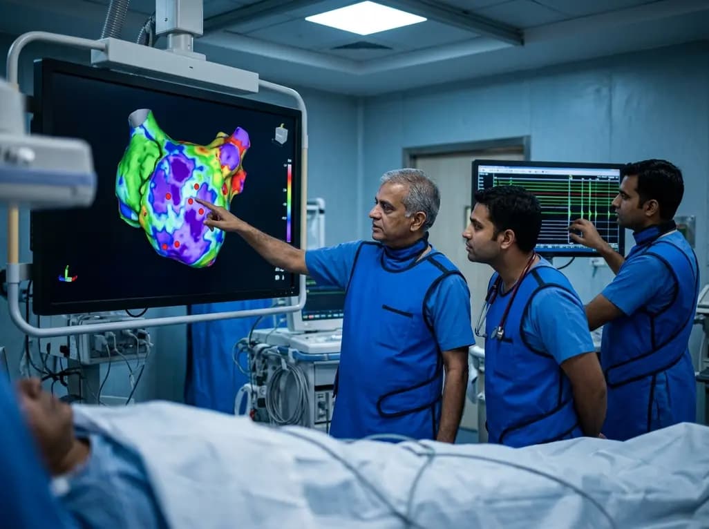
A study of 1,428 AF ablation patients reveals that over 60% of left atrial non-PV triggers arise within low-voltage areas, with anterior and roof triggers colocalizing entirely within fibrotic substrate. Identifying and ablating these sites is critical for long-term arrhythmia freedom.
Today

A comprehensive meta-analysis of over 278,000 women demonstrates that maternal atopic dermatitis significantly increases risks of premature rupture of membranes and low birth weight. Discover essential clinical insights on disease mechanisms, serial antenatal growth monitoring, and safe therapeutic interventions.
Today

A pioneering study highlights the first United States experience using the FARAPOINT focal pulsed-field ablation catheter for refractory ventricular tachycardia. The catheter achieved complete acute non-inducibility across tested patients without procedural complications, marking a milestone in ventricular ablation.
Today

A biomechanical study shows that augmenting thin fascia lata grafts with a reinforced polyester patch doubles ultimate tensile strength and stiffness, matching the performance of thick grafts for superior capsule reconstruction while reducing donor-site morbidity.
Today

A cross-sectional study reveals that elevated METS-IR is independently associated with higher myopia odds in adolescents, showing biological synergy with obesity and highlighting the need for metabolic screening in pediatric eye care.
Today
